Journal List > Blood Res > v.58(1) > 1516082118

This article has been corrected. See "Erratum" in Volume 58 on page 124.
Song, Kang, Lee, Ryu, Lee, Yun, and Jo: Acquired von willebrand syndrome in patients with Philadelphia-negative myeloproliferative neoplasm

Abstract

Background

Acquired von Willebrand syndrome (AVWS) has not been investigated in Korean patients with Philadelphia chromosome-negative myeloproliferative neoplasm.

Methods

This study analyzed the prevalence at diagnosis and clinical features of AVWS in patients with essential thrombocythemia (ET), polycythemia vera (PV), prefibrotic/early primary myelofibrosis (pre-PMF), or overt PMF (PMF) diagnosed between January 2019 and December 2021 at Chungam National University Hospital, Daejeon, Korea. AVWS was defined as below the lower reference limit (56%) of ristocetin cofactor activity (VWFRCo).

Results

Sixty-four consecutive patients (36 with ET, 17 with PV, 6 with pre-PMF, and 5 with PMF; 30 men and 34 women) with a median age of 67 years (range, 18‒87 yr) were followed for a median of 25.1 months (range, 2.6‒46.4 mo). AVWS was detected in 20 (31.3%) patients at diagnosis and was most frequent in ET patients (41.4%), followed by patients with pre-PMF (33.3%) and PV (17.6%) patients. VWFRCo was negatively correlated with the platelet count (r=0.937; P=0.002). Only one episode of minor bleeding occurred in a patient with ET and AVWS. Younger age (<50 yr) [odds ratio (OR), 7.08; 95% confidence interval (CI), 1.27‒39.48; P=0.026] and thrombocytosis (>600×109/L) (OR, 13.70; 95% CI, 1.35‒138.17; P=0.026) were independent risk factors for developing AVWS.

Conclusion

AVWS based on VWFRCo was common in patients with ET and pre-PMF, but less common in patients with PV in the Korean population. Clinically significant bleeding is rare in these patients.

INTRODUCTION

Philadelphia chromosome-negative myeloproliferative neoplasms (Ph- MPNs) include essential thrombocythemia (ET), polycythemia vera (PV), prefibrotic primary myelofibrosis (pre-PMF) and overt primary myelofibrosis (PMF). These clonal hematologic disorders are characterized by thrombotic vascular events [1] and myelofibrotic and leukemic transformations [2]. In addition to thrombosis, hemorrhagic vascular events often occur in these patients, and some cases are associated with acquired von Willebrand syndrome (AVWS) [3].
Von Willebrand disease (VWD) is a common genetic bleeding disorder that affects men and women equally and often manifests itself as mucosal bleeding. This can be due to quantitative (Types 1 and 3) or qualitative (Type 2) defects in the von Willebrand factor (VWF) [4]. Acquired VWD (AVWD) or AVWS can occur in many settings, such as MPN, plasma cell dyscrasia, other lymphoproliferative disorders, autoimmune disorders, and causes of increased shear forces, such as aortic stenosis or other structural heart diseases and mechanical circulatory support [5]. Depletion of VWF, particularly high molecular weight multimers, can cause mucocutaneous bleeding and arteriovenous malformations, especially in the gastrointestinal tract [5, 6]. Increased platelet count in patients with MPN is believed to be associated with AVWS. This is most frequently reported in patients with ET, but also in patients with PV [5, 7]. Due to the risk of bleeding, many physicians hesitate to prescribe low-dose aspirin in patients with extreme thrombocytosis and AVWS [8].
AVWS has rarely been addressed in Korean patients with Ph- MPN. Only one case of AVWS has been reported in a patient with PV [9]. In 2020, the Korean Society of Hematology revised the guidelines for diagnosing and managing MPN based on published evidence and the experience of an expert panel. However, AVWS was not addressed [10]. Therefore, the prevalence and clinical features of AVWS were based on reports from Western countries. The incidence of MPN is increasing in Korea [11-13], which is mainly attributable to changes in diagnostic criteria and studies on driver gene mutations, so there is a need for information on AVWS in these patients. In this prospective observational study, we analyzed the prevalence at diagnosis and clinical features of AVWS in Ph- MPN patients in a Korean population.

MATERIALS AND METHODS

Patients

Patients diagnosed with ET, PV, pre-PMF, or PMF between January 2019 and December 2021 at the Chungnam National University Hospital, Daejeon, Korea, were enrolled in this study. At the time of diagnosis, the patients underwent tests for the following parameters: complete blood count (CBC), prothrombin time (PT), activated partial thromboplastin time (aPTT), VWF factor VIII-related antigen (VWF:Ag), VWF ristocetin cofactor activity (VWF:RCo), blood chemistry, driver gene mutations and bone marrow examination. In patients with low VWF:RCo at diagnosis, VWF:Ag and VWF:RCo were followed up after initiating cytoreductive treatment at weeks 2 and 6, and then every 1–3 months. The International Prognostic Score for Essential Thrombocythemia (IPSET) [14] and the International Prognostic Scoring System (IPSS) [15] were used for the prognostic stratification of patients with ET and PMF, respectively. Hydroxyurea or anagrelide was used for cytoreduction according to standard recommendations, drug availability, and compliance. Patients with PV patients underwent regular phlebotomy, so their hematocrit was <45%. Low-dose aspirin (100 mg/day) was prescribed to prevent thrombosis, except in low- and very low-risk patients. Based on risk stratification, aspirin was not administered to patients with VWF:RCo <30% who did not require cytoreduction. This study was approved by the Institutional Review Board of the Chungnam National University Hospital.

Definition of AVWS

Based on previous studies on AVWS in patients with MPN [16, 17], AVWS was diagnosed when all four criteria were met: VWF:RCo <56% (lower reference limit), VWF:RCo/VWF:Ag ratio <0.7, no personal or family history of bleeding disorders, and no history of frequent or abnormally prolonged episodes of bleeding. VWF:Ag and VWF:RCo were determined using an enzyme-linked immunosorbent assay and a fixed platelet aggregation assay, respectively (GC Labs, Yongin, Korea).

Definition and classification of splenomegaly

Splenomegaly was defined using previously described criteria [18]. Briefly, 'palpable splenomegaly' indicated that the spleen was palpable below the left costal margin, and 'volumetric splenomegaly' indicates that the volume of the spleen was greater than the mean plus three standard deviations of the reference volumes based on both age and body surface area.

Driver gene mutation analyses

The Janus kinase 2 mutation (JAK2V617F) was identified using a quantitative allele-specific real-time polymerase chain reaction (PCR). A calreticulin (CALR) mutation was detected in exon 9 by fragment analysis and Sanger sequencing. The myeloproliferative leukemia gene mutation (MPLW515K/L) was evaluated using PCR and Sanger sequencing.

Definitions of thrombotic and hemorrhagic events

Thrombotic events included cerebrovascular (ischemic stroke, transient ischemic attack, and venous sinus thrombosis), coronary (any ischemic heart disease, including acute coronary syndrome), splanchnic and peripheral thrombo-embolism. Bleeding was defined based on guidelines proposed by a group of VWD experts [19]. Major bleeding included episodes that required hospital admission, surgical intervention, blood transfusion, hemoglobin drop ≥2 g/dL, bleeding involving critical areas (e.g., intracranial, intraspinal, intraocular, retroperitoneal, intraarticular, pericardial, or intramuscular with compartment syndrome), or recurrent bleeding that affects the ability to attend normal school, work, or social activities. Minor bleeding included episodes other than major bleeding.

Statistical analysis

Descriptive data are presented as means±standard deviations (SD), medians (range), or percentages, and were compared using Student’s t-test, the chi-square test, or Fisher exact test. Correlations between VWF:RCo and various parameters were assessed using Pearson correlation analysis. The risk factors for the development of AVWS were determined using logistic regression analysis. Statistical analyzes were performed using SPSS software (version 24.0; IBM Corp., Armonk, NY, USA). P<0.05 was considered statistically significant.

RESULTS

Patient characteristics

During the study period, 64 patients were diagnosed with MPN. All 64 consecutive patients (36 with ET, 17 with PV, 6 with pre-PMF and 5 with PMF; 30 men and 34 women) with a median age of 67 years (range, 18–87 yr) were enrolled. The patients were followed for a median of 25.1 months (range, 2.6 to 46.4 mo). Palpable splenomegaly was not observed in any of the patients. Volumetric splenomegaly was detected in all patients with pre-PMF and PMF , while it was found in 88.2% of patients with PV and 66.7% of patients with ET. WBC counts and lactate dehydrogenase (LDH) levels of patients with pre-PMF were higher than those of patients with ET [15.9±3.3×10⁹/L and 10.9±4.7×10⁹/L; 2.0±0.9 and 1.3±0.4× upper normal limit (UNL), respectively; all P<0.05]. Monocyte counts and LDH levels in patients with PMF were higher than those of patients with ET (1.4±1.2×10⁹/L and 0.7±0.6×10⁹/L; 1.9±0.3× UNL and 1.3±0.4× UNL, respectively; all P<0.05). The platelet counts of PV patients were lower than those of ET patients (870.3±314.0 and 614.2±381.3×10⁹/L; P =0.013). Platelet counts of patients with pre-PMF and PMF did not differ from those of patients with ET. JAK2V617F was detected more frequently in patients with PV (100%), followed by ET (83.3%), pre-PMF (66.7%), and PMF (60.0%). The patients with ET were evenly distributed among the IPSET risk groups. Cytoreductive therapy was prescribed most frequently for patients with PMF (100%), followed by PV (88.2%), pre-PMF (83.4%) and ET (63.9%). Most of the patients received low-dose aspirin (Table 1).

Prevalence of AVWS at the time of diagnosis

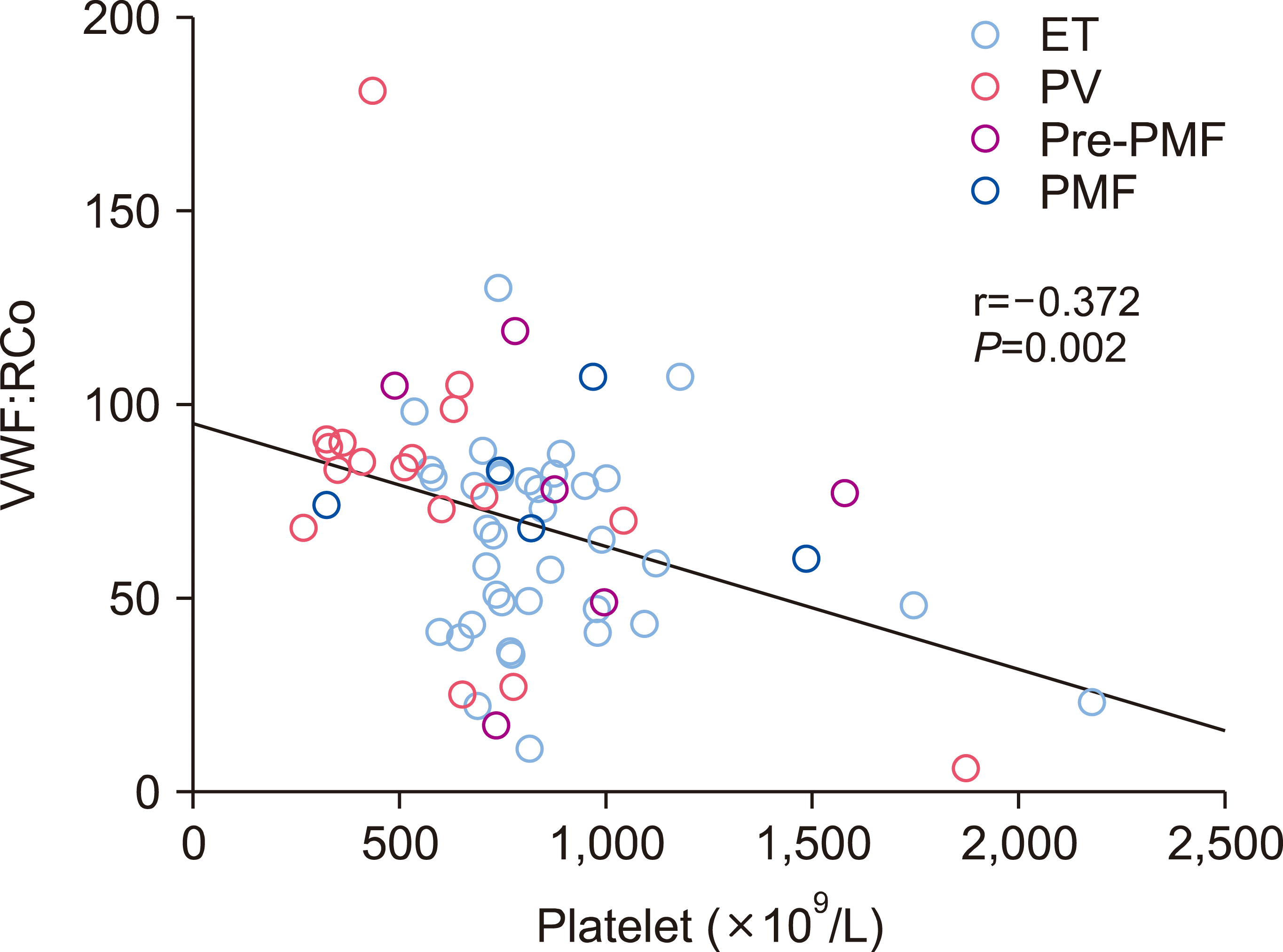
AVWS was detected in 20 (31.3%) of the 64 patients at the time of MPN diagnosis, and was more frequent in patients with ET (41.4%), followed by patients with pre-PMF (33.3%) and PV (17.6%). No AVWS was found in patients with PMF. AVWS with VWF:RCo <30% was found most frequently in patients with PV (17.6%), followed by patients with pre-PMF (16.7%), and ET (8.3%) (Table 2). VWF:RCo was negatively correlated with platelet count (r=-0.372; P=0.002) (Fig. 1). However, VWF:RCo did not correlate with age, WBC count, monocyte count, hemoglobin level, LDH level, ferritin level, splenic volume, JAK2V617F positivity, or allele burden (data not shown).

Clinical characteristics of patients with AVWS

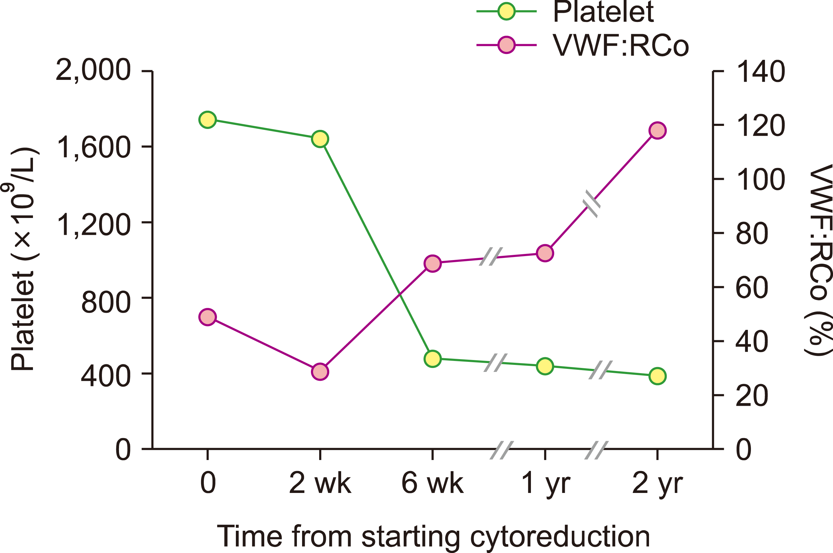
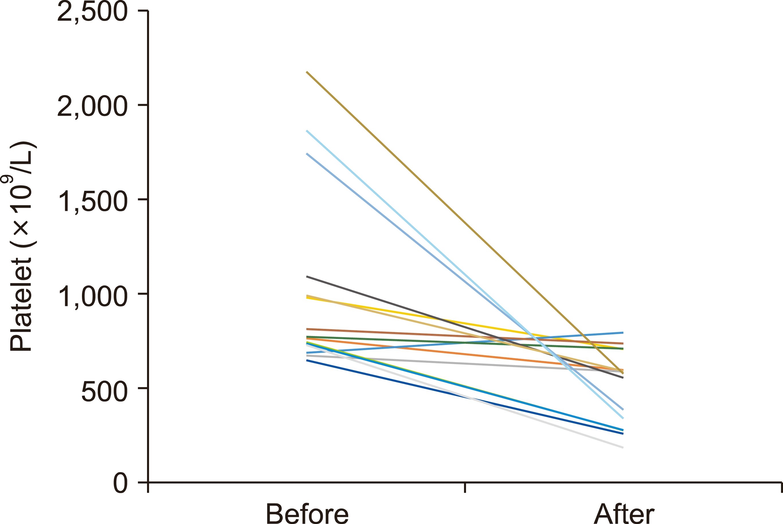
The clinical characteristics at the time of diagnosis and the clinical course of patients with AVWS were compared with patients without AVWS. Patients with ET with AVWS (N=15) were younger than those without AVWS (N=21) [56 (18–83) yr and 65 (40–84) yr; P=0.035]. Volumetric splenomegaly, WBC count, monocyte count, hemoglobin level, LDH, PT, and aPTT did not differ between the two groups. The platelet counts of ET patients with AVWS were higher than those without. However, the difference was not significant (949.7±440.0×10⁹/L and 813.7±170.7×10⁹/L; P=0.205). The positivity for JAK2V617F positivity and the burden of alleles did not differ between the two groups. Thrombotic vascular events were detected in 13.3% of ET patients with AVWS and 19.0% of those without AVWS (P=0.650). No major bleeding was observed in any of the patients. Only one episode of minor bleeding (hematoma at the intramuscular injection site) occurred in a patient with ET and AVWS. Of the 15 patients with ET with AVWS, 11 underwent cytoreductive therapy. VWF:RCo normalized in all 11 patients after a median of 8 weeks (range, 2–18 wk) (Fig. 2, Table 3). Three patients with ET had VWF:RCo <30% at the time of diagnosis. These patients had significantly higher platelet counts than those with VWF:RCo ≥30% (1,228.3±824.8×10⁹/L and 837.8±228.8×10⁹/L; P=0.037) (Table 3). Patients with PV with AVWS (N=3) had significantly higher platelet counts than those without AVWS (N=14) (1,120.7±671.7×10⁹/L and 510.0±206.7×10⁹/L; P=0.009). VWF:RCo normalized after cytoreductive therapy in the three patients in 6–10 weeks. No bleeding episodes were observed in any of the patients with PV (Table 4). The low VWF:RCo in the two patients with pre-PMF normalized after initiating cytoreductive treatment. No bleeding episodes were observed in the patients with pre-PMF (data not shown). Thrombocytosis persisted in many patients when the VWF:RCo ratio was normalized (≥450×109/L in 60.0%; ≥600×109/L in 40.0% of the patients) (Fig. 3).

Risk factors for developing AVWS

Logistic regression analysis was performed to determine risk factors for AVWS in all enrolled patients with MPN. Univariate analysis showed that younger age (<50 yr) [odds ratio (OR), 4.29; 95% confidence interval (CI), 1.05–17.45; P=0.042], ET diagnosis (OR, 3.29; 95% CI, 1.02–10.62; P=0.047) and thrombocytosis (>600×109/L) (OR, 8.87; 95% CI, 1.07–73.03; P=0.043) at the time of diagnosis were risk factors. Multivariate analysis revealed that younger age (OR, 7.08; 95% CI, 1.27–39.48; P=0.026) and thrombocytosis (OR, 13.70; 95% CI, 1.35–138.17; P=0.026) were independent risk factors. The WBC count, hemoglobin level, volumetric splenomegaly, and the JAK2V617F mutation or its allele burden did not affect the development of AVWS (Table 5).

DISCUSSION

This study investigated the frequency of low VWF activity at the time of diagnosis and its clinical relevance in 64 newly diagnosed patients with Ph- MPN. To our knowledge, this is the first study to address Ph- MPN-related AVWS in the Korean population. Ph- MPN are characterized by frequent thrombotic vascular events and, to a lesser degree, bleeding. The clinical features of thrombotic vascular events in Korean patients with Ph- MPN differ somewhat from those in Western cases regarding the time of occurrence and sites involved [1]. Therefore, it is reasonable to obtain information on VWF abnormalities in Korean patients with Ph- MPN. Many previous reports on AVWS include the results of VWF tests not only at the time of MPN diagnosis, but also during follow-up [16, 17, 20]. Cytoreductive treatment strongly affects VWF activity [21]; therefore, testing VWF after treatment initiation could lead to an erroneous prevalence rate of AVWS. The present study enrolled only newly diagnosed patients to determine the exact prevalence of AVWS. AVWS was detected in 41.7% of patients with ET and 17.6% of patients with PV, and clinically significant bleeding was rarely observed. Individuals with type O generally have lower plasma levels of VWF than those with other blood types [22]. The present study did not incorporate blood type into defining AVWS; therefore, the prevalence of AVWS may have been overestimated. Estimates of AVWS in Western studies are 20% to 70% in ET and 12% to 30% in patients with PV according to laboratory criteria, but clinically significant bleeding is much less common, at 4% to 7% in some studies, affecting less than half of those with a laboratory diagnosis [5, 7]. Therefore, the prevalence of AVWS and bleeding in Korean patients did not differ from those in cases of Western patients. AVWS has seldom been reported in patients with PMF. We also did not diagnose AVWS in any of the five patients with PMF enrolled in the study. However, our data cannot be considered conclusive due to the limited number of patients.
We found the abnormal VWF:RCo ratio normalized after starting cytoreductive treatment in all patients, although platelet count remained high. This correction of VWF:RCo after cytoreduction could be a reason why bleeding was uncommon, as most of the patients with ET and PV in this study were placed on cytoreductive treatment according to standard risk stratification. Most reports of major bleeding in patients with MPN with AVWS are based on anecdotes, mainly in patients with unrecognized AVWS [23-26]. These observations indicate that most hemorrhagic events induced by AVWS occur before or at the time of MPN diagnosis and that proper cytoreduction can minimize the risk of bleeding. Taken together, it remains to be determined whether AVWS screening should be performed in all patients with Ph- MPN at the time of diagnosis.
A subpopulation of patients with ET present with a platelet count ≥1,000×109/L, which is arbitrarily called extreme thrombocytosis [27]. Extreme thrombocytosis is believed to be a prerequisite for AVWS [28]. Bleeding diathesis in ET or PV is currently believed to be multifactorial in etiology [29]. AVWS in patients with ET or PV is characterized by the loss of large VWF multimers, related to their increased adsorption to platelets and thus increased proteolysis by ADAMTS13 in a platelet count-dependent manner [21, 30-32]. Therefore, the guidelines recommend using aspirin with caution in both ET and PV in the presence of extreme thrombocytosis, as it promotes the development of AVWS [29, 33]. However, AVWS can occur even when the platelet count is <1,000×109/L [16, 17, 34]. In the present study, only 16.7% of patients with ET and 11.8% of patients with PV had platelet counts ≥1,000×109/L. Therefore, many patients with AVWS had platelet counts <1,000×109/L. These data support the suggestion that evaluating the laboratory AVWS is recommended in the presence of abnormal bleeding, regardless of platelet count [29].
In a previous study, younger age, platelet count, hemoglobin level and JAK2V617F mutation independently predicted the development of AVWS among patients with ET , while only platelet count predicted its development among patients with PV [17]. In the present study, younger age (<50 yr) and high platelet counts (≥600×109/L) were independent risk factors for developing AVWS in patients with Ph- MPN. AVWS has been reported even in reactive thrombocytosis [28]. On the contrary, of the 21 patients with chronic myeloid leukemia and platelet count ≥450×109/L (≥600×109/L in 10 patients), only one (4.8%) had a low VWF:RCo (personal observation). As mentioned above, most of the patients with AVWS in the present study had platelet counts <1,000× 109/L, and cytoreductive treatment normalized VWF:RCo even when platelet count was not normalized. Taken together, thrombocytosis is the mainstay for developing AVWS; however, more is involved. Platelet activation has been suggested to be involved in this diathesis [35]. The JAK2V617F mutation per se or its allele burden did not affect the occurrence of AVWS in the present study. However, these observations need to be validated in further studies with more patients.
In conclusion, AVWS defined on VWF:RCo was common in patients with ET and pre-PMF, and less common in patients with PV in the Korean population. No major bleeding was observed in any of the patients. To determine the role of AVWS screening at the time of MPN diagnosis, further characterization of this thesis is warranted in future studies that recruit many patients.

Notes

Authors’ Disclosures of Potential Conflicts of Interest

No potential conflicts of interest relevant to this article were reported.

REFERENCES

1. Song IC, Yeon SH, Lee MW, et al. 2021; Thrombotic and hemorrhagic events in 2016 World Health Organization-defined Philadelphia- negative myeloproliferative neoplasm. Korean J Intern Med. 36:1190–203. DOI: 10.3904/kjim.2020.634. PMID: 34289585. PMCID: PMC8435504. PMID: 1171ae3cdf6c42e4a204252610a84058.
crossref
2. Song IC, Yeon SH, Lee MW, et al. 2022; Myelofibrotic and leukemic transformation in 2016 WHO-defined Philadelphia-negative myeloproliferative neoplasm. Blood Res. 57:59–68. DOI: 10.5045/br.2021.2021209. PMID: 35256550. PMCID: PMC8958372.
crossref
3. van Genderen PJ, Leenknegt H, Michiels JJ, Budde U. 1996; Acquired von Willebrand disease in myeloproliferative disorders. Leuk Lymphoma. 22(Suppl 1):79–82. DOI: 10.3109/10428199609074364. PMID: 8951776.
crossref
4. Weyand AC, Flood VH. 2021; Von Willebrand disease: current status of diagnosis and management. Hematol Oncol Clin North Am. 35:1085–101. DOI: 10.1016/j.hoc.2021.07.004. PMID: 34400042. PMCID: PMC8919990.
5. Langer AL, Connell NT. 2021; Acquired von Willebrand syndrome. Hematol Oncol Clin North Am. 35:1103–16. DOI: 10.1016/j.hoc.2021.07.005. PMID: 34391604.
crossref
6. Biguzzi E, Siboni SM, Peyvandi F. 2020; How I treat gastrointestinal bleeding in congenital and acquired von Willebrand disease. Blood. 136:1125–33. DOI: 10.1182/blood.2019003702. PMID: 32584960.
crossref
7. Awada H, Voso MT, Guglielmelli P, Gurnari C. 2020; Essential thrombocythemia and acquired von Willebrand syndrome: the shadowlands between thrombosis and bleeding. Cancers (Basel). 12:1746. DOI: 10.3390/cancers12071746. PMID: 32629973. PMCID: PMC7407619. PMID: f848ec192cf64638bc5639cee75ebecb.
crossref
8. Koren-Michowitz M, Lavi N, Ellis MH, Vannucchi AM, Mesa R, Harrison CN. 2017; Management of extreme thrombocytosis in myeloproliferative neoplasms: an international physician survey. Ann Hematol. 96:87–92. DOI: 10.1007/s00277-016-2826-4. PMID: 27734130.
crossref
9. Choi KS, Kim JM, Ryu JI, Oh YH. 2015; Concurrent ruptured pseudoaneurysm of the internal carotid artery and cerebral infarction as an initial manifestation of polycythemia vera. J Korean Neurosurg Soc. 58:137–40. DOI: 10.3340/jkns.2015.58.2.137. PMID: 26361530. PMCID: PMC4564746.
crossref
10. Kim SY, Bae SH, Bang SM, et al. 2021; The 2020 revision of the guidelines for the management of myeloproliferative neoplasms. Korean J Intern Med. 36:45–62. DOI: 10.3904/kjim.2020.319. PMID: 33147902. PMCID: PMC7820646. PMID: 9e4b5446bcec4caa9c9db822e52ba48a.
crossref
11. Choi CW, Bang SM, Jang S, et al. 2015; Guidelines for the management of myeloproliferative neoplasms. Korean J Intern Med. 30:771–88. DOI: 10.3904/kjim.2015.30.6.771. PMID: 26552452. PMCID: PMC4642006.
crossref
12. Lim Y, Lee JO, Bang SM. 2016; Incidence, survival and prevalence statistics of classical myeloproliferative neoplasm in Korea. J Korean Med Sci. 31:1579–85. DOI: 10.3346/jkms.2016.31.10.1579. PMID: 27550486. PMCID: PMC4999400.
crossref
13. Byun JM, Kim YJ, Youk T, Yang JJ, Yoo J, Park TS. 2017; Real world epidemiology of myeloproliferative neoplasms: a population based study in Korea 2004-2013. Ann Hematol. 96:373–81. DOI: 10.1007/s00277-016-2902-9. PMID: 28028559.
crossref
14. Passamonti F, Thiele J, Girodon F, et al. 2012; A prognostic model to predict survival in 867 World Health Organization-defined essential thrombocythemia at diagnosis: a study by the International Working Group on Myelofibrosis Research and Treatment. Blood. 120:1197–201. DOI: 10.1182/blood-2012-01-403279. PMID: 22740446.
crossref
15. Cervantes F, Dupriez B, Pereira A, et al. 2009; New prognostic scoring system for primary myelofibrosis based on a study of the International Working Group for Myelofibrosis Research and Treatment. Blood. 113:2895–901. DOI: 10.1182/blood-2008-07-170449. PMID: 18988864.
crossref
16. Mital A, Prejzner W, Świątkowska-Stodulska R, Hellmann A. 2015; Factors predisposing to acquired von Willebrand syndrome during the course of polycythemia vera - retrospective analysis of 142 consecutive cases. Thromb Res. 136:754–7. DOI: 10.1016/j.thromres.2015.07.029. PMID: 26269222.
crossref
17. Rottenstreich A, Kleinstern G, Krichevsky S, Varon D, Lavie D, Kalish Y. 2017; Factors related to the development of acquired von Willebrand syndrome in patients with essential thrombocythemia and polycythemia vera. Eur J Intern Med. 41:49–54. DOI: 10.1016/j.ejim.2016.11.011. PMID: 27919526.
crossref
18. Lee MW, Yeon SH, Ryu H, et al. 2021; Volumetric splenomegaly in patients with essential thrombocythemia and prefibrotic/early primary myelofibrosis. Int J Hematol. 114:35–43. DOI: 10.1007/s12185-021-03121-x. PMID: 33704663.
crossref
19. Connell NT, James PD, Brignardello-Petersen R, et al. 2021; von Willebrand disease: proposing definitions for future research. Blood Adv. 5:565–9. DOI: 10.1182/bloodadvances.2020003620. PMID: 33496750. PMCID: PMC7839375.
20. Kander EM, Raza S, Zhou Z, et al. 2015; Bleeding complications in BCR-ABL negative myeloproliferative neoplasms: prevalence, type, and risk factors in a single-center cohort. Int J Hematol. 102:587–93. DOI: 10.1007/s12185-015-1871-4. PMID: 26440973.
crossref
21. Lancellotti S, Dragani A, Ranalli P, et al. 2015; Qualitative and quantitative modifications of von Willebrand factor in patients with essential thrombocythemia and controlled platelet count. J Thromb Haemost. 13:1226–37. DOI: 10.1111/jth.12967. PMID: 25876231.
crossref
22. Nitu-Whalley IC, Lee CA, Griffioen A, Jenkins PV, Pasi KJ. 2000; Type 1 von Willebrand disease - a clinical retrospective study of the diagnosis, the influence of the ABO blood group and the role of the bleeding history. Br J Haematol. 108:259–64. DOI: 10.1046/j.1365-2141.2000.01830.x. PMID: 10691852.
crossref
23. Goyal J, Reddy VV, Marques MB. 2013; Acquired von Willebrand's disease in myelofibrosis and essential thrombocythemia. Haemophilia. 19:e256–7. DOI: 10.1111/hae.12152. PMID: 23590694.
crossref
24. Adam R, Priglinger M, Harrington T, Gottlieb D, Krause M. 2014; An unusual cause of cerebellar hemorrhage in a young patient: essential thrombocythemia. J Stroke Cerebrovasc Dis. 23:e373–4. DOI: 10.1016/j.jstrokecerebrovasdis.2013.12.013. PMID: 24582788.
crossref
25. Rupa-Matysek J, Lewandowski K, Lewandowska M, et al. 2015; Bleeding complications after arthroscopy in a JAK2V617F- positive patient with essential thrombocythemia and acquired von Willebrand syndrome (AVWS). Int J Hematol. 101:405–10. DOI: 10.1007/s12185-014-1707-7. PMID: 25432436.
crossref
26. Kanderi T, Puthenpura M, Shrimanker I, Sapna F, Felter SC. 2020; Triple-negative essential thrombocythemia complicated by thrombosis and acquired von Willebrand disease in a young man. Am J Case Rep. 21:e924560. DOI: 10.12659/AJCR.924560.
crossref
27. Tefferi A, Szuber N, Pardanani A, et al. 2021; Extreme thrombocytosis in low-risk essential thrombocythemia: retrospective review of vascular events and treatment strategies. Am J Hematol. 96:E182–4. DOI: 10.1002/ajh.26137. PMID: 33617676.
crossref
28. Budde U, van Genderen PJ. 1997; Acquired von Willebrand disease in patients with high platelet counts. Semin Thromb Hemost. 23:425–31. DOI: 10.1055/s-2007-996119. PMID: 9387201.
crossref
29. Tefferi A, Barbui T. 2020; Polycythemia vera and essential thrombo-cythemia: 2021 update on diagnosis, risk-stratification and management. Am J Hematol. 95:1599–613. DOI: 10.1002/ajh.26008. PMID: 32974939.
crossref
30. Tatewaki W, Takahashi H, Shibata A. 1988; Multimeric composition of plasma von Willebrand factor in chronic myeloproliferative disorders. Clin Lab Haematol. 10:417–25. DOI: 10.1111/j.1365-2257.1988.tb01190.x. PMID: 3150698.
crossref
31. Tatewaki W, Shibata A. 1989; Acquired von Willebrand disease in patients with chronic myeloproliferative disorders. Leuk Lymphoma. 1:51–7. DOI: 10.3109/10428198909042459. PMID: 27456408.
crossref
32. Kubo M, Sakai K, Hayakawa M, et al. 2022; Increased cleavage of von Willebrand factor by ADAMTS13 may contribute strongly to acquired von Willebrand syndrome development in patients with essential thrombocythemia. J Thromb Haemost. 20:1589–98. DOI: 10.1111/jth.15717. PMID: 35352474.
crossref
33. Barbui T, Tefferi A, Vannucchi AM, et al. 2018; Philadelphia chromosome-negative classical myeloproliferative neoplasms: revised management recommendations from European LeukemiaNet. Leukemia. 32:1057–69. DOI: 10.1038/s41375-018-0077-1. PMID: 29515238. PMCID: PMC5986069.
crossref
34. Tefferi A, Smock KJ, Divgi AB. 2010; Polycythemia vera-associated acquired von Willebrand syndrome despite near-normal platelet count. Am J Hematol. 85:545. DOI: 10.1002/ajh.21730. PMID: 20575028.
crossref
35. Michiels JJ, Berneman Z, Schroyens W, Finazzi G, Budde U, van Vliet HH. 2006; The paradox of platelet activation and impaired function: platelet-von Willebrand factor interactions, and the etiology of thrombotic and hemorrhagic manifestations in essential thrombocythemia and polycythemia vera. Semin Thromb Hemost. 32:589–604. DOI: 10.1055/s-2006-949664. PMID: 16977569.
crossref

Fig. 1
Correlation between von Willebrand factor ristocetin cofactor activity (VWF:RCo) and platelet count.
br-58-1-42-f1.tif
Fig. 2
Representative changes with time in platelet count and von Willebrand factor ristocetin cofactor activity (VWF:RCo) following hydroxyurea treatment in a patient with essential thrombocythemia.
br-58-1-42-f2.tif
Fig. 3
Changes in platelet count after normalization of von Willebrand factor ristocetin cofactor activity following cytoreductive treatment in patients with acquired von Willebrand syndrome.
br-58-1-42-f3.tif
Table 1
Patient characteristics (N=64).
ET (N=36) PV (N=17) pre-PMF (N=6) PMF (N=5)
Age (yr), median (range) 65 (18–84) 72 (17–86) 60.5 (34–87) 77 (42–86)
Female, N (%) 20 (55.6) 10 (58.8) 3 (50.0) 1 (20.0)
Palpable splenomegaly, N (%) 0 (0.0) 0 (0.0) 0 (0.0) 0 (0.0)
Volumetric splenomegaly, N (%) 24 (66.7) 15 (88.2) 6 (100) 5 (100)
Laboratory findings
WBC, ×109/L 10.9±4.7 14.6±4.6a) 15.9±3.3a) 12.9±9.0
Monocyte, ×109/L 0.7±0.6 0.6±0.3 0.6±0.3 1.4±1.2a)
Hemoglobin, g/dL 14.0±1.4 17.9±2.5a) 12.7±2.5 10.7±1.7a)
Platelet, ×109/L 870.3±314.0 614.2±381.3a) 909.5±369.4 868.4±420.8
Platelet, ×109/L, N (%)
450–599 4 (11.1) 2 (11.8) 1 (16.7) 4 (80.0)
600–999 26 (72.2) 6 (35.3) 4 (66.7) 0 (0.0)
1,000–1,449 4 (11.1) 0 (0.0) 0 (0.0) 0 (0.0)
≥1,500 2 (5.6) 2 (11.8) 1 (16.7) 1 (20.0)
LDH, ×UNL 1.3±0.4 1.5±0.4 2.0±0.9a) 1.9±0.3a)
Driver gene mutation, N (%)
JAK2V617F 30 (83.3) 17 (100) 4 (66.7) 3 (60.0)
CALR 3 (8.3) - 1 (16.7) 1 (20.0)
MPL 1 (2.8) - 0 (0.0) 0 (0.0)
JAK2V617F VAF (%) 26.3±11.1 63.6±19.9a) 47.6±19.4a) 50.5±37.0a)
IPSET, N (%)
Low 9 (25.0) - - -
Intermediate 12 (33.3) - - -
High 15 (41.7) - - -
IPSS, N (%)
Low - - 2 (33.3) 1 (20.0)
Intermediate-1 - - 2 (33.3) 1 (20.0)
Intemediate-2 - - 1 (16.7) 3 (60.0)
High - - 1 (16.7) 0 (0.0)
Comorbidity, N (%)
Hypertension 15 (41.7) 13 (76.5) 1 (16.7) 3 (60.0)
Diabetes mellitus 4 (11.1) 6 (35.3) 1 (16.7) 1 (20.0)
Chronic kidney disease 6 (16.7) 7 (41.2) 2 (33.3) 2 (40.0)
Smoking 7 (19.4) 3 (17.6) 2 (33.3) 1 (20.0)
Dyslipidemia 5 (13.9) 3 (17.6) 1 (16.7) 1 (20.0)
Treatments, N (%)
Cytoreductive treatment
Hydroxyurea 23 (63.9) 17 (88.2) 6 (66.7) 4 (80.0)
Anagrelide 0 (0.0) 0 (0.0) 1 (16.7) 1 (20.0)
Aspirin 33 (91.7) 17 (100) 6 (100) 6 (100)
Thrombosis, N (%)b) 6 (16.7) 3 (17.6) 3 (33.3) 3 (60.0)
FU (yr), median (range) 1.8 (0.1–3.9) 2.5 (1.1–3.8) 1.3 (0.2–2.9) 2.5 (1.3–3.1)

a)P<0.05 compared with ET. b)Thrombosis occurred before and at the time of diagnosis.

Abbreviations: ET, essential thrombocythemia; FU, follow-up; IPSET, International Prognostic Score for Essential Thrombocythemia; IPSS, International Prognostic Scoring System; LDH, lactate dehydrogenase; pre-PMF, prefibrotic/early myelofibrosis; PV, polycythemia vera; UNL, upper normal limit; VAF, variant allele frequency.

Table 2
Prevalence of acquired von Willebrand syndrome in patients with myeloproliferative neoplasm (N=64).
ET (N=36) PV (N=17) pre-PMF (N=6) PMF (N=5)
VWF:RCo <56%, N (%) 15 (41.7) 3 (17.6) 2 (33.3) 0 (0.0)
VWF:Rco <30%, N (%) 3 (8.3) 3 (17.6) 1 (16.7) 0 (0.0)

Abbreviations: ET, essential thrombocythemia; pre-PMF, prefibrotic/early primary myelofibrosis; PV, polycythemia vera; VWF:Rco, von Willebrand factor ristocetin cofactor activity.

Table 3
Clinical features of patients with essential thrombocythemia, according to von Willebrand factor ristocetin cofactor activity (N=36).
≥56%
(N=21)
<56%
(N=15)
Pa) ≥30%
(N=33)
<30%
(N=3)
Pa)
Age (yr), median (range) 65 (40–84) 56 (18–83) 0.035 65 (18–84) 69 (56–72) 0.560
Volumetric splenomegaly, N (%) 13 (61.9) 11 (73.3) 0.473 23 (69.7) 1 (33.3) 0.201
Laboratory findings
WBC, ×109/L 10.5±3.2 11.5±6.4 0.527 11.0±4.8 10.0±2.5 0.718
Monocyte, ×109/L 0.7±0.4 0.7±0.3 0.971 0.7±0.6 0.5±0.1 0.562
Hemoglobin, g/dL 14.0±1.5 13.9±1.5 0.821 14.0±1.2 13.2±0.8 0.406
Platelet, ×109/L 813.7±170.7 949.7±440.0 0.205 837.8±228.8 1,228.3±824.8 0.037
Platelet ≥600×109/L, N (%) 18 (85.7) 14 (93.3) 0.473 29 (87.9) 3 (100) 0.522
Platelet ≥1,000×109/L, N (%) 3 (14.3) 3 (20.0) 0.650 5 (15.2) 1 (33.3) 0.418
Platelet ≥1,500×109/L, N (%) 0 (0.0) 2 (13.2) 0.085 1 (3.0) 1 (33.3) 0.028
LDH, ×UNL 1.4±0.4 1.2±0.3 0.141 1.3±0.4 1.5±0.4 0.488
PT, sec 12.1±1.0 11.5±0.9 0.057 11.9±0.9 11.0±1.6 0.139
aPTT, sec 35.6±3.8 35.8±3.6 0.829 35.9±3.6 32.9±3.7 0.168
Driver gene mutation, N (%)
JAK2V617F 19 (90.5) 12 (80.0) 0.370 29 (87.9) 2 (66.7) 0.309
JAK2V617F VAF >30% 7 (33.3%) 3 (20.0%) 0.379 9 (27.3) 1 (33.3) 0.825
CALR 1 (4.8) 2 (13.3) 0.625 2 (6.1) 1 (33.3) 0.249
Thrombosis, N (%)b) 4 (19.0) 2 (13.3) 0.650 6 (18.2) 0 (0.0) 0.418
Bleeding, N (%)
Major bleeding 0 (0.0) 0 (0.0) - 0 (0.0) 0 (0.0) -
Minor bleeding 0 (0.0) 1 (6.7) 0.230 1 (3.0) 0 (0.0) 0.760
Overall 0 (0.0) 1 (6.7) 0.230 1 (3.0) 0 (0.0) 0.760
Normalization of VWF:RCo after cytoreduction, N (%)c) - 11/11 (100) - - - -
Time to normalization of VWF:RCo (wk), median (range) - 8 (2–18) - - - -

a)Data presented as mean±SD were analyzed using Student t-test for pared samples; data presented as percentages were analyzed using the Chi-square test. b)Thrombosis occurred before, at the time of, and after diagnosis. c)Platelet counts were 556.5±171.8×10⁹/L at the time of initial normalization of VWF:RCo.

Abbreviations: aPTT, activated partial thromboplastin time; LDH, lactate dehydrogenase; PT, prothrombin time; UNL, upper normal limit; VAF, variant allele frequency; VWF:RCo, von Willabrand factor ristocetin cofactor activity.

Table 4
Clinical features of patients with polycythemia vera according to von Willebrand factor ristocetin cofactor activity (N=17).
≥30% (N=14) <30% (N=3) Pa)
Age (yr), median (range) 73.5 (44–86) 61 (55–72) 0.377
Volumetric splenomegaly, N (%) 13 (92.9) 2 (66.7) 0.201
Laboratory findings
WBC, ×109/L 15.2±5.0 16.6±2.7 0.656
Monocyte, ×109/L 00.6±0.2 0.8±0.2 0.075
Hemoglobin, g/dL 17.9±2.6 17.7±2.6 0.879
Platelet, ×109/L 510.0±206.7 1,120.7±671.7 0.009
Platelet >600×109/L, N (%) 5 (35.7) 3 (100) 0.043
Platelet >1,000×109/L, N (%) 1 (7.1) 1 (33.3) 0.201
Platelet >1,500×109/L, N (%) 1 (7.1) 1 (33.3) 0.201
LDH, ×UNL 1.5±0.5 1.6±0.1 0.728
JAK2V617F, N (%) 14 (100) 3 (100) -
JAK2V617F VAF >50%, N (%) 2 (14.3) 0 (0.0) 0.661
Thrombosis, N (%)b) 3 (21.4) 0 (0.0) 0.377
Bleeding, N (%) 0 (0.0) 0 (0.0) -
Normalization of VWF:RCo after cytoreduction, N (%)c) - 3 (100) -

a)Data presented as mean±SD were analyzed using Student t-test for pared samples; data presented as percentages were analyzed using the Chi-square test. b)Thrombosis occurred before, at the time of, and after diagnosis. c)Platelet counts were 444.0±194.6×10⁹/L at the time of initial normalization of VWF:RCo.

Abbreviations: LDH, lactate dehydrogenase; UNL, upper normal limit; VAF, variant allele frequency; VWF:RCo; von Willabrand factor, ristocetin cofactor activity.

Table 5
Logistic regression analysis of the risk factors for developing acquired von Willebrand syndrome at diagnosis in patients with myeloproliferative neoplasm (N=64).
Univariate analysis Multivariate analysis
OR 95% CI P OR 95% CI P
Age <50 yr 4.29 1.05–17.45 0.042 7.08 1.27–39.48 0.026
Male 0.73 0.73–2.81 0.651 - - -
ET 3.29 1.02–10.62 0.047 2.94 0.76–11.36 0.117
IPSET higha) 0.28 0.05–1.62 0.155 - - -
R-IPSET-T higha) 0.47 0.03–8.60 0.603 - - -
WBC >11.0×10⁹/L 0.89 0.23–3.42 0.864 - - -
Monocyte >1.0×10⁹/L 0.30 0.03–3.04 0.310 - - -
Platelet >600×109/L 8.87 1.07–73.03 0.043 13.70 1.35–138.17 0.026
Platelet >800×109/L 1.23 0.45–3.79 0.631 - - -
Platelet >1,000×109/L 1.58 0.39–6.38 0.518 - - -
LDH >1.5×UNL 1.25 0.30–5.23 0.760 - - -
Serum ferritin 0.99 0.98–1.00 0.201 - - -
Positive JAK2V617F 0.90 0.20–4.01 0.884 - - -
JAK2V617F VAF >50% 0.67 0.19–2.40 0.535 - - -
JAK2V617F VAF >70% 0.43 0.08–2.22 0.314 - - -
Positive CALR mutation 1.52 0.23–9.88 0.662 - - -
Chronic kidney disease 1.83 0.68–5.83 0.306 - - -
Diabetes mellitus 1.23 0.30–4.28 0.863 - - -
Hypertension 0.56 0.19–1.63 0.283 - - -
Smoking 0.60 0.15–2.47 0.479 - - -
Dyslipidemia 0.93 0.22–4.05 0.926 - - -
Volumetric splenomegaly 0.77 0.22–2.70 0.684 - - -
Thrombosis before or at diagnosis 0.65 0.10–4.14 0.652 - - -

a)Among ET patients.

Abbreviations: CI, confidence interval; ET, essential thrombocythemia; IPSET, International Prognostic Score for Essential Thrombocythemia; LDH, lactate dehydrogenase; OR, odds ratio; R-IPSET-T, revised IPSET-thrombosis; UNL, upper normal limit; VAF, variant allele frequency.

TOOLS
Similar articles