Journal List > Korean J Gastroenterol > v.81(3) > 1516082090

Venous Thromboembolism in Patients with Advanced Pancreatic Cancer Receiving Palliative Chemotherapy: Incidence and Effect on Prognosis

Abstract

Background/Aims

This study evaluated the incidence of venous thromboembolism (VTE) in patients with advanced pancreatic ductal adenocarcinoma (PDAC) at the authors’ institution and analyzed the risk factors associated with VTE and the overall survival (OS).

Methods

One hundred and seventy patients with locally advanced or metastatic PDAC who received palliative chemotherapy at Daegu Catholic University Medical Center from January 2011 to December 2020 were included.

Results

During a median follow-up period of 341 days, 24 patients (14.1%) developed VTE. Cumulative incidence values of VTE were 4.7% (95% confidence interval [CI], 2.39-9.22) at 90 days, 9.9% (95% CI, 6.14-15.59) at 180 days, and 16.9% (95% CI, 11.50-24.36) at 360 days. Multivariate analysis showed that a carbohydrate antigen 19-9 (CA 19-9) level over 1,000 U/mL (hazard ratio [HR], 2.666; 95% CI, 1.112-6.389; p=0.028) and a history of alcohol consumption (HR, 0.327; 95% CI, 0.109-0.981; p=0.046) were significant factors associated with VTE. Patients with VTE showed a shorter median survival (347 days vs. 556 days; p=0.041) than those without VTE. Multivariate analysis revealed VTE (HR, 1.850; 95% CI, 1.049-3.263; p=0.033) and CA 19-9 level over 1,000 U/mL (HR, 1.843; 95% CI, 1.113-3.052; p=0.017) to be significant risk factors associated with OS.

Conclusions

The cumulative incidence of VTE in patients with advanced PDAC was 16.9% at 360 days. While a history of alcohol consumption was a protective factor, a high CA19-9 level was a risk factor for VTE. In addition, the occurrence of VTE was associated with poor prognosis.

INTRODUCTION

Although pancreatic ductal adenocarcinoma (PDAC) is the fourteenth most common cancer worldwide, it is the seventh leading cause of cancer death.1 In Korea, it is the eighth most common cancer and the fourth leading cause of cancer death.2 A surgical resection is the only way to cure the disease, but the number of patients eligible for surgery at the initial diagnosis is less than 15%.3 Thus, most patients are treated with systemic chemotherapy for palliative purposes.3
Venous thromboembolism (VTE) is a frequent but under-recognized complication in patients with PDAC,4 particularly in those receiving chemotherapy.5,6 The incidence of VTE in patients with PDAC varies depending on the study population, follow-up duration, definition of VTE, and the methods used for diagnosing VTE.4 The incidence ranged from 18% to 41.3% in retrospective Western cohorts.5,7-10 On the other hand, several studies reported that it was low in Asians (5.3-18%).11-15 In addition, it is unclear if the occurrence of VTE affects the prognosis. Many studies showed that VTE was associated with a poor overall survival (OS).7,9,10,12,16. On the contrary, several studies reported that the occurrence of VTE was not associated with OS.5,8,11,13,14
On the other hand, there are only a small number of studies on Korean patients, especially those with advanced PDAC who received palliative chemotherapy.12,14,15 Therefore, this study examined the incidence of VTE in patients with advanced PDAC who received palliative chemotherapy. The risk factors associated with VTE and OS were also examined.

SUBJECTS AND METHODS

1. Study population and data acquisition

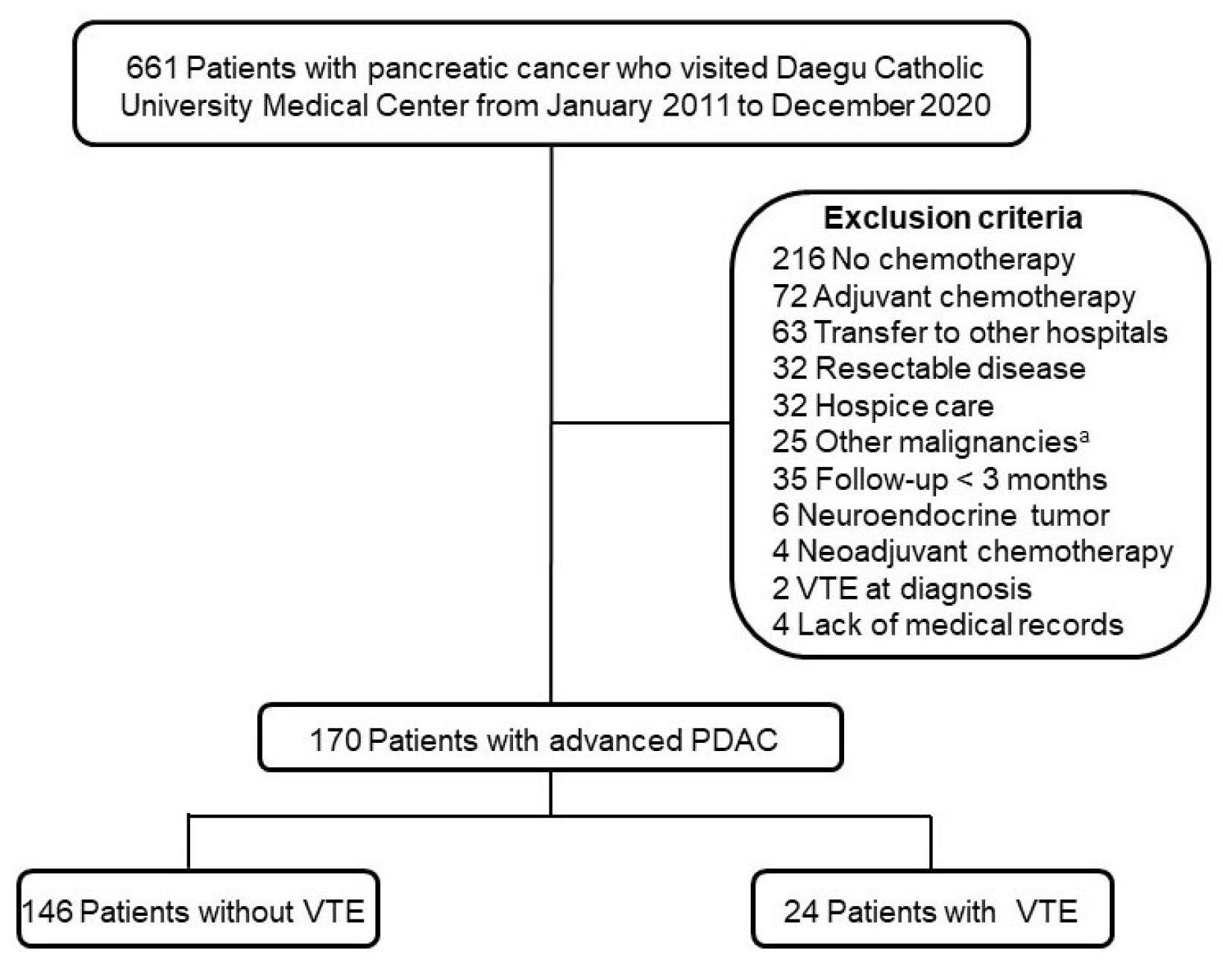
Six hundred sixty-one patients with pancreatic cancer who visited the Daegu Catholic University Medical Center from January 2011 to December 2020 were eligible (Fig. 1). All cases were retrieved using the diagnostic code for pancreatic cancer (C250, C251, C252, C253, C257, and C259) based on the Korean Standard Classification of Diseases, 8th edition. The exclusion criteria were as follows: patients who did not receive palliative chemotherapy, those with resectable disease, those with other malignancies, the presence of VTE at diagnosis, follow-up period <three months, those transferred to other hospitals, those with a neuroendocrine tumor, and a lack of medical records. The medical records were reviewed, and the following data were collected: demographics, laboratory findings, comorbidities, and radiologic findings.

2. Study design

This study was a retrospective, observational cohort study. The primary outcome was the incidence of VTE in patients with advanced PDAC who received palliative chemotherapy. The secondary outcomes were the risk factors associated with VTE and OS. The study protocol was reviewed and approved by the Institutional Review Board of Daegu Catholic University Medical Center (IRB number: CR-22-069-L). The need for informed consent was waived because this study was performed retrospectively.

3. Definitions

Advanced PDAC included locally advanced and metastatic disease, defined according to National Comprehensive Cancer Network Guideline (Version 1. 2022).17 Metastatic disease was defined as a distant metastasis, including non-regional lymph node metastasis; locally advanced disease was defined as solid tumor contact of >180申 with the superior mesenteric artery or celiac axis, or unreconstructible superior mesenteric vein or portal vein due to tumor involvement.17 The OS was defined as the time from the diagnosis to the date of death from any causes. Alcohol consumption was confirmed by the medical records at the time of the PDAC diagnosis. A patient was considered a drinker if their drinking history was marked ‘yes’ on the medical records. A former drinker was defined as a patient who did not consume alcohol in the last 12 months.
VTE included deep vein thrombosis (DVT), pulmonary thromboembolism (PTE), and visceral venous thrombosis (VVT). DVT was detected through ultrasonography or CT. It was performed when patients were diagnosed with PTE or had lower leg pain, swelling, or both. DVT of the upper extremity was not included in this study. PTE was diagnosed via chest CT performed for the response evaluation according to the chemotherapy schedule or when patients had symptoms, such as dyspnea and chest discomfort. VVT was defined as thrombosis in the abdominal veins, such as the portal vein, mesenteric vein, splenic vein, and inferior vena cava. The condition was detected through abdomen and pelvis CT. The Khorana score was calculated from the results of laboratory tests at the diagnosis of PDAC.18

4. Statistical analysis

Statistical analysis was performed using IBM SPSS statistics for Windows version 26.0 (IBP Corp., Armonk, NY, USA). The continuous variables were described as the median with interquartile ranges. A student t-test or a Mann–Whitney U-test was used for them. The chi-square test or Fisher’s exact test was used to compare the categorical variables. The Wilcoxon signed-rank test and McNemar’s test were used to compare the changes in the variables in patients with VTE. The cumulative incidence of VTE was estimated using the Kaplan–Meier method. OS was also evaluated using the Kaplan–Meier method. A log-rank test was used to compare the OS between VTE and non-VTE group. The Cox-proportional hazard model was used to identify the risk factors associated with VTE and OS. The variables with p<0.2 were used in multivariate analysis. The results are presented as the hazard ratio (HR) and the 95% confidence interval (CI). The designated level of statistical significance was p<0.05 (two-tailed).

RESULTS

1. Baseline characteristics of patients

Of 170 patients with advanced PDAC, 24 (14.1%) developed VTE during the median follow-up period of 341 days. Table 1 lists the baseline characteristics of the patients according to the occurrence of VTE. The median age of the patients was 64 years. The proportion of males was 58.2%, and the median BMI was 21.9 kg/m2. The most common tumor location was the head (n=83, 48.8%), followed by the body (n=63, 37.1%) and tail (n=19, 11.2%). One hundred and one patients (59.4%) had metastatic disease, and 69 (40.6%) had locally advanced disease. One hundred and seven patients (62.9%) were diagnosed by endoscopic ultrasound-guided fine needle aspiration (EUS-FNA) and endoscopic ultrasound-guided fine needle biopsy (EUS-FNB); ERCP and liver biopsy were used in 28 (16.5%) and 12 (7.1%) patients, respectively. The tumor differentiation was unknown in most of the patients (n=108, 63.5%). Twenty-seven patients (15.9%) had moderately differentiated cancer, and 11 (6.5%) had poorly differentiated cancer. Only four patients (2.4%) had a well-differentiated cancer. Fifty-six (32.9%), 61 (35.9%) 12 (7.1%) patients had hypertension, diabetes mellitus, and hypercholesterolemia, respectively. One hundred and thirty-six patients (80.0%) received gemcitabine-based chemotherapy, and 31 patients (18.2%) had 5-FU-based chemotherapy. The median level of the carbohydrate antigen 19-9 (CA 19-9) was 369 U/mL, and that of CEA was 5.45 ng/mL at the diagnosis of PDAC. The proportion of patients with a Khorana score over 2 was 18.8%. Patients with VTE were older than those without VTE (68 years vs. 62 years, p=0.039). Other than age, there was no significant difference between the two groups with respect to demographics, clinical features, and laboratory findings.

2. Clinical features of patients with venous thromboembolism

Of 24 patients with VTE, 17 (70.8%) had metastatic disease, and seven (29.2%) had locally advanced disease at the time of the VTE diagnosis. Table 2 lists the clinical features of the patients with VTE. Eleven patients (45.8%) had progressive disease, while 10 (41.7%) had stable disease. Twenty patients (83.3%) were treated with direct oral anticoagulants, and four patients (16.7%) were not treated. The median time from PDAC diagnosis to death was 164 days, and there was no immediate death from VTE. The hemoglobin level was lower at the time of the VTE diagnosis than at the PDAC diagnosis (9.6 g/L vs. 12.6 g/L, p<0.001). The PT was prolonged at the time of VTE diagnosis than that at the time of PDAC diagnosis (INR 1.15 vs. 1.05, p<0.001).

3. Cumulative incidence and risk factors for venous thromboembolism

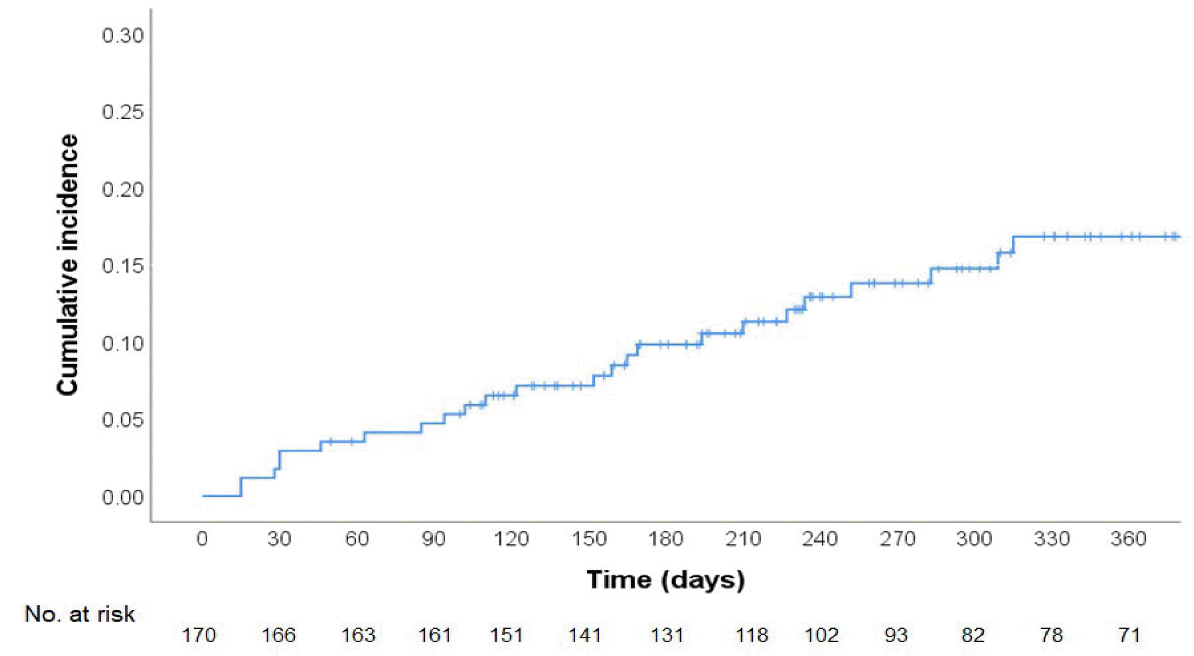
The median period from PDAC diagnosis to the occurrence of VTE was 152 days. Fig. 2 shows the cumulative incidence. The cumulative incidence of VTE was 4.7% (95% CI, 2.39-9.22) at 90 days, 9.9% (95% CI, 6.14-15.59) at 180 days, and 16.9% (95% CI, 11.50-24.36) at 360 days. Table 3 lists the risk factors for VTE. In univariate analysis, age over 65 (HR, 2.413; 95% CI, 1.070-5.443) was the only significant factor. On the other hand, in multivariate analysis, CA 19-9 level over 1,000 U/mL (HR, 2.666; 95% CI, 1.112-6.389; p=0.028) and a history of alcohol consumption (HR, 0.327; 95% CI, 0.109-0.981; p=0.046) were statistically significant factors associated with VTE.

4. Types of venous thromboembolism and overall survival

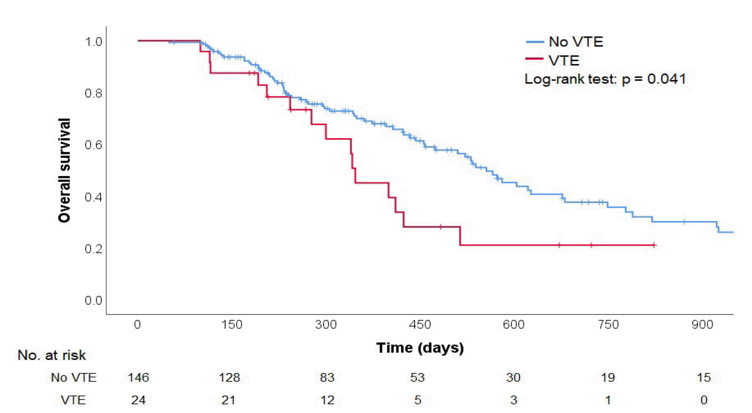
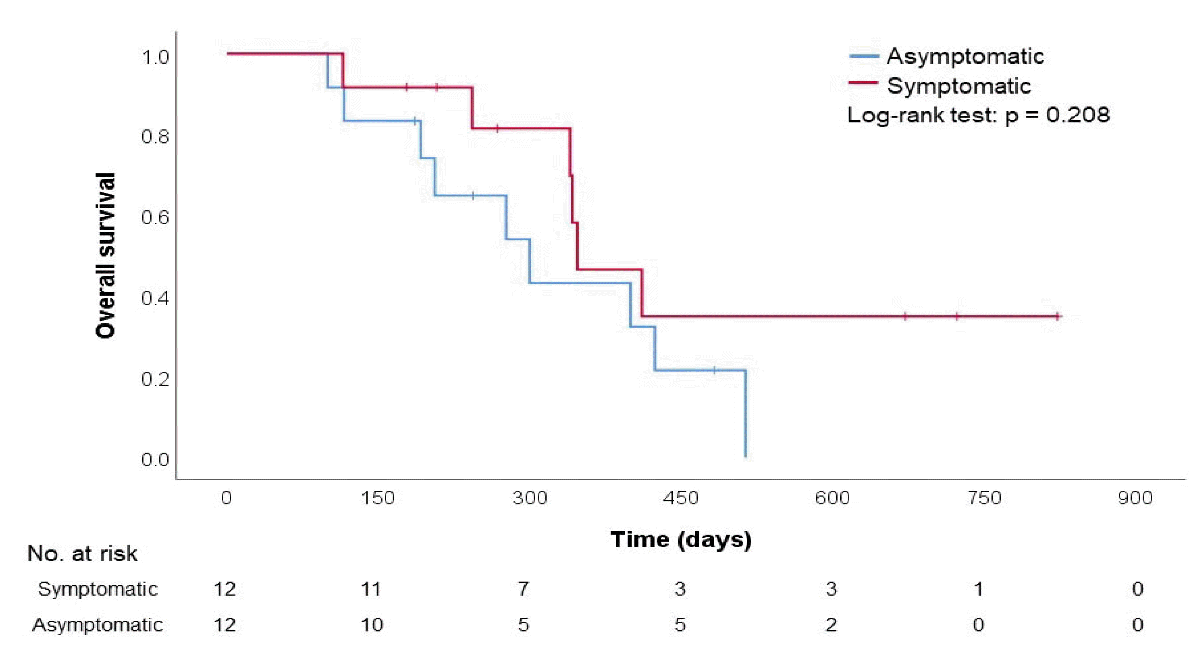
Of the 24 patients with VTE, 12 (50.0%) were symptomatic, and 12 (50.0%) were asymptomatic. Table 4 lists the types of VTE. More than half of patients with PTE or DVT had symptoms, whereas none of the patients with VVT had symptoms. The patients with VTE showed shorter median survival than those without VTE (347 vs. 556 days, p=0.041), as shown in Fig. 3. On the other hand, there was no significant difference in OS according to the presence or absence of symptoms (Fig. 4).

5. Risk factors associated with overall survival

Table 5 lists the risk factors associated with OS. Univariate analysis revealed metastatic disease (HR, 1.781; 95% CI, 1.140-2.780; p=0.011), pancreatic tail cancer (HR, 1.951; 95% CI, 1.111-3.428; p=0.030), CA 19-9 level over 1,000 U/mL (HR, 2.191; 95% CI, 1.352-3.548; p=0.001), and VTE (HR, 1.786; 95% CI, 1.017-3.135; p=0.044) to be statistically significant factors associated with OS. Multivariate analysis showed that a CA 19-9 level over 1,000 U/mL (HR, 1.843; 95% CI, 1.113-3.052; p=0.017) and VTE (HR, 1.850; 95% CI, 1.049-3.263; p=0.033) were significant factors. Metastatic disease (HR, 1.591; 95% CI, 0.996-2.541; p=0.052) was an important factor, but it was not statistically significant.

DISCUSSION

The cumulative incidence of VTE in patients with advanced PDAC who received palliative chemotherapy was 16.86% (95% CI, 11.50-24.36) at 360 days in this study. This result was lower than in previous studies in Western countries, where the incidence of VTE ranged from 18% to 41.3%.5,7-10 On the other hand, a recent large prospective study reported that the cumulative incidence of VTE was 19.21% at 12 months, which was not significantly higher than the present result.19 To the best of the authors’ knowledge, there have been no prospective studies on the incidence of VTE in patients with PDAC in Korea. Moreover, there have been only three retrospective studies. The reported incidences were 5.3%, 9.2%, and 18.6%, respectively.12,14,15 VTE was not associated with a poor prognosis in these studies.12,14 These are summarized in Table 6. The difference in the results among the studies was probably attributed to the difference in the study population, such as the proportion of metastatic disease, surgical resection, and chemotherapy.
Cancer has been demonstrated to be an independent risk factor for VTE. Moreover, PDAC carries the highest risk for VTE.4,18 One possible explanation is that tissue factor-positive microvesicles released from cancer provide a surface for assembling different coagulation factor complexes.20 The endocrine function of the pancreas provides an easy route for transporting tissue factor-positive microvesicles from a tumor to the blood.20 In this study, a CA 19-9 level over 1,000 U/mL was a significant risk factor for VTE. The CA 19-9 level is a well-known prognostic factor for PDAC and reflects the disease burden.21 CA 19-9 was associated with thrombin generation in treatment-naïve patients with PDAC.22 The location of the primary tumor is also a well-known risk factor for VTE, which has been commonly identified in several previous studies.7,8,14,19 Most reported that body or tail cancer had a higher risk of VTE than head cancer.7,8,19 On the other hand, in this study, the tumor location was not a statistically significant factor, even though pancreatic body cancer had a marginally protective effect according to multivariate analysis. Frere et al.19 reported that the tumor location was not a statistically significant factor when VVT was excluded from the analysis. Hence, it is probably because the proportion of VVT in this study was only 16.7%, which contrasts with other studies: 29.6-58.3%.7,8,14,19
Interestingly, a history of alcohol consumption was a protective factor for VTE in this study. Several studies reported that moderate alcohol consumption is associated with a decreased risk of VTE.23-25 Light-to-moderate alcohol consumption was associated with lower levels of coagulation factors, including fibrinogen, von Willebrand factor, and factor VII.26 On the other hand, a detailed history of alcohol consumption could not be obtained because of its retrospective design, and alcohol consumption probably decreased after the diagnosis of PDAC. Thus, further prospective studies are needed to investigate the potential protective role of alcohol against VTE in patients with advanced PDAC.
In this study, although the disease stage of patients did not change significantly at VTE diagnosis, 45.8% of patients had been diagnosed with disease progression. This rate was comparable to the result of previous studies, which reported that the proportion of patients with progressive disease at the time of VTE diagnosis ranged from 29.4% to 57.1%.12,16,27 In addition, the hemoglobin level was lower, and the PT was prolonged during VTE diagnosis. Nevertheless, anemia is a common hematologic adverse event in patients with PDAC who are undergoing chemotherapy.28 Moreover, prolonged PT reflects the biosynthetic capacity of the liver; hepatic insufficiency caused by disease progression may lead to a decreased coagulation factor.29 PT prolongation was associated with disease progression in other cancers, particularly liver metastasis.29,30 Therefore, it is difficult to attribute these changes solely to the development of VTE.
Patients with VTE showed significantly reduced OS in this study. In addition, CA 19-9 level and VTE were independent prognostic factors in multivariate analysis. The elevation of the CA 19-9 level is a well-known prognostic factor in previous studies.14,16 In contrast, VTE has shown inconsistent results in previous studies. Several studies have shown that the occurrence of VTE is a predictor of a poor prognosis,10,12,19 particularly in early VTE, which was defined as within 1.5 months after the initiation of chemotherapy or within 3 months after diagnosis of PDAC.7,9,13,16 Other studies reported that the occurrence of VTE does not significantly affect the prognosis.5,8,11,14 As mentioned above, this is probably explained by the difference in the study population among studies. Berger et al.5 reported that VTE was not associated with a poor prognosis in patients with advanced PDAC who received palliative chemotherapy. On the other hand, the study population had a higher proportion of metastatic disease than the present study (92.0% vs. 59.4%) and had shorter median survival in the no VTE group (9.9 months vs. 347 days).5
Regarding symptoms, the proportion of incidental VTE was 50% in this study, similar to the 52.4% to 72% reported elsewhere.7,8,10,12,14,19 In contrast, there have been conflicting results as to whether symptomatic VTE has a poorer progress than incidental VTE.7,10,14,19 The data showed no significant difference with respect to OS according to the presence or absence of symptoms.
To the best of the authors’ knowledge, there have been five large prospective studies to evaluate the efficacy of prophylactic anticoagulation. Among them, two studies were conducted on only patients with advanced PDAC.31,32 Although thromboprophylaxis decreased the risk of VTE in these studies significantly, there was no survival gain. Therefore, extensive prospective studies on thromboprophylaxis in patients with the risk factors for VTE are needed.
This study had some limitations. First, it was a retrospective single-center study. Thus, there was selection bias. In addition, it is difficult to generalize the results of this study. Second, the history of alcohol consumption, an important protective factor for VTE in this study, was not obtained in detail because of a lack of medical records. Third, 48.2% of patients were diagnosed by EUS-FNA because EUS-FNB had not been performed before 2017 at the authors’ institution. Therefore, it was not possible to obtain enough samples to evaluate the tumor differentiation. Fourth, the risk factors affecting the occurrence of VTE were not evaluated thoroughly because of the retrospective design and lack of medical records. These factors included recent surgery, neurological diseases, performance status, and medications, such as oral contraceptives, anticoagulants, and antiplatelet agents. On the other hand, because the patients in this study included those who had received chemotherapy, they were neither bedridden nor in a poor performance status. Finally, a chest CT scan was performed only when needed. Thus, the incidence of VTE might be underestimated. Nevertheless, regular follow-up was performed during chemotherapy because the study was conducted on only patients who received palliative chemotherapy. Therefore, the incidence of VTE in patients in this study with PDAC is probably close to the actual value.
In conclusion, the cumulative incidence of VTE in patients with advanced PDAC who received palliative chemotherapy was 16.9% (95% CI, 11.50-24.36) at 360 days. Although a history of alcohol consumption was a protective factor, a high CA 19-9 level was a risk factor for VTE. Furthermore, the occurrence of VTE was associated with a poor prognosis. On the other hand, because there has been no prospective study on Asian patients, a further large prospective study will be needed to investigate the incidence and risk factors associated with VTE.

Notes

Financial support

None.

Conflict of interest

None.

REFERENCES

1. Sung H, Ferlay J, Siegel RL, et al. Global Cancer Statistics 2020: GLOBOCAN Estimates of incidence and mortality worldwide for 36 cancers in 185 countries. CA Cancer J Clin. 2021; 71:209–249. DOI: 10.3322/caac.21660. PMID: 33538338.
2. Jung KW, Won YJ, Kang MJ, Kong HJ, Im JS, Seo HG. 2022; Prediction of cancer incidence and mortality in Korea, 2022. Cancer Res Treat. 54:345–351. DOI: 10.4143/crt.2022.179. PMID: 35313101. PMCID: PMC9016306.
3. Sung MJ, Bang S. 2020; Updates of chemotherapy and radiotherapy for pancreatic cancer. Korean J Pancreas Biliary Tract. 25:72–82. DOI: 10.15279/kpba.2020.25.2.72.
4. Frere C. 2021; Burden of venous thromboembolism in patients with pancreatic cancer. World J Gastroenterol. 27:2325–2340. DOI: 10.3748/wjg.v27.i19.2325. PMID: 34040325. PMCID: PMC8130043.
5. Berger AK, Singh HM, Werft W, et al. 2017; High prevalence of incidental and symptomatic venous thromboembolic events in patients with advanced pancreatic cancer under palliative chemotherapy: A retrospective cohort study. Pancreatology. 17:629–634. DOI: 10.1016/j.pan.2017.04.012. PMID: 28462862.
6. Lyman GH, Eckert L, Wang Y, Wang H, Cohen A. 2013; Venous thromboembolism risk in patients with cancer receiving chemotherapy: a real-world analysis. Oncologist. 18:1321–1329. DOI: 10.1634/theoncologist.2013-0226. PMID: 24212499. PMCID: PMC3868427.
7. García Adrián S, González AR, de Castro EM, et al. 2022; Incidence, risk factors, and evolution of venous thromboembolic events in patients diagnosed with pancreatic carcinoma and treated with chemotherapy on an outpatient basis. Eur J Intern Med. 105:30–37. DOI: 10.1016/j.ejim.2022.07.020. PMID: 35931614.
8. Hanna-Sawires RG, Groen JV, Hamming A, et al. 2021; Incidence, timing and risk factors of venous thromboembolic events in patients with pancreatic cancer. Thromb Res. 207:134–139. DOI: 10.1016/j.thromres.2021.08.002. PMID: 34628229.
9. Barrau M, Maoui K, Le Roy B, et al. 2021; Early venous thromboembolism is a strong prognostic factor in patients with advanced pancreatic ductal adenocarcinoma. J Cancer Res Clin Oncol. 147:3447–3454. DOI: 10.1007/s00432-021-03590-x. PMID: 33715087.
10. Kruger S, Haas M, Burkl C, et al. 2017; Incidence, outcome and risk stratification tools for venous thromboembolism in advanced pancreatic cancer - A retrospective cohort study. Thromb Res. 157:9–15. DOI: 10.1016/j.thromres.2017.06.021. PMID: 28675831.
11. Suzuki T, Hori R, Takeuchi K, et al. 2021; Venous thromboembolism in Japanese patients with pancreatic cancer. Clin Appl Thromb Hemost. 27:10760296211051766. DOI: 10.1177/10760296211051766. PMID: 34730013. PMCID: PMC8573688.
12. Yoon SY, Lee MY, Yun J, et al. 2018; The incidence of venous thromboembolism is not lowin Korean patients with advanced pancreatic cancer [corrected]. Blood Res. 53:227–232. Erratum in: Blood Res 2018;53:335-336. DOI: 10.5045/br.2018.53.3.227. PMID: 30310790. PMCID: PMC6170311.
13. Chen JS, Hung CY, Chang H, et al. 2018; Venous thromboembolism in Asian patients with pancreatic cancer following palliative chemotherapy: low incidence but a negative prognosticator for those with early onset. Cancers (Basel). 10:501. DOI: 10.3390/cancers10120501. PMID: 30544670. PMCID: PMC6315992.
14. Lee JC, Ro YS, Cho J, et al. 2016; Characteristics of venous thromboembolism in pancreatic adenocarcinoma in East Asian ethnics: a large population-based observational study. Medicine (Baltimore). 95:e3472. DOI: 10.1097/MD.0000000000003472. PMID: 27124043. PMCID: PMC4998706.
15. Oh SY, Kim JH, Lee KW, et al. 2008; Venous thromboembolism in patients with pancreatic adenocarcinoma: lower incidence in Asian ethnicity. Thromb Res. 122:485–490. DOI: 10.1016/j.thromres.2007.12.015. PMID: 18234292.
16. Yamai T, Ikezawa K, Hiraga E, et al. 2022; Early detection of venous thromboembolism after the initiation of chemotherapy predicts a poor prognosis in patients with unresectable metastatic pancreatic cancer who underwent first-line chemotherapy with gemcitabine plus nab-paclitaxel. PLoS One. 17:e0264653. DOI: 10.1371/journal.pone.0264653. PMID: 35231078. PMCID: PMC8887762.
17. Pancreatic Adenocarcionma (Version 1. 2022). 2022 [Internet]. 2022. National Comprehensive Cancer Network;Plymouth Meeting (PA): Available from: https://www.nccn.org/professionals/physician_gls/pdf/pancreatic.pdf. cited 2022 Nov 2.
18. Khorana AA, Kuderer NM, Culakova E, Lyman GH, Francis CW. 2008; Development and validation of a predictive model for chemotherapy-associated thrombosis. Blood. 111:4902–4907. DOI: 10.1182/blood-2007-10-116327. PMID: 18216292. PMCID: PMC2384124.
19. Frere C, Bournet B, Gourgou S, et al. BACAP Consortium. Incidence of venous thromboembolism in patients with newly diagnosed pancreatic cancer and factors associated with outcomes. Gastroenterology. 2020; 158:1346–1358.e4. DOI: 10.1053/j.gastro.2020.03.004.
20. Hisada Y, Mackman N. 2017; Cancer-associated pathways and biomarkers of venous thrombosis. Blood. 130:1499–1506. DOI: 10.1182/blood-2017-03-743211. PMID: 28807983. PMCID: PMC5620413.
21. Poruk KE, Gay DZ, Brown K, et al. 2013; The clinical utility of CA 19-9 in pancreatic adenocarcinoma: diagnostic and prognostic updates. Curr Mol Med. 13:340–351. DOI: 10.2174/1566524011313030003. PMID: 23331006. PMCID: PMC4419808.
22. Mattila N, Hisada Y, Przybyla B, et al. 2021; Levels of the cancer biomarker CA 19-9 are associated with thrombin generation in plasma from treatment-naïve pancreatic cancer patients. Thromb Res. 199:21–31. DOI: 10.1016/j.thromres.2020.12.018. PMID: 33385797.
23. Hanna K, Khalid A, Hamidi M, et al. 2019; Chronic alcohol consumption and risk of deep venous thrombosis: a propensity-matched analysis. J Surg Res. 244:251–256. DOI: 10.1016/j.jss.2019.06.067. PMID: 31301481.
24. Pomp ER, Rosendaal FR, Doggen CJ. 2008; Alcohol consumption is associated with a decreased risk of venous thrombosis. Thromb Haemost. 99:59–63. DOI: 10.1160/TH07-07-0470. PMID: 18217135.
25. Gaborit FS, Overvad K, Nørgaard M, Kristensen SR, Tjønneland A, Severinsen MT. 2013; Alcohol intake and risk of venous thromboembolism. A Danish follow-up study. Thromb Haemost. 110:39–45. DOI: 10.1160/TH12-10-0790. PMID: 23595624.
26. Mukamal KJ, Jadhav PP, D'Agostino RB, et al. 2001; Alcohol consumption and hemostatic factors: analysis of the Framingham Offspring cohort. Circulation. 104:1367–1373. DOI: 10.1161/hc3701.096067. PMID: 11560851.
27. Ouaissi M, Frasconi C, Mege D, et al. 2015; Impact of venous thromboembolism on the natural history of pancreatic adenocarcinoma. Hepatobiliary Pancreat Dis Int. 14:436–442. DOI: 10.1016/S1499-3872(15)60397-6. PMID: 26256090.
28. Conroy T, Desseigne F, Ychou M, et al. Groupe Tumeurs Digestives of Unicancer; PRODIGE Intergroup. FOLFIRINOX versus gemcitabine for metastatic pancreatic cancer. N Engl J Med. 2011; 364:1817–1825. DOI: 10.1056/NEJMoa1011923. PMID: 21561347.
29. Kwon HC, Oh SY, Lee S, et al. 2008; Plasma levels of prothrombin fragment F1+2, D-dimer and prothrombin time correlate with clinical stage and lymph node metastasis in operable gastric cancer patients. Jpn J Clin Oncol. 38:2–7. DOI: 10.1093/jjco/hym157. PMID: 18258711.
30. Wang XP, Mao MJ, He ZL, et al. 2017; A retrospective discussion of the prognostic value of combining prothrombin time(PT) and fibrinogen( Fbg) in patients with Hepatocellular carcinoma. J Cancer. 8:2079–2087. DOI: 10.7150/jca.19181. PMID: 28819409. PMCID: PMC5559970.
31. Pelzer U, Opitz B, Deutschinoff G, et al. 2015; Efficacy of prophylactic low-molecular weight heparin for ambulatory patients with advanced pancreatic cancer: outcomes from the CONKO-004 trial. J Clin Oncol. 33:2028–2034. DOI: 10.1200/JCO.2014.55.1481. PMID: 25987694.
32. Maraveyas A, Waters J, Roy R, et al. 2012; Gemcitabine versus gemcitabine plus dalteparin thromboprophylaxis in pancreatic cancer. Eur J Cancer. 48:1283–1292. DOI: 10.1016/j.ejca.2011.10.017. PMID: 22100906.

Fig. 1
Flowchart of the study population. PDAC, pancreatic ductal adenocarcinoma; VTE, venous thromboembolism. aOther malignancies included six colorectal cancers, four thyroid cancers, three renal cell carcinomas, three hematologic malignancies, two gallbladder cancers, two breast cancer, one hepatocellular carcinoma, one lung cancer, one Klatskin tumor, one cervical cancer, and one prostate cancer.
kjg-81-3-109-f1.tif
Fig. 2
Cumulative incidence of venous thromboembolism in patients with pancreatic cancer.
kjg-81-3-109-f2.tif
Fig. 3
Overall survival of patients with pancreatic cancer according to venous thromboembolism. VTE, venous thromboembolism.
kjg-81-3-109-f3.tif
Fig. 4
Overall survival of patients with venous thromboembolism according to symptom.
kjg-81-3-109-f4.tif
Table 1
Baseline Characteristics of the Study Population
Variables Total (n=170) Patients with VTE (n=24, 14.1%) Patients without VTE (n=146, 85.9%) p-value
Age (yr) 64 (56-72) 68 (64-74) 62 (55-70) 0.039
Male 99 (58.2) 13 (54.2) 86 (58.9) 0.824
BMI (kg/m2) 21.9 (19.8-24.2) 23.8 (19.5-25.7) 21.7 (19.9-23.8) 0.190
Tumor location 0.269
Head 83 (48.8) 13 (54.2) 70 (47.9)
Body 63 (37.1) 6 (25.0) 57 (39.0)
Tail 19 (11.2) 5 (20.8) 14 (9.6)
Overlap 5 (2.9) 0 (0.0) 5 (3.4)
Stage 0.825
Locally advanced 69 (40.6) 9 (37.5) 60 (41.1)
Metastatic 101 (59.4) 15 (62.5) 86 (58.9)
Differentiation 0.228
Well 4 (2.4) 1 (4.2) 3 (2.1)
Moderately 27 (15.9) 7 (29.2) 20 (13.7)
Poorly 11 (6.5) 2 (8.3) 9 (6.2)
Unknown 108 (63.5) 12 (50.0) 96 (65.8)
Atypical cell 20 (11.8) 2 (8.3) 18 (12.3)
Diagnostic method 0.163
EUS-FNA 82 (48.2) 6 (25.0) 76 (52.1)
EUS-FNB 25 (14.7) 5 (20.8) 20 (13.7)
Liver biopsy 12 (7.1) 3 (12.5) 9 (6.2)
ERCP 28 (16.5) 6 (25.0) 22 (15.1)
Othersa 23 (13.5) 4 (16.7) 19 (13.0)
Alcohol consumption 53 (31.2) 4 (16.7) 40 (33.6) 0.152
Smoking 60 (35.3) 6 (25.0) 54 (37.0) 0.357
Comorbidity
Hypertension 56 (32.9) 11 (45.8) 45 (30.8) 0.164
Diabetes mellitus 61 (35.9) 11 (45.8) 50 (34.2) 0.358
Hypercholesterolemia 12 (7.1) 1 (4.2) 11 (7.5) 1.000
CVA 7 (4.1) 1 (4.2) 6 (4.1) 1.000
Arrythmia 4 (2.4) 0 (0.0) 4 (2.7) 1.000
IHD 4 (2.4) 0 (0.0) 4 (2.7) 1.000
First line therapy 0.395
Gemcitabine-based 136 (80.0) 20 (83.3) 116 (79.5)
5-FU based 31 (18.2) 3 (12.5) 28 (19.2)
TS-1 3 (1.8) 1 (4.2) 2 (1.4)
Laboratory test
WBC (103/µL) 6,350 (5,413-8,260) 6,700 (5,413-7,875) 6,300 (5,425-8,300) 0.683
Hemoglobin (g/L) 12.6 (11.2-13.7) 12.6 (10.1-13.3) 12.7 (11.2-13.8) 0.159
Platelet (103/µL) 213 (167-269) 214 (151-264) 213 (174-275) 0.361
AST (U/L) 26 (18-80) 35 (20-79) 25 (18-83) 0.228
ALT (U/L) 25 (14-103) 38 (17-174) 24 (14-100) 0.176
Total bilirubin (U/L) 0.7 (0.4-3.1) 0.7 (0.3-6.5) 0.7 (0.4-2.9) 0.893
BUN (mg/dL) 12.6 (10.1-16.2) 12.6 (10.9-16.2) 12.6 (9.9-16.2) 0.605
Creatinine (mg/dL) 0.7 (0.6-0.9) 0.9 (0.6-1.0) 0.7 (0.6-0.8) 0.101
PT, INR 1.03 (0.97-1.09) 1.05 (1.01-1.11) 1.03 (0.97-1.09) 0.285
aPTT (sec) 35.4 (33.3-38.4) 35.7 (33.3-39.2) 35.3 (33.2-38.4) 0.584
Tumor marker
CA 19-9 (U/mL) 369 (73-1,818) 714 (137-8,282) 356 (72-1,540) 0.174
CEA (ng/mL) 5.45 (3.10-14.14) 9.43 (3.70-14.77) 5.20 (3.09-13.75) 0.394
Khorana score >2 32 (18.8) 7 (29.2) 25 (17.1) 0.167
Follow-up period 341 (209-534) 289 (196-421) 347 (215-549) 0.228

Data are presented as number (%) or median (interquartile range).

VTE, venous thromboembolism; EUS-FNA, endoscopic ultrasound-guided fine needle aspiration; EUS-FNB, endoscopic ultrasound-guided fine needle biopsy; CVA, cerebrovascular accident; IHD, ischemic heart disease; 5-FU, 5-fluorouracil; TS-1, tegafur/gimetacil/oteracil potassium; WBC, white blood cell; CA 19-9, carbohydrate antigen 19-9.

aOthers included 15 endoscopic duodenal biopsies, 3 open biopsies, 2 unknown, and 1 percutaneous transhepatic biliary drainage.

Table 2
Clinical Features of Patients with Pancreatic Cancer and Venous Thromboembolism
Variables At PDAC diagnosis (n=24) At VTE diagnosis (n=24) p-value
Stage 0.500
Locally advanced 9 (37.5) 7 (29.2)
Metastatic 15 (62.5) 17 (70.8)
Disease statusa NA
Stable disease NA 10 (41.7)
Progressive disease NA 11 (45.8)
Partial response NA 3 (12.5)
Therapy for VTE NA
None NA 4 (16.7)
Warfarin NA 0 (0.0)
DOAC NA 20 (83.3)
Laboratory findings
WBC (103/µL) 6,700 (5,413-7,875) 7,650 (4,775-13,850) 0.277
Hemoglobin (g/L) 12.6 (10.1-13.3) 9.6 (9.1-11.0) <0.001
Platelet (103/µL) 214 (151-264) 160 (98.8-310) 0.541
PT, INR 1.05 (1.01-1.11) 1.15 (1.09-1.32) <0.001
aPTT (sec) 35.7 (33.3-39.2) 34.8 (32.8-41.0) 0.819

Data are presented as number (%) or median (interquartile range).

PDAC, pancreatic ductal adenocarcinoma; VTE, venous thromboembolism; NA, not available; DOAC, direct oral anticoagulants; WBC, white blood cell.

aDisease status was evaluated according to Response Evaluation Criteria in Solid Tumor (RECIST) version 1.1.

Table 3
Risk Factors Associated with VTE in Patients with Pancreatic Cancer
Variables Univariate analysis Multivariate analysis


HR (95% CI) p-value HR (95% CI) p-value
Disease stage
Locally advanceda
Metastatic 1.250 (0.546-2.859) 0.597
Sex
Femalea
Male 0.880 (0.394-1.965) 0.755
Tumor location
Head or taila
Body 0.495 (0.196-1.247) 0.136 0.398 (0.156-1.013) 0.053
Body or taila
Head 1.242 (0.556-2.774) 0.596
Head or bodya
Tail 1.921 (0.717-5.146) 0.194
Age
≤65a
>65 2.413 (1.070-5.443) 0.034
CA 19-9 (U/mL)
≤1,000a
>1,000 1.901 (0.829-4.361) 0.129 2.666 (1.112-6.389) 0.028
CEA (ng/mL)
≤5.2a
>5.2 1.384 (0.622-3.081) 0.426
BMI (kg/m2)
≤25a
>25 1.999 (0.829-4.822) 0.123
Chemoport
Noa
Yes 0.514 (0.153-1.727) 0.282
Chemotherapy
Othersa
5-FU based 2.802 (0.372-21.110) 0.317
Khorana score
≤2a
>2 1.959 (0.812-4.727) 0.135
Hypertension
Noa
Yes 1.946 (0.871-4.347) 0.105 2.241 (0.977-5.139) 0.057
Diabetes mellitus
Noa
Yes 1.628 (0.729-3.638) 0.234
Alcohol consumption
Noa
Yes 0.447 (0.153-1.309) 0.142 0.327 (0.109-0.981) 0.046
Smoking
Noa
Yes 0.635 (0.252-1.601) 0.336

HR, hazard ratio; CI, confidence interval; CA 19-9, carbohydrate antigen 19-9; 5-FU, 5-fluorouracil.

aReference category.

Table 4
Types of Venous Thromboembolisms
Event Total Symptomatic Incidental
VTEa 24 (100.0) 12 (50.0) 12 (50.0)
DVT 15 (62.5) 12 (80.0) 3 (20.0)
PTE 16 (66.7) 8 (50.0) 8 (50.0)
VVT 4 (16.7) 0 (0.0) 4 (100.0)
DVT+PTE 11 (45.8) 8 (72.7) 3 (27.3)

Data are presented as number (%).

VTE, venous thromboembolism; DVT, deep vein thrombosis; PTE, pulmonary thromboembolism; VVT, visceral venous thrombosis.

aPatients with at least one of deep vein thrombosis, pulmonary thromboembolism, and visceral venous thrombosis.

Table 5
Risk Factors Associated with the Overall Survival of Patients with Pancreatic Cancer
Variables Univariate analysis Multivariate analysis


HR (95% CI) p-value HR (95% CI) p-value
Disease stage
Locally advanceda
Metastatic 1.781 (1.140-2.780) 0.011 1.591 (0.996-2.541) 0.052
Sex
Femalea
Male 1.295 (0.843-1.990) 0.237
Tumor location
Head or taila
Body 0.965 (0.634-1.468) 0.866
Body or taila
Head 0.791 (0.507-1.234) 0.301
Head or bodya
Tail 1.951 (1.111-3.428) 0.030
Age
≤65a
>65 1.268 (0.817-1.968) 0.289
CA 19-9 (U/mL)
≤1,000a
>1,000 2.191 (1.352-3.548) 0.001 1.843 (1.113-3.052) 0.017
CEA (ng/mL)
≤5.2a
>5.2 1.442 (0.936-2.223) 0.097
VTE
Noa
Yes 1.786 (1.017-3.136) 0.044 1.850 (1.049-3.263) 0.033
Chemotherapy
Othersa
5-FU based 1.102 (0.152-7.991) 0.924
Hypertension
Noa
Yes 0.822 (0.518-1.305) 0.406
Diabetes mellitus
Noa
Yes 1.148 (0.736-1.790) 0.543
Alcohol consumption
Noa
Yes 1.048 (0.671-1.638) 0.836
Smoking
Noa
Yes 1.202 (0.770-1.877) 0.419

HR, hazard ratio; CI, confidence interval; CA 19-9, carbohydrate antigen 19-9; VTE, venous thromboembolism; 5-FU, 5-fluorouracil.

aReference category.

Table 6
Studies on the Incidence of Thromboembolism in Patients with Pancreatic Cancer in Korea
Parameters Lee et al.14 Yoon et al.12 Oh et al.15 Current study
Study design Single center Single center Single center Single center
Retrospective Retrospective Retrospective Retrospective
Study period 2005-2010 2006-2012 2003-2005 2011-2020
Incidence of VTE 9.2%a 18.60% 5.30% 16.86%b
Disease stage
Resectable 316 (28.3) 0 (0.0) 0 (0.0) 0 (0.0)
Locally advanced 191 (17.1) 231 (45.7) 25 (33.3) 69 (40.6)
Metastatic 608 (54.5) 252 (49.9) 50 (66.7) 101 (59.4)
Recurrence 0 (0.0) 22 (4.4) 0 (0.0) 0 (0.0)
Chemotherapy
None 216 (19.4) 177 (34.3) 35 (46.7) 0 (0.0)
Adjuvant 173 (15.5) 0 (0.0) 0 (0.0) 0 (0.0)
Palliative 689 (61.8) 332 (65.7) 40 (53.3) 170 (100.0)
VTE 132 94 4 24
DVT 17c 18 2 15
PTE 38 19 0 16
SVT 77 38 2 4
OS with or without VTE, months (p-value) 9.5 vs. 9.3 (p=0.649) 9.0 vs. 8.2 (p=0.237) NA 11.6 vs. 18.5 (p=0.041)

Data are presented as number (%), number, or %

VTE, venous thromboembolism; DVT, deep vein thrombosis; PTE, pulmonary thromboembolism; SVT, splanchnic vein thrombosis; NA, not available; OS, overall survival.

aCumulative incidence at 2 years. bCumulative incidence at 360 days. cPatients had only deep vein thrombosis without pulmonary thromboembolism.

TOOLS
Similar articles