See the reply "READER’S FORUM" in Volume 53 on page 217.
Abstract
Objective
The study aimed to evaluate the changes in mandibular width after sagittal split ramus osteotomy (SSRO) in patients with mandibular asymmetric prognathism using cone-beam computed tomography (CBCT).
Methods
Seventy patients who underwent SSRO for mandibular setback surgery were included in two groups, symmetric (n = 35) and asymmetric (n = 35), which were divided according to the differences in their right and left setback amounts. The mandibular width was evaluated three-dimensionally using CBCT images taken immediately before surgery (T1), 3 days after surgery (T2), and 6 months after surgery (T3). Repeated measures analysis of variance was applied to verify the differences in mandibular width statistically.
Enhancement of the facial profile has become a significant priority in orthognathic surgery due to increasing social concerns about esthetics. Currently, a symmetric and tapered facial profile in the frontal view is preferred, and a straight facial profile is preferred over a convex or concave profile in the lateral view.1 However, Asians tend to have a large percentage of prognathic and squared facial profiles.2,3
Orthodontic treatment must be accompanied by orthognathic surgery to improve severe facial asymmetry and mandibular prognathism. In practice, two main methods, internal vertical ramus osteotomy (IVRO) and sagittal split ramus osteotomy (SSRO) are used for mandibular orthognathic surgery.4 Unlike IVRO, SSRO can be used for various mandibular deformities, including prognathism, retrognathism, and asymmetry.
There have been many studies on the stability of SSRO since its introduction.5,6 Factors influencing the stability of SSRO include presurgical orthodontic treatment, internal fixation method, quality of postsurgical occlusion, and location of the proximal segment.6 The site of the proximal segment is crucial not only for stability but also for esthetics. The shape of the mandibular ramus, especially the gonial area, may influence patient esthetics and, thus, patient satisfaction.
Previous studies7-9 using two-dimensional (2D) images have shown changes in mandibular width after mandibular setback surgery using SSRO. However, a 2D image can be distorted depending on the magnification and orientation of the image. To overcome these limitations, there have been increasing attempts to assess the changes after mandibular setback surgery using three-dimensional (3D) cone-beam computed tomography (CBCT). Most studies have focused on evaluating condylar stability and not the mandibular width. A study10 assessing the change in mandibular width after SSRO in patients without mandibular asymmetry has been published. However, for patients with asymmetry of the mandible, when the mandibular midline shifts towards the facial midline, the distal segment of the non-deviated side tends to move toward the inside and the deviated side toward the outside.11 This may increase the mandibular width due to flaring or a gap between segments. Therefore, assessing the mandibular width in patients with mandibular asymmetry is more clinically meaningful.
In this study, our objective was to evaluate the 3D changes in mandibular width after SSRO in patients with mandibular asymmetric prognathism using CBCT.
This study was reviewed by the Institutional Review Board of Pusan National University Dental Hospital (PNUDH -2019037).
Seventy patients who underwent SSRO for mandibular setback surgery between 2013 and 2018 at the Pusan National University Dental Hospital were included in this retrospective study. Patients were divided into two groups according to the differences in their right and left setback amounts. The asymmetric group (ASG; n = 35) included patients with a setback difference of 2 mm or greater. The symmetric group (SYG; n = 35) included patients with a setback difference of less than 2 mm (Table 1).9,12 All patients met the following criteria: 1) mandibular prognathism, 2) pre-operative orthodontic treatment, 3) no mandibular angle osteotomy, 4) no history of temporomandibular joint disorder, 5) no maxillofacial syndromes, and 6) no maxillary asymmetry. The anatomic landmarks used to determine maxillary asymmetry were based on a previous study.13 For 2-jaw surgery, only Le Fort I maxillary impaction or advancement surgery was included. The sample size of 35 subjects in each group was calculated based on the following: β error = 0.20, a error = 0.05, and effect size = 0.5.14
Every patient was treated with modified bilateral SSRO surgery15 by a single oral maxillofacial surgeon with more than 16 years of experience in orthognathic surgery. The medial pterygoid muscle was detached to minimize muscle extension. The mandible was located using a pre-manufactured surgical stent. Early contact between segments was removed using a surgical round bur. Internal fixation was performed by using mini-plates with monocortical screws. Postsurgical orthodontic treatment began one month after surgery.
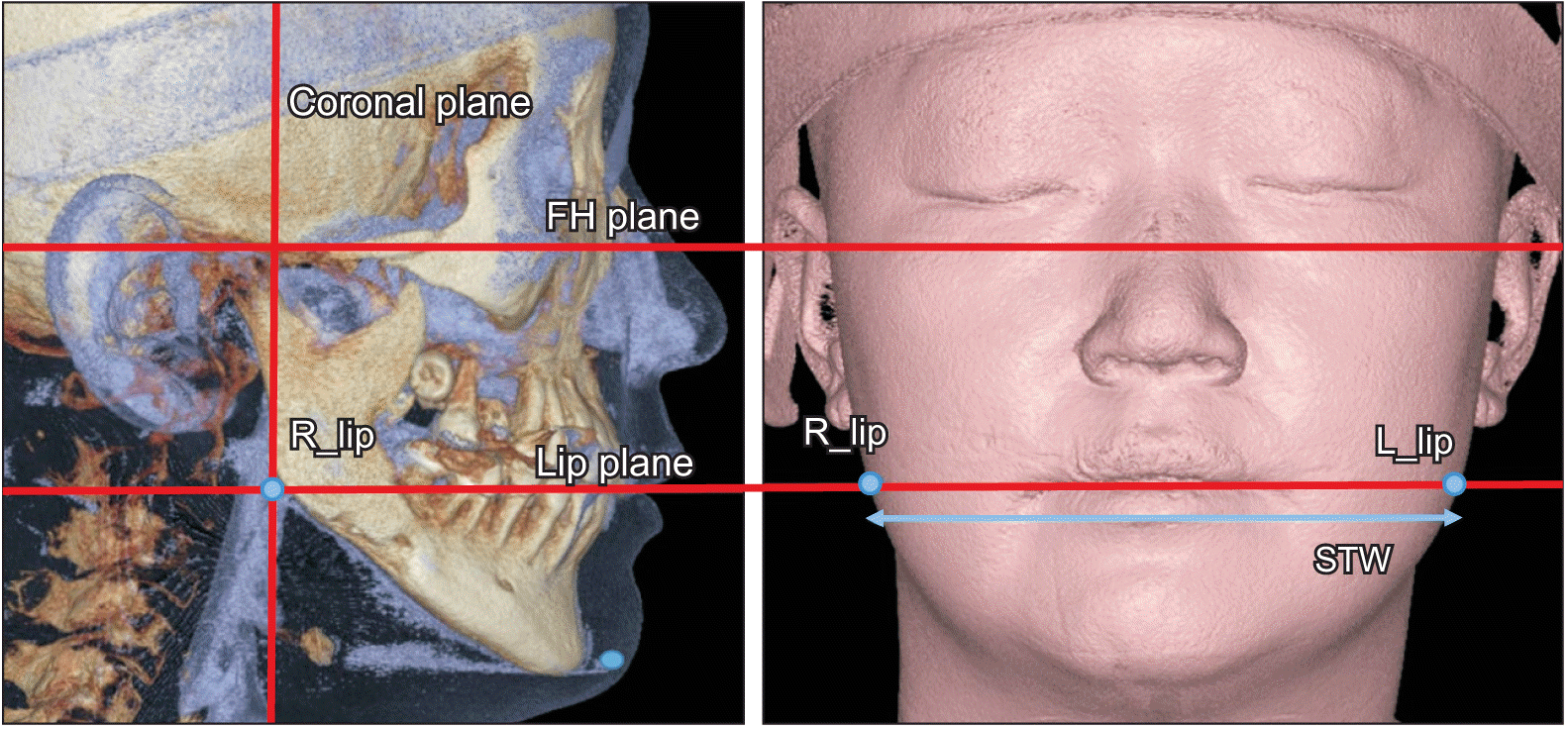
CBCT (Pax-Zenith3D; Vatech Co., Seoul, Korea) images taken immediately before surgery (T1), 3 days after surgery (T2), and 6 months after surgery (T3) were used to evaluate changes in mandibular width after SSRO. The following CBCT settings were used: tube voltage of 90 kVp; scanning time of 24 seconds; tube current of 4 mA; voxel size of 0.3 mm; and field of view of 20 × 19 cm. The Ondemand3D (Cybermed, Seoul, Korea) software was used to convert the CBCT data into a 3D model. To re-orient the transformed 3D models, the horizontal reference plane (HRP, passing through both orbitale and right porion) and vertical reference plane (VRP, perpendicular to HRP and connecting the nasion and basion) were used. Based on the anterior cranial base, CBCT images at T2 and T3 were superimposed on those at T1. The landmarks are based on previous studies (Figures 1 and 2, Table 2).9 The measurements of the hard tissue were as follows: (1) inter-gonial width (IGW), a straight line connecting the left and right gonion points (R_go and L_go); (2) inter-ramal width (IRW), a straight line connecting the landmarks (R_ra and L_ra) of the ramus located on the mastoid process line; and (3) unilateral gonial width (UGW), the distance from VRP to the unilateral gonion point (R_go or L_go). The soft tissue was measured using (4) the soft tissue width (STW), a straight line connecting the soft tissue landmarks (R_lip and L_lip) located on the lip commissure line. The menton was used as the landmark for measuring the amount of mandibular setback. The dental midline of the mandible and the menton were well aligned in all subjects through preoperative orthodontic treatments. The amount of mandibular setback was evaluated by the following procedure.16
1. Using the facebow transfer, the cast is mounted to an articulator.
2. Horizontal markings, parallel to the baseplate, are created. Vertical markings are then placed at the facial midline and first molar cusp. The distance from the mandibular dental midline to the menton on the CBCT is measured. Then, the horizontal position of the menton is drawn on the mandibular cast using this measured distance.
3. The planned movement, which rotates the menton to match the facial midline, is carried out, and the segments are reassembled in the postoperative position.
4. The setback amount is measured using vertical reference lines at the first molar cusp.
The average chin deviation of SYG and ASG, which was the distance from the VRP to the menton on the CBCT, was 1.1 and 4.58 mm, respectively. All patients' chin deviations were successfully corrected through orthognathic surgery (Table 1).
All measurements were recorded by a single researcher. Dahlberg's formula and intraclass correlation coefficient (ICC) were used to assess intra-examiner reliability. Twenty-one patients were randomly selected and the measurements were re-done 1 month later. The average Dahlberg error was 0.32 mm, and all ICC values were greater than 0.913. The Shapiro–Wilk and Levene's tests were used to determine whether the data were normal and homogeneous. Repeated measures analysis of variance was applied to statistically verify the differences in mandibular width at T1, T2, and T3 for each group. The correlation between the setback amount and the mandibular width was determined using Pearson correlation analysis. The SPSS program (version 19.0; IBM Corp., Armonk, NY, USA) was used for statistics.
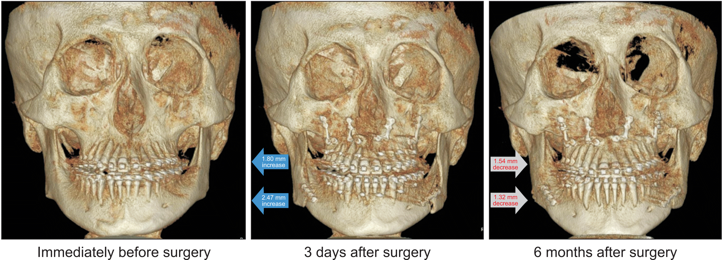
The changes in IGW, IRW, and STW at T1, T2, and T3 are listed in Table 3. IGW, IRW, and STW values showed a statistically significant increase in both groups postoperatively (T2). However, after 6 months (T3), the values decreased significantly (Figure 3). In particular, no significant differences were noted in IRW and STW between T1 and T3. Moreover, a statistical difference between ASG and SYG was not found (p > 0.05).
Table 4 shows UGW in the ASG. On the deviated side, UGW showed a significant increase at T2 and a decrease at T3. The deviated side had a more substantial change compared to the non-deviated side. The asymmetry of the deviated and non-deviated sides for UGW remained after surgery. However, these differences were not statistically significant (p > 0.05). The amount of setback had no statistically significant correlation with IGW, IRW, or STW values (Table 5).
This study showed that immediately after SSRO, both groups showed an increase in mandibular width. These results were consistent with those of previous 2D studies.7,9 However, a statistical difference between ASG and SYG was not found. This may be due to the removal of the early contact between the segments with a surgical round bur during surgery. In addition, since the arch form of the mandible is wider toward the posterior and the width of the distal segment in the gonial area decreases when the distal segment goes setback, mandibular rotation is assumed to be less affected during rotational setback surgery.
The mandibular width decreased at T3 compared to that at T2. Although IGW was statistically different between T1 and T3, the difference was about 1 mm. In particular, no significant differences were identified between T1 and T3 for IRW and STW, which could be because of remodeling caused by the separation of the medial pterygoid muscle, which reduced muscle extension in the gonial region.17 Alternatively, the proximal segment may return to its original position with semi-rigid fixation and neuromuscular adaption. The distal segment can easily change postoperatively, whereas the proximal segment does not change as easily because of its adaptation to the preoperative environment.
The STW of the gonial region almost returned to its original width at T3 after a significant increase at T2. The increase in STW after surgery is mainly due to edema, whereas the subsequent decrease is due to soft tissue adaptation. However, since changes in the soft tissue are influenced by various parameters, such as body weight, facial expressions, and muscle mass, the results should be interpreted cautiously.
The deviated side of the asymmetric patient showed a considerable increase in UGW than the non-deviated side. When the distal segment is shifted toward the facial midline, the rotation causes the proximal segment to flare more on the deviated than on the non-deviated side. Although not statistically significant, the results were similar to previous 2D studies.12,18 If the asymmetry of the deviated and non-deviated sides remains significant after surgery, additional surgery, such as mandibular angle osteotomy, is required.
No statistically significant correlation between the mandibular width and the amount of setback was found, which is also consistent with the previous results.7 The mandibular width does not change proportionally, even when the setback amount is larger.
The head's orientation must be kept constant to compare 3D data at different times accurately. To ensure identical head orientation, CBCT images taken at T2 and T3 were superimposed with those taken at T1 based on the anterior cranial base.19 Furthermore, to reduce measurement inaccuracies, this study selected landmarks such as the gonion point, mastoid process, and lip commissures that can be easily identified.
Despite the valuable findings of this study, there are some limitations. First, the results may lead to bias since a single oral maxillofacial surgeon from a single center treated patients. A multicenter study with a large sample size is required to overcome these limitations. Second, the arch form of the mandible was not fully considered. Third, mandibular asymmetry is not sufficiently categorized in detail. The number of canting-, translation-, and yawing-dominant types in the ASG was 0, 15, and 20, respectively.20 The change in mandibular width may vary depending on the type.
Regardless of the mandibular asymmetry, the mandibular width expanded immediately after mandibular setback surgery using SSRO. Six months after surgery, the mandibular width was similar to its original width.
A video presentation of this article is available at https://youtu.be/xg30oaGH0G0 or www.e-kjo.org.
Notes
AUTHOR CONTRIBUTIONS
Conceptualization: SHK. Data curation: YIK. Formal analysis: YIK. Investigation: YIK. Methodology: SSK. Project administration: SHK. Resources: SSK. Software: SBP. Supervision: SHK. Validation: SSK. Visualization: SBP. Writing–original draft: SSK. Writing–review & editing: all authors.
REFERENCES
1. Jang KS, Bayome M, Park JH, Park KH, Moon HB, Kook YA. 2017; A three-dimensional photogrammetric analysis of the facial esthetics of the Miss Korea pageant contestants. Korean J Orthod. 47:87–99. DOI: 10.4041/kjod.2017.47.2.87. PMID: 28337418. PMCID: PMC5359635.
2. Kim SK, Han JJ, Kim JT. 2001; Classification and treatment of prominent mandibular angle. Aesthetic Plast Surg. 25:382–7. DOI: 10.1007/s002660010150. PMID: 11692255.
3. Yang WS. 1995; The study on the orthodontic patients who visited Department of Orthodontics, Seoul National University Hospital during last 10 years (1985-1994). Korean J Orthod. 25:497–509.
4. Bell WH. 1993. Modern practice in orthognathic and reconstructive surgery. 3rd ed. Saunders;Philadelphia: DOI: 10.1097/00006534-199308000-00030.
5. Busby BR, Bailey LJ, Proffit WR, Phillips C, White RP Jr. 2002; Long-term stability of surgical class III treatment: a study of 5-year postsurgical results. Int J Adult Orthodon Orthognath Surg. 17:159–70. PMID: 12353934.
6. Proffit WR, Phillips C, Dann C 4th, Turvey TA. 1991; Stability after surgical-orthodontic correction of skeletal Class III malocclusion. I. Mandibular setback. Int J Adult Orthodon Orthognath Surg. 6:7–18. PMID: 1940541.
7. Choi HS, Rebellato J, Yoon HJ, Lund BA. 2005; Effect of mandibular setback via bilateral sagittal split ramus osteotomy on transverse displacement of the proximal segment. J Oral Maxillofac Surg. 63:908–16. DOI: 10.1016/j.joms.2004.06.064. PMID: 16003615.
8. Choi WH, Kim JH, Park YW. 2003; A clinical study on the effects of bilateral sagittal split ramus osteotomy on the postoperative condylar positional changes in the mandibular prognathism. J Korean Assoc Maxillofac Plast Reconstr Surg. 25:525–32.
9. Yoo JY, Kwon YD, Suh JH, Ko SJ, Lee B, Lee JW, et al. 2013; Transverse stability of the proximal segment after bilateral sagittal split ramus osteotomy for mandibular setback surgery. Int J Oral Maxillofac Surg. 42:994–1000. DOI: 10.1016/j.ijom.2013.01.021. PMID: 23538214.
10. Yeo BR, Han JJ, Jung S, Park HJ, Oh HK, Kook MS. 2018; Horizontal changes of the proximal mandibular segment after mandibular setback surgery using 3-dimensional computed tomography data. Oral Surg Oral Med Oral Pathol Oral Radiol. 125:14–9. DOI: 10.1016/j.oooo.2017.07.002. PMID: 28958899.
11. Lim SY, Jiang T, Oh MH, Kook MS, Cho JH, Hwang HS. 2017; Cone-beam computed tomography evaluation on the changes in condylar long axis according to asymmetric setback in sagittal split ramus osteotomy patients. Angle Orthod. 87:254–9. DOI: 10.2319/043016-349. PMID: 28253453. PMCID: PMC8384354.
12. Song SH, Kim JY, Lee SH, Park JH, Jung HD, Jung YS. 2019; Three-dimensional analysis of transverse width of hard tissue and soft tissue after mandibular setback surgery using intraoral vertical ramus osteotomy: a retrospective study. J Oral Maxillofac Surg. 77:407.e1–6. DOI: 10.1016/j.joms.2018.10.008. PMID: 30439330.
13. Ha SW, Kim SJ, Choi JY, Baek SH. 2022; Characterization of facial asymmetry phenotypes in adult patients with skeletal Class III malocclusion using three-dimensional computed tomography and cluster analysis. Korean J Orthod. 52:85–101. DOI: 10.4041/kjod.2022.52.2.85. PMID: 35321948. PMCID: PMC8964472.
14. Cohen J. 1992; A power primer. Psychol Bull. 112:155–9. DOI: 10.1037/0033-2909.112.1.155. PMID: 19565683.
15. Dal Pont G. 1961; Retromolar osteotomy for the correction of prognathism. J Oral Surg Anesth Hosp Dent Serv. 19:42–7.
16. Frost D, Bays R, Marciani R, Betts N, Williams T, Powers M, et al. 2000. Oral and maxillofacial surgery. Saunders;Philadelphia: DOI: 10.1016/s0278-2391(00)80047-6.
17. Pan JH, Lee JJ, Lin HY, Chen YJ, Jane Yao CC, Kok SH. 2013; Transverse and sagittal angulations of proximal segment after sagittal split and vertical ramus osteotomies and their influence on the stability of distal segment. J Formos Med Assoc. 112:244–52. DOI: 10.1016/j.jfma.2012.02.013. PMID: 23660219. PMID: fa7aa9c9cded405e9a93471b75d75f96.
18. Kim JW, Son WS, Kim SS, Kim YI. 2015; Proximal segment changes after bilateral sagittal split ramus osteotomy in facial asymmetry patients. J Oral Maxillofac Surg. 73:1592–605. DOI: 10.1016/j.joms.2015.02.021. PMID: 25976689.
19. Choi JH, Mah J. 2010; A new method for superimposition of CBCT volumes. J Clin Orthod. 44:303–12. PMID: 20831099.
20. Kim KA, Lee JW, Park JH, Kim BH, Ahn HW, Kim SJ. 2017; Targeted presurgical decompensation in patients with yaw-dependent facial asymmetry. Korean J Orthod. 47:195–206. DOI: 10.4041/kjod.2017.47.3.195. PMID: 28523246. PMCID: PMC5432441.
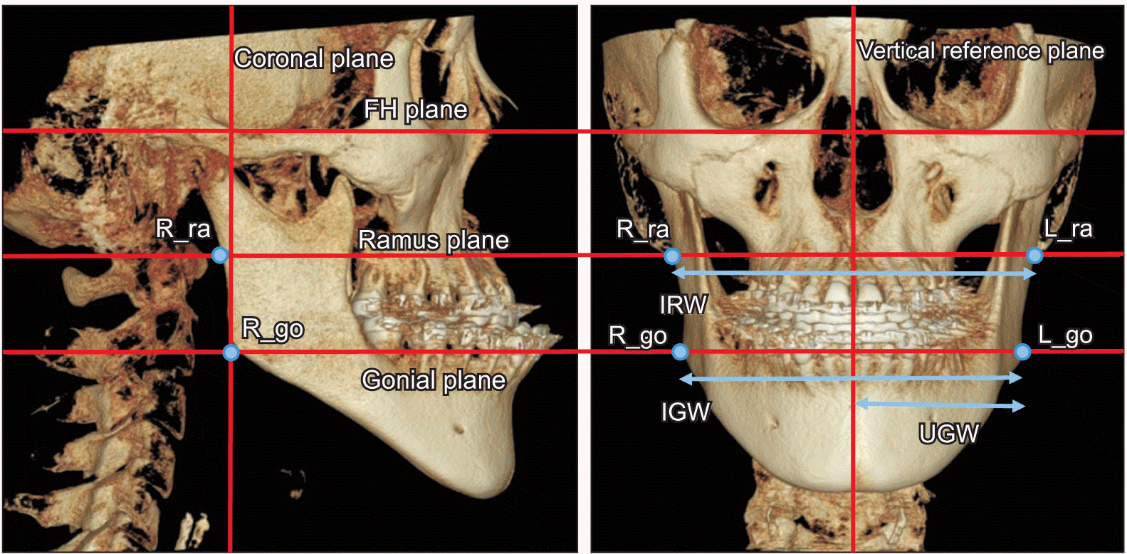
Table 1
Variable | Symmetrical group (n = 35) | Asymmetrical group (n = 35) | p-value |
---|---|---|---|
Sex (Male/Female) | 18/17 | 19/16 | 0.811† |
Age (yr) | 22.7 ± 2.4 | 23.1 ± 2.7 | 0.887* |
1-jaw (SSRO only)/2-jaw | 17/18 | 15/20 | 0.631† |
Chin deviation before surgery (mm) | 1.10 ± 0.74 | 4.58 ± 2.10 | < 0.001* |
Chin deviation after surgery (mm) | 0.01 ± 0.02 | 0.03 ± 0.02 | 0.571* |
Amount of setback (mm) | |||
Both sides | 8.52 ± 2.73 | 8.10 ± 4.28 | 0.623* |
Deviated side | 5.80 ± 3.68 | ||
Non-deviated side | 10.39 ± 3.58 |
Table 2
Table 3
Variable | T1 | T2 | T3 | T2-T1 | T3-T2 | T3-T1 | ||||||||
---|---|---|---|---|---|---|---|---|---|---|---|---|---|---|
Mean ± SD | Mean ± SD | Mean ± SD | Mean ± SD | p-value | Mean ± SD | p-value | Mean ± SD | p-value | ||||||
IGW (mm) | ||||||||||||||
SYG | 99.49 ± 6.19 | 102.40 ± 6.42 | 100.77 ± 6.29 | 2.91 ± 1.67 | < 0.001* | –1.62 ± 1.30 | < 0.001* | 1.29 ± 1.69 | < 0.001* | |||||
ASG | 100.38 ± 6.40 | 102.85 ± 6.36 | 101.53 ± 6.09 | 2.47 ± 1.88 | < 0.001* | –1.32 ± 1.08 | < 0.001* | 1.15 ± 1.85 | 0.002* | |||||
IRW (mm) | ||||||||||||||
SYG | 109.85 ± 4.90 | 112.49 ± 4.72 | 109.95 ± 4.95 | 2.64 ± 1.77 | < 0.001* | –2.54 ± 1.38 | < 0.001* | 0.10 ± 1.58 | 0.674 | |||||
ASG | 110.38 ± 5.81 | 112.19 ± 6.10 | 110.64 ± 6.003 | 1.80 ± 1.85 | < 0.001* | –1.54 ± 1.65 | < 0.001* | 0.26 ± 1.67 | 0.386 | |||||
STW (mm) | ||||||||||||||
SYG | 131.57 ± 10.15 | 143.26 ± 11.89 | 131.33 ± 9.84 | 11.68 ± 7.60 | < 0.001* | –11.92 ± 7.28 | < 0.001* | –0.24 ± 4.53 | 0.713 | |||||
ASG | 133.00 ± 12.29 | 143.40 ± 14.08 | 132.70 ± 13.52 | 10.39 ± 4.87 | < 0.001* | –10.70 ± 5.21 | < 0.001* | –0.30 ± 3.17 | 0.592 |
Table 4
Variable | T1 | T2 | T3 | T2-T1 | T3-T2 | T3-T1 | ||||||||
---|---|---|---|---|---|---|---|---|---|---|---|---|---|---|
Mean ± SD | Mean ± SD | Mean ± SD | Mean ± SD | p-value | Mean ± SD | p-value | Mean ± SD | p-value | ||||||
UGW (mm) | ||||||||||||||
Deviated | 51.05 ± 3.25 | 52.72 ± 3.03 | 51.78 ± 3.52 | 1.67 ± 2.01 | 0.003* | –0.96 ± 1.56 | 0.003* | 0.73 ± 2.39 | 0.554 | |||||
Non-deviated | 49.30 ± 4.37 | 50.12 ± 4.38 | 49.76 ± 3.73 | 0.82 ± 2.10 | 0.105 | –0.36 ± 1.69 | 0.370 | 0.46 ± 2.44 | 0.416 |