

 PDF
PDF Citation
 Print
Print


INTRODUCTION
The concept of occlusion in dentistry pertains to the relationship between all components of the masticatory system in normal and abnormal functions.1-3 The bone and muscle components may dynamically affect each other’s function, resulting in morphological changes of the bone.4,5 Owing to greater masticatory forces, males show greater mandibular growth and remodeling than do females.6 Morphological changes in the temporomandibular joint (TMJ) of young adults may result in malocclusion.7 Morphological alterations occur based on simple developmental variability, such as remodeling of the condyle to adapt to developmental variations, malocclusion, trauma, and other developmental abnormalities and diseases.
The mandibular condylar morphology varies significantly between individuals8,9 and is associated with age, sex, facial type, occlusal force, functional load, malocclusion type, and right and left sides.10 In young adults, the mandibular condyle plays an essential role in the stability of long-term orthodontic and orthognathic treatments.11,12 Many orthodontic studies have been conducted on TMJ spaces, morphological shapes, and volumetric size.11,13-17 Anthropological studies have analyzed the mandibular condyle shape in different populations.18-23 However, conventional two-dimensional imaging methods, such as panoramic radiography, are inadequate to accurately examine the three-dimensional (3D) mandibular condyle morphology.10,24 Therefore, this study aimed to morphologically and morphometrically investigate the mandibular condyle based on sex and different sagittal and vertical skeletal malocclusions using cone-beam computed tomography (CBCT). Each orthodontist must understand the normal variations of the mandibular condyle to avoid misdiagnosis and provide more efficient orthodontic treatment.7,11,12
Go to : 
MATERIALS AND METHODS
Sample-size calculation
A power analysis using G*Power software (Power version 3.1.9.7; University of Dusseldorf, Dusseldorf, Germany) was used to estimate the required sample size to detect differences between group means using analysis of variance (ANOVA), with an effect size f = 0.40; 102 participants were required to achieve a power exceeding 0.90, p = 0.05.
Participants
This study was approved by the Nanjing Medical University Research Ethical Committee, Jiangsu province-affiliated hospital (PJ2018-059-001). All procedures followed in this experiment were in accordance with the ethical standards of the responsible committee on human experimentation (institutional and national) and the Helsinki Declaration of 1964 and its later versions. Informed consent was obtained from all patients for inclusion in the study.
The study sample comprised 135 patients, including 54 males (23.7 ± 4.39 years) and 81 females (21 ± 3.89 years) who visited our institution seeking various dental treatments. They were classified according to the sagittal skeletal relationship (A point-nasion-B point [ANB] angle): skeletal Class I (1° ≤ ANB ≤ 4°), skeletal Class II (ANB > 4°), and skeletal Class III (ANB < 1°). They were also classified according to the vertical skeletal relationship based on the sella-nasion and mandibular plane (SN-MP angle) as follows: hypodivergent (SN-MP < 27°), normodivergent (27° ≤ SN-MP ≤ 37°), and hyperdivergent (SN-MP > 37°). According to age, the sample was classified into three groups (group 1 ≤ 20 years; 21 years ≤ group 2 < 30 years; and group 3 ≥ 30 years) (Table 1). CBCT images were obtained using NewTom VGi Evo (Cefla S.C., Imola, Italy) with the following exposure parameter settings: 17 seconds scan time, 18 × 16-cm field of view, and 0.5-mm voxel size. The patients were instructed to sit upright, bite in centric occlusion (CO), and look forward to maintain the Frankfort horizontal plane parallel to the floor. The CBCT data were saved in Digital Imaging and Communications in Medicine (DICOM) files. Patients were excluded if they had a history of previous orthodontic treatment, CO-centric relationship discrepancy, trauma to the dentofacial region, TMJ disorders, and diseases affecting bone metabolism.
Table 1
Sample distribution
![]()
Study design
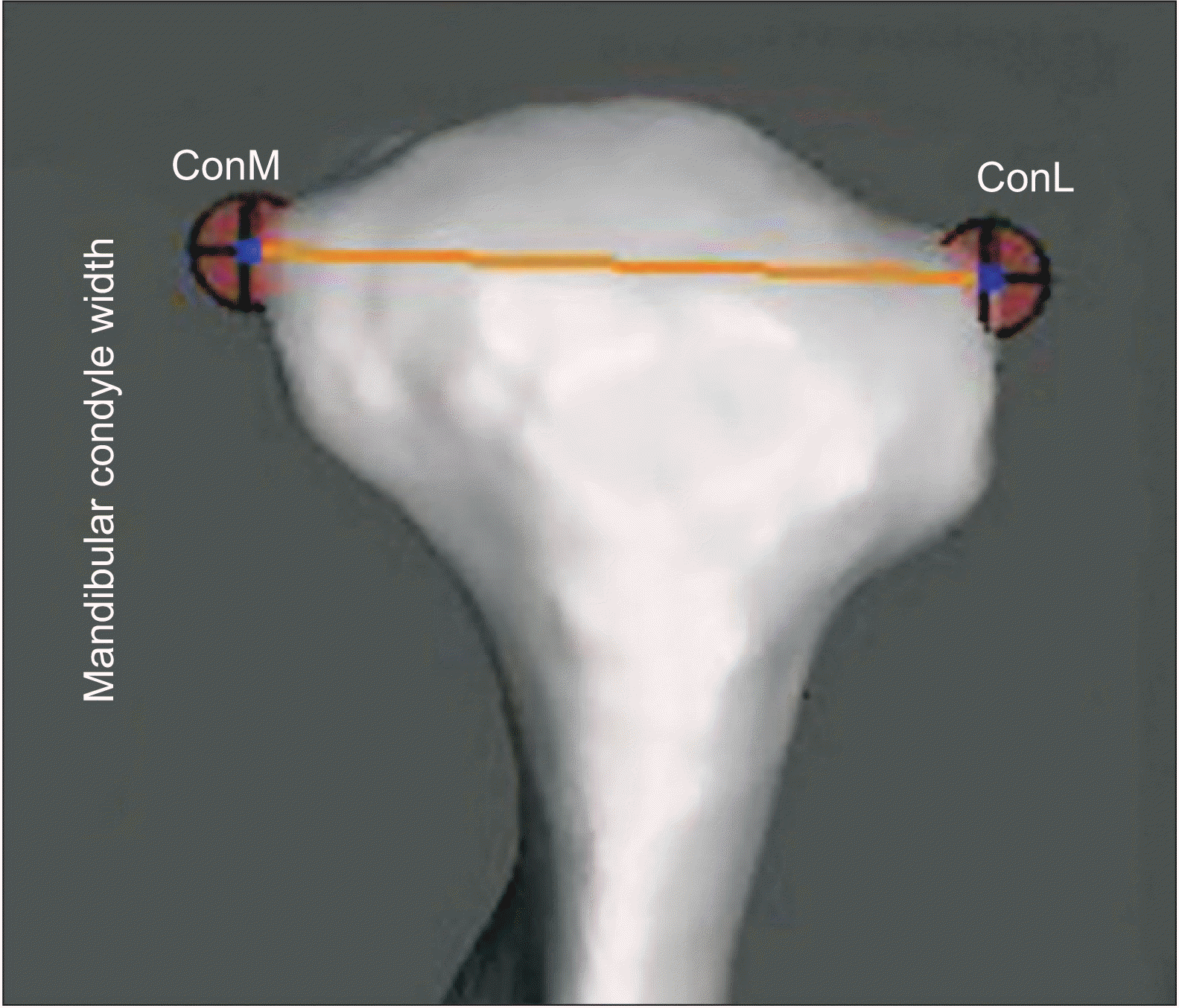
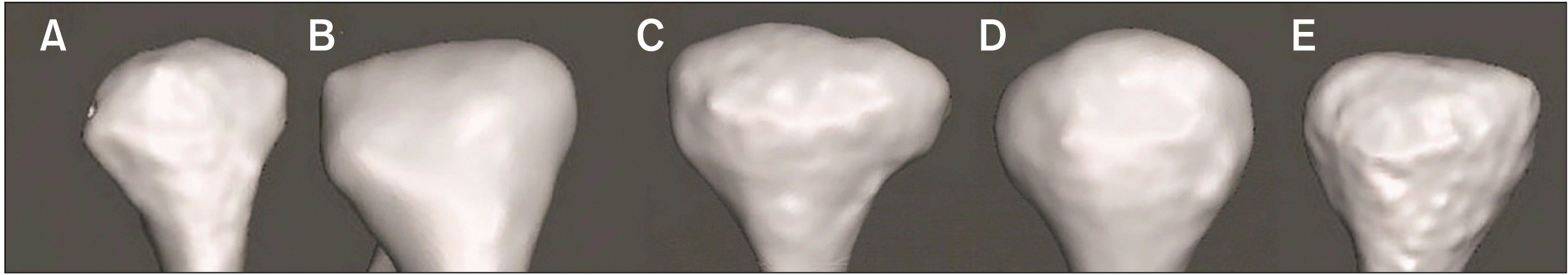
Morphological and morphometric variables were blindly investigated in each patient group. Dolphin© (version 11.9.20; Dolphin Imaging & Management Solutions, Chatsworth, CA, USA) was used for cephalometric analysis. The ITK-SNAP software (version 3.8; Penn Image Computing and Science Laboratory at the University of Pennsylvania, the Scientific Computing and Imaging Institute at the University of Utah) was used for mandible segmentation by outlining the boundaries using semiautomated discrimination procedures to create a 3D standard tessellation language (STL) model of the area of interest. The Meshmixer software (version 3.5 Autodesk open-source) was used to determine the variable locations, shapes, and measurements, and all variables on both sides were evaluated separately. The Frankfort and orbital planes were parallel to the floor (Figure 1). Based on previous studies,18,22,23 the landmarks used for analysis are illustrated in Figures 2 and 3. Condylion (Con) is the most superior point of the head of the mandibular condyle in the sagittal section; condylion lateral (ConL) is the most lateral point of the condyle process in the coronal section; condylion medial (ConM) is the most medial point of the condylion process in the coronal section; sigmoid point (Sig) is the deepest point of the sigmoid notch of the mandible, which represents the sigmoid plane; the line passing from the condylion to the mandibular notch plane along the long axis of the condylar process is the ConH line; and the condylar width (ConL-ConM) line connects the most lateral and medial points of the condyle. Figures 4 and 5 show the morphological shapes of the mandibular condyle in sagittal and coronal sections.
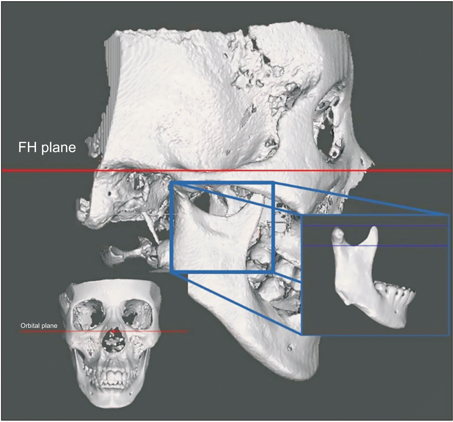 | Figure 1Three-dimensional model orientation: the Frankfort horizontal (FH) and orbital planes parallel to the floor and the sigmoid plane in the box parallel to the Frankfort plane.
|
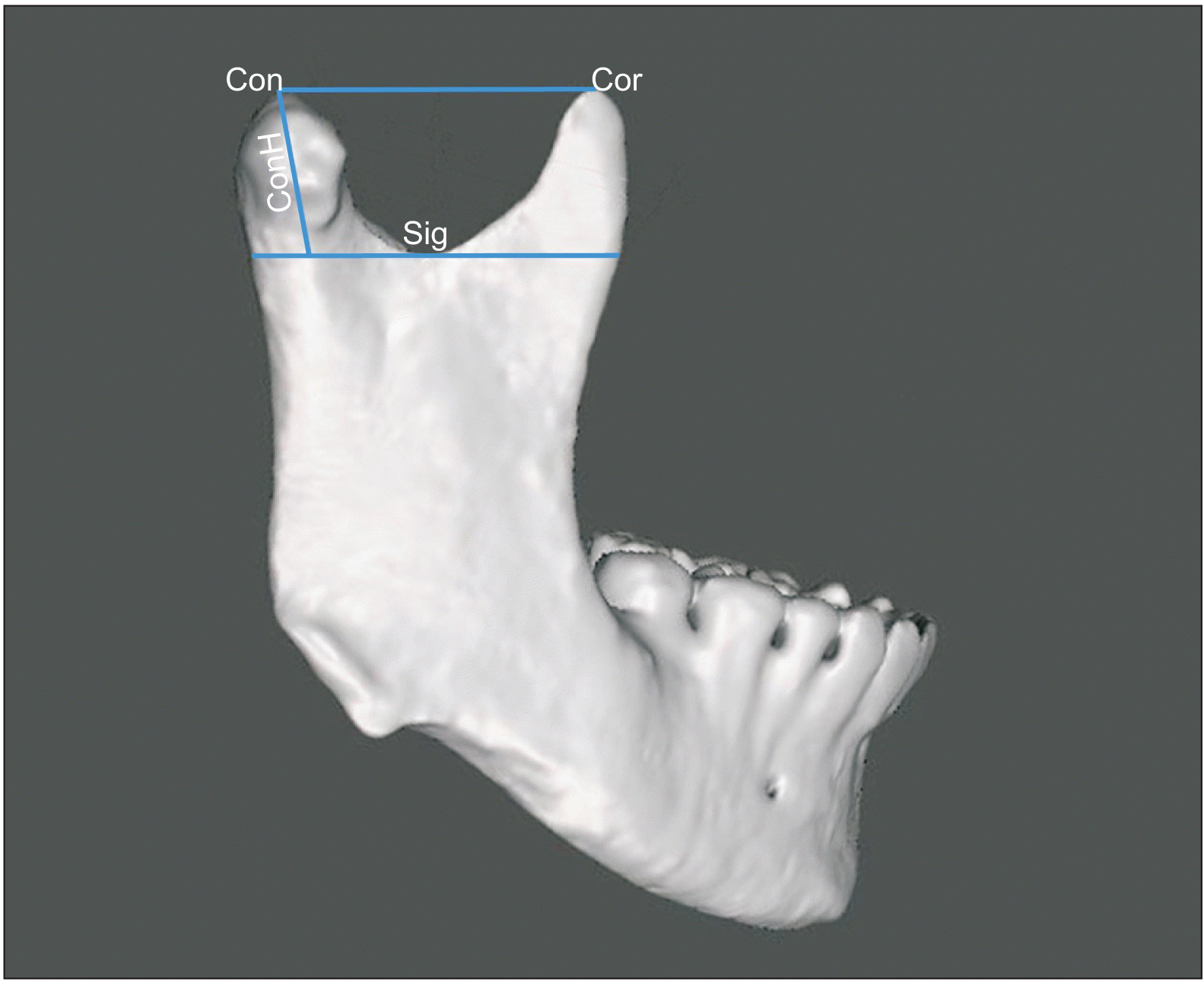 | Figure 2Sagittal view showing the landmarks used for measurements of the mandibular condyle.
Con, the most superior point of the mandibular condyle; Cor, the most superior point of the coronoid process; ConH, a line extending from Con to the sigmoid plane, intersecting the long axis of the mandibular condyle; Sig, sigmoid notch.
|
Statistical analysis
The sample distributions are listed in Table 1. One investigator collected all measurements from the 135 participants. To evaluate intraexaminer reliability, the same examiner re-analyzed 20 randomly selected participants within a 3-week interval. The measures were evaluated using intraclass correlation coefficients. The results show good intraexaminer repeatability (Table 2). SPSS software (version 24.0 for Windows; IBM Corp., Armonk, NY, USA) was used for statistical analysis. The Shapiro–Wilk normality test was performed on continuous variables with a normal distribution pattern. We used an independent sample t-test to compare the participants’ mean condylar height and width by sex (Table 3), while an ANOVA test was used to compare the mean values of the mandibular condyle height and width in various skeletal patterns and age groups (SN-MP, ANB) (Tables 4–6); Scheff's post hoc test was used when the ANOVA test was significant (Tables 4–6). The chi-square test was used to evaluate the prevalence of condyle shape in coronal and sagittal sections and determine whether there is a statistical association among different sexes, skeletal patterns, and age groups (Tables 7–10). Statistical significance was set at p < 0.05.
Table 2
Intraexaminer repeatability test
![]()
Table 3
Independent t-test among male and female
| Variable | Sex | Mean | SD | T-value | p-value |
|---|---|---|---|---|---|
| LCW | M | 19.40 | 2.42 | 1.73 | 0.08 |
| F | 18.64 | 2.55 | |||
| LCH | M | 19.08 | 3.38 | 0.67 | 0.50 |
| F | 18.69 | 3.23 | |||
| RCW | M | 19.14 | 2.44 | 0.79 | 0.42 |
| F | 18.80 | 2.39 | |||
| RCH | M | 18.97 | 3.01 | 0.27 | 0.78 |
| F | 18.82 | 3.14 |
![]()
Table 4
ANOVA of the mandibular condylar height and width in different sagittal skeletal patterns
| Variables | ANB groups | Mean | SD | p-value† | Mean difference | p-value‡ | ||
|---|---|---|---|---|---|---|---|---|
| LCW | Class I | 18.85 | 2.36 | 0.454 | Class I | Class II | 0.18 | 0.942 |
| Class II | 18.66 | 2.71 | Class III | –0.46 | 0.683 | |||
| Class III | 19.32 | 2.48 | Class II | Class I | –0.18 | 0.942 | ||
| Class III | –0.65 | 0.476 | ||||||
| LCH | Class I | 18.49 | 3.17 | 0.000*** | Class I | Class II | 0.84 | 0.438 |
| Class II | 17.65 | 3.07 | Class III | –1.92 | 0.015 | |||
| Class III | 20.41 | 3.05 | Class II | Class I | –0.84 | 0.438 | ||
| Class III | –2.76 | 0.000*** | ||||||
| RCW | Class I | 18.84 | 2.20 | 0.510 | Class I | Class II | 0.14 | 0.963 |
| Class II | 18.70 | 2.64 | Class III | –0.42 | 0.703 | |||
| Class III | 19.27 | 2.38 | Class II | Class I | –0.14 | 0.963 | ||
| Class III | –0.56 | 0.538 | ||||||
| RCH | ClassI | 18.74 | 2.72 | 0.000*** | Class I | Class II | 1.15 | 0.168 |
| Class II | 17.58 | 3.11 | Class III | –1.58 | 0.037 | |||
| Class III | 20.32 | 2.81 | Class II | Class I | –1.15 | 0.168 | ||
| Class III | –2.74 | 0.000*** | ||||||
![]()
Table 5
ANOVA of the mandibular condylar height and width in different vertical skeletal patterns
| Variables | SN-MP groups | Mean | SD | p-value† | Mean difference | p-value‡ | ||
|---|---|---|---|---|---|---|---|---|
| LCW | Hypodivergent | 19.25 | 2.03 | 0.553 | Hypodivergent | Normodivergent | 0.28 | 0.861 |
| Normodivergent | 18.96 | 2.51 | Hyperdivergent | 0.66 | 0.555 | |||
| Hyperdivergent | 18.59 | 2.98 | Normodivergent | Hypodivergent | –0.28 | 0.861 | ||
| Hyperdivergent | 0.37 | 0.784 | ||||||
| LCH | Hypodivergent | 18.87 | 3.23 | 0.986 | Hypodivergent | Normodivergent | 0.06 | 0.995 |
| Normodivergent | 18.80 | 3.29 | Hyperdivergent | –0.04 | 0.998 | |||
| Hyperdivergent | 18.92 | 3.44 | Normodivergent | Hypodivergent | –0.06 | 0.995 | ||
| Hyperdivergent | –0.11 | 0.987 | ||||||
| RCW | Hypodivergent | 19.31 | 2.01 | 0.276 | Hypodivergent | Normodivergent | 0.30 | 0.837 |
| Normodivergent | 19.01 | 2.45 | Hyperdivergent | 0.91 | 0.295 | |||
| Hyperdivergent | 18.40 | 2.67 | Normodivergent | Hypodivergent | –0.30 | 0.837 | ||
| Hyperdivergent | 0.60 | 0.489 | ||||||
| RCH | Hypodivergent | 18.87 | 2.88 | 0.999 | Hypodivergent | Normodivergent | –0.01 | 1.000 |
| Normodivergent | 18.89 | 2.90 | Hyperdivergent | –0.01 | 1.000 | |||
| Hyperdivergent | 18.88 | 3.65 | Normodivergent | Hypodivergent | 0.01 | 1.000 | ||
| Hyperdivergent | 0.00 | 1.000 | ||||||
![]()
Table 6
ANOVA of the mandibular condyle height and width in different age groups
| Variables | SN-MP groups | Mean | SD | p-value† | Mean difference | p-value‡ | ||
|---|---|---|---|---|---|---|---|---|
| LCW | Group 1 ≤ 20 yr | 18.31 | 2.28 | 0.233 | Group 1 ≤ 20 yr | 21 yr ≤ Group 2 < 30 yr | –0.88 | 0.238 |
| 21 yr ≤ Group 2 < 30 yr | 19.2 | 2.38 | Group 3 ≥ 30 yr | –0.72 | 0.550 | |||
| Group 3 ≥ 30 yr | 19.04 | 3.13 | 21 yr ≤ Group 2 < 30 yr | Group 1 ≤ 20 yr | 0.88 | 0.238 | ||
| Group 3 ≥ 30 yr | 0.15 | 0.963 | ||||||
| LCH | Group 1 ≤ 20 yr | 18.57 | 3 | 0.844 | Group 1 ≤ 20 yr | 21 yr ≤ Group 2 < 30 yr | –0.39 | 0.844 |
| 21 yr ≤ Group 2 < 30 yr | 18.97 | 3.21 | Group 3 ≥ 30 yr | –0.3 | 0.942 | |||
| Group 3 ≥ 30 yr | 18.87 | 3.95 | 21 yr ≤ Group 2 < 30 yr | Group 1 ≤ 20 yr | 0.39 | 0.844 | ||
| Group 3 ≥ 30 yr | 0.09 | 0.991 | ||||||
| RCW | Group 1 ≤ 20 yr | 18.14 | 2.07 | 0.084 | Group 1 ≤ 20 yr | 21 yr ≤ Group 2 < 30 yr | –1.06 | 0.099 |
| 21 yr ≤ Group 2 < 30 yr | 19.21 | 2.32 | Group 3 ≥ 30 yr | –1.04 | 0.257 | |||
| Group 3 ≥ 30 yr | 19.18 | 2.9 | 21 yr ≤ Group 2 < 30 yr | Group 1 ≤ 20 yr | 1.06 | 0.099 | ||
| Group 3 ≥ 30 yr | 0.02 | 0.990 | ||||||
| RCH | Group 1 ≤ 20 yr | 18.44 | 3.04 | 0.501 | Group 1 ≤ 20 yr | 21 yr ≤ Group 2 < 30 yr | –0.46 | 0.767 |
| 21 yr ≤ Group 2 < 30 yr | 18.91 | 2.96 | Group 3 ≥ 30 yr | –0.95 | 0.504 | |||
| Group 3 ≥ 30 yr | 19.4 | 3.48 | 21 yr ≤ Group 2 < 30 yr | Group 1 ≤ 20 yr | 0.46 | 0.767 | ||
| Group 3 ≥ 30 yr | –0.49 | 0.789 | ||||||
![]()
Table 7
Chi-square test; prevalence of mandibular condyle shapes by sex
![]()
Table 8
Chi-square test; prevalence of mandibular condyle shapes by the sagittal skeletal pattern
![]()
Table 9
Chi-square test; prevalence of mandibular condyle shapes by the vertical skeletal pattern
![]()
Table 10
Chi-square test; prevalence of the mandibular condyle shapes by age
![]()
Go to :
RESULTS
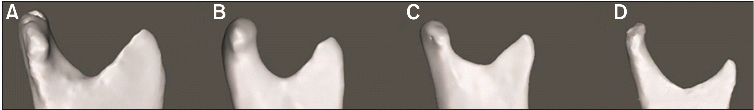
The mean values of the mandibular condyle height and width among males were more prominent than those among females; there was a significant difference in condylar height (p < 0.05) in different (ANB) groups (Table 4), and the mean condylar height values in Class III malocclusion were more prominent than those in Class I and II malocclusions (Table 4). There were no differences between the mean condylar height and width values in the SN-MP groups (Table 5). Table 6 presents the mean condylar height and width values among different age groups, with no significant differences among the age groups. The chi-square test results based on sex (Table 7), skeletal pattern (Tables 8 and 9), and age (Table 10) showed that the most prevalent shape of the condyle in the coronal view was convex (35.1%); the convex shape was also the most prevalent among males (44.4%) and in the Class I (33.3%), Class II (35.6%), Class III (36.7%), hypodivergent (31.4%), normodivergent (35.6%), and hyperdivergent (38.2%) groups. The most prevalent condylar shape among females was round (33.3%). In the sagittal view, the prevalence of the oval condyle shape was 53.7% overall, 52.7% among males, 54.3% among females, 50% in the Class I, 45.5% in the Class II, 65% in the Class III, 58.5% in the hypodivergent, 53% in the normodivergent, and 50% in the hyperdivergent groups. Chi-square test results showed no statistically significant associations of the mandibular condyle shapes in different sections with sex or skeletal pattern (p > 0.05).
Go to :
DISCUSSION
Our study investigated the morphology and morphometrics of the mandibular condyle. Several previous anthropological studies18,20,22,23,25 have investigated the morphological shapes of the mandibular condyle based on sex; however, no study has attempted to identify the morphological variations in the condyle between different sagittal and vertical skeletal malocclusions. Lopez et al.25 and Ishwarkumar et al.20 found that the mandibular condyle height and width were more prominent in males than in females. Previous studies16,26 have reported a greater mandibular condyle volume among skeletal Class III malocclusion patients. Noh et al.17 observed that the condylar height and width were greater in Class III malocclusion than in Class II malocclusion, and the condylar width of hypodivergent patients was greater than that of hyperdivergent patients. In line with previous studies, the current research indicates that the condylar height of patients with Class III malocclusion is greater than that of patients with Class I and II malocclusion. We found that the condylar width was more prominent in Class III malocclusion than in Class I and II malocclusions and greater in the hypodivergent group than in the normodivergent and hyperdivergent groups. These findings may indicate that there is excessive vertical development of the mandibular ramus in patients with Class III malocclusion. It is likely that condylar height plays an essential role during the development of Class III malocclusion but has no impact on vertical skeletal malocclusions. In the current study, patients ≤ 20 years had a lower condylar height than those > 20 years. Although this difference was not statistically significant, it might be related to late mandibular condyle growth. Wolff’s law27 states that bone morphology and internal architecture depend on the load applied to the bone. Previous studies have found that hypodivergent patients have higher maximum bite forces.28,29 In contrast, hyperdivergent patients have weaker bite forces during clenching and chewing because of decreased muscle tonicity;30 thus, it may affect the mandibular condyle shape viewed in sagittal or coronal sections because of different loads applied by the masticatory muscles. Our findings contradicted the hypothesis as no statistical association was found between the mandibular condyle shape and different groups of sex, age, and sagittal and vertical skeletal patterns. Tassoker et al.18 and Yale et al.31 observed the that the condylar shape was predominantly convex in the coronal view. Similarly, we found that the most prominent shape of the mandibular condyle is convex and has no clinical association with sex, age, or sagittal and vertical skeletal malocclusions. Previous anthropological studies18,21,23,32-34 reported that the round/oval shape of the mandibular condyle is the most common in sex from the sagittal view. Vahanwala et al.35 and Anisuzzaman et al.36 reported that most common condylar shape was oval (> 60%) and the least common was the crooked-finger shape (2%). Our study found that the most common condylar shape from the sagittal view was oval shape (53.7%) and the least common was the crooked finger shape (8.1%). The morphological shape of the condyle showed no statistical association with sex, age, and sagittal and vertical skeletal malocclusions. However, it might be related to TMJ spaces where the crooked finger-shaped condyle had the smallest size in comparison with that of condyles with other shapes. The differences in the measurements might be attributed to racial diversity, sample size, and study design. Our findings suggest no association between mandibular condyle shape variations and sex, age, and sagittal and vertical skeletal malocclusion. At the same time, there was a statistically significant relationship between the mandibular condyle height and skeletal sagittal malocclusion, suggesting that the mandibular condyle growth potential in Class III malocclusion was higher than that in Class I and II malocclusion patients.
A potential limitation of our study is an insufficient age group distribution, which might have affected the accuracy of our results. Future studies on the morphological shapes of the mandibular condyle and their relation to the TMJ and dysfunctions are necessary.
Go to :
CONCLUSIONS
This study morphologically and morphometrically evaluated the relationship between mandibular condyles and various skeletal malocclusion patterns in adults, using CBCT.
There were no statistical associations between the morphological shapes of the mandibular condyle and sex and sagittal and vertical skeletal malocclusions.
The mandibular condyle height was greater in Class III malocclusion than in Class I and II malocclusions.
The mandibular condyle width of males was greater than that of females.
Go to :
 XML Download