

 PDF
PDF Citation
 Print
Print


INTRODUCTION
Canine impaction is two to three times more frequent in female than in male.1-3 Following its normal eruption pathway, the maxillary canine moves in the occlusal direction in a close relationship with the neighboring teeth. The canine angulation to the midline reaches its maximum tipping around 9 years of age.4 From this development stage, the eruption path of the maxillary canine presents a gradual uprighting due to distal movement of its cusp tip.4 Maxillary canines with excessive mesial angulation and displacement, exceeding the limits of adjacent roots and overlapping them, have been considered potentially impacted.5-7
The sequelae related to the ectopic eruption of maxillary canines can be esthetically and functionally devastating for the patient, especially when considering the risk of maxillary incisor loss and the possibility of failure of orthodontic traction of a severely impacted canine. These clinical conditions end up determining the need for complex, long-term, and costly orthodontic treatments.
Although relevant benefits of interceptive treatment in early mixed dentition have already been shown, the early diagnosis of potentially impacted canines is not always performed on time,8 possibly because canine ectopic eruption is a silent developmental problem that may have associated genetic or environmental risk factors.3,9 A previous study reported that, in a sample of palatally displaced canines, 48% of patients had some maxillary lateral incisor anomaly, including peg-shaped, small, or absent lateral incisor, while 52% did not.10 Thus, even when risk factors cannot be identified, maxillary canine ectopic eruption should not be discounted, and local clinical signs produced by canine ectopic position should be prioritized to evaluate the need for a supplementary radiographic examination.11-14 This is especially true for young patients with no objective treatment need, who represent a significant percentage of the population.15 The systematic indication of a radiographic examination at this age just to exclude an eventual canine eruption deviation can be considered controversial, since the prevalence of canine impaction is around 0.2–2.4%.3,16
Despite its great diagnostic relevance, the clinical signs associated with potentially impacted canines at an early age may be subtle and less well known than the widely reported radiographic findings. From radiographic studies, it is known that maxillary lateral incisor characteristics can both influence and be influenced by the maxillary canine eruption pathway.6,17,18 According to Ericson and Kurol’s criteria,12 8% of children over 10 years of age may require a supplementary radiographic examination. However, when known genetic and environmental risk factors are not present, patients may be at lower risk for canine displacement and may have more restricted clinical signals. As far as we know, no other non-radiographic study has excluded known genetic and environmental risk factors to evaluate whether clinical signs may still be able to predict mesially displaced canines in low-risk patients. Given this scenario, the objective of this study was to evaluate the null hypothesis that there is no difference in a set of clinical predictors represented by the position, size, and shape of the clinical crown of the maxillary lateral incisor, as well as the dental arch dimensions between low-risk patients with and without potentially impacted canines.
MATERIALS AND METHODS
This observational cross-sectional investigation complies with the STROBE (Strengthening the Reporting of Observational Studies in Epidemiology) statement and was based on retrospective data obtained from orthodontic records of patients who sought treatment at the School of Dentistry, Federal University of Rio Grande do Sul, and the Orthodontic Service of the Military Polyclinic at Porto Alegre, Brazil. It was approved by the corresponding institutional review board, under number CAAE28116920.4.0000.5347. The sample was selected from a pool of 1,534 orthodontic records of patients treated at two research centers. Orthodontic records were retrospectively selected from June 2019 to January 2020. The parents of all participants at each research center signed informed consent forms. The sample size calculation was performed assuming values of 5% and 20% for α (Type I error) and β (Type II error), respectively. The minimum difference of the angulation of the maxillary lateral incisors to be detected in patients with and without potentially impacted canines was 5°. The standard deviation (8°) was taken from a previous study.19 The sample size calculation indicated that a minimum of 32 canines was needed in each group.
Sample selection was based on the following inclusion criteria: patients who have good quality of orthodontic records with panoramic radiographs and dental casts taken on the same date, showing mixed dentition stage, ectopically or normally erupting bilateral maxillary canines in Nolla's developmental stages 7 or 8,20 erupted permanent maxillary lateral incisors in Nolla's developmental stages 8 to 10.
Exclusion criteria were patients with restoration or reshaping of the maxillary lateral incisors; maxillary anterior crowding greater than 2 mm; syndromes; cyst or any other periapical lesion in the maxillary anterior segment; dental anomalies of size, number, or shape; previous orthodontic treatment; history of prolonged sucking habits and dental arch narrowing; history of dental trauma; and early loss of deciduous maxillary canines or molars. All orthodontic records that met these criteria were included in this study. Thus, low-risk patients were selected by eliminating known genetic and environmental risk factors for canine displacement, such as crowding, dental anomalies, pathological lesions, maxillary arch narrowing, dental trauma, and early tooth loss.
The normal canine position group (NC) consisted of 30 patients with 60 normally erupting canines with the crown positioned in sector I, while the mesially displaced canine group (DC) consisted of 30 patients with 41 potentially impacted canines with the crown positioned in sectors II, III, or IV.5 Canines positioned in sector II should also have an angle between the canine long axis and the midline (α angle) equal to or greater than 30°,21 and/or cusp tip positioned in the apical radicular third of the adjacent maxillary lateral incisor as additional severity aggravating factors. The NC group had a mean age of 9.30 years (16 female and 14 male participants) and the DC group had a mean age of 9.46 years (23 female and 7 male participants), with a total of 101 evaluated canines that were selected from consecutive orthodontic records from two centers.
Maxillary dental casts were digitized using a 3Shape 3D scanner (3Shape A/S, Copenhagen, Denmark). Measurements of the positional and dimensional characteristics of the lateral incisor and dental arch were performed on the digital dental models using OrthoAnalyzer 3D software (3Shape A/S), as shown in Figures 1 and 2. In addition, the clinical crown of the maxillary lateral incisor was classified according to its morphological characteristic. A square shape was defined as a tooth with parallel interproximal lines, a triangular shape was defined as a tooth with interproximal lines that flared from the gingival margin to the incisal edge, and an oval shape was defined as a tooth with interproximal lines that curved toward each other incisally and cervically.
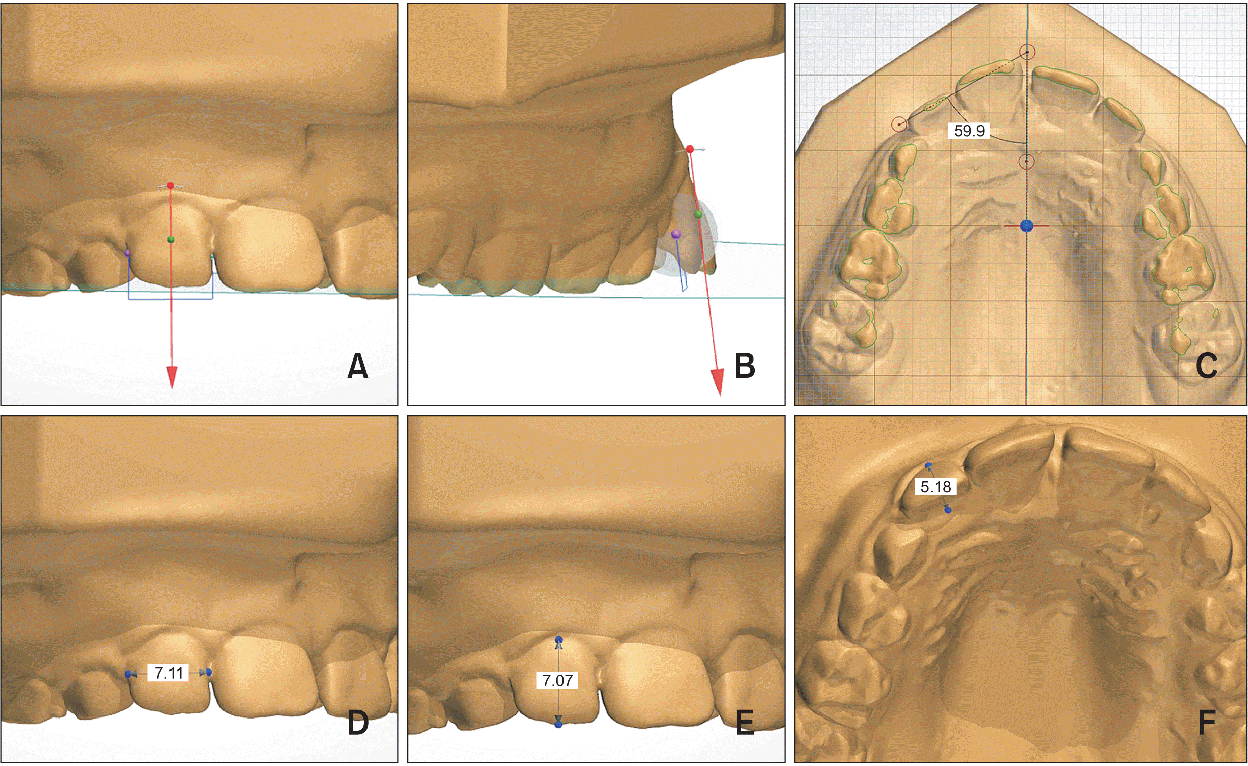
Angulation, inclination, and rotation of the lateral incisor were evaluated by using the occlusal plane passing through the right and left mesiobuccal cusp tips of the permanent maxillary first molars and a point located at the mesioincisal angle of the left central incisor as a reference. The long axis of the maxillary lateral incisors was represented by an arrow in the virtual setup module of OrthoAnalyzer (Figures 1A and 1B). To determine tooth angulation, this arrow was mesiodistally manipulated on the buccal side, adjusting it to the facial axis of the clinical crown (FACC) defined by Andrews (Figure 1A). From a distal view of the maxillary lateral incisor, the arrow was buccolingually manipulated at Andrews' facial axis point to determine the crown tipping (Figure 1B). The mesiodistal and buccolingual angular position of the maxillary lateral incisor was determined by the angle between the arrow and the occlusal plane, which was automatically calculated by the software (Figures 1A and 1B). The axial rotation of the maxillary lateral incisor was determined by the angle between the line passing through the incisal edge of the lateral incisor and the mid-palatal raphe, which were projected on the occlusal plane (Figure 1C).
The mesiodistal width of the maxillary lateral incisor was represented by the greatest distance between the mesial and distal contact points parallel to the incisal surface (Figure 1D). The clinical crown height was the distance between the incisal and cervical limits of the FACC (Figure 1E). The distance between the cervical limit of the FACC and the corresponding cervical limit on the palatal side was the buccolingual width of the maxillary lateral incisor (Figure 1F).
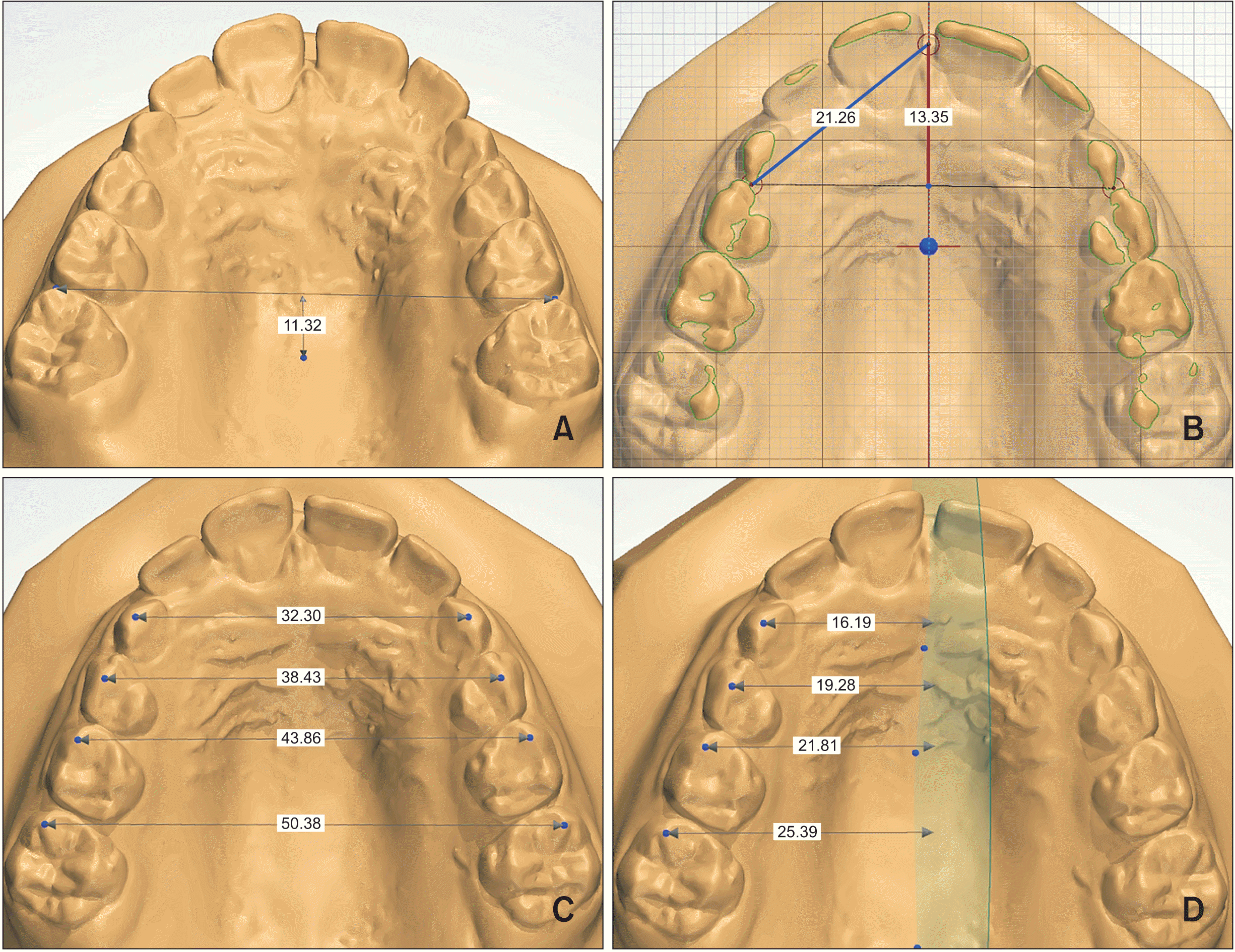
Palatal depth was measured from a line passing through the mesial gingival papillae of the right and left permanent first molars to the deepest point on the palatal surface (Figure 2A). The maxillary anterior arch length was measured perpendicularly in the occlusal plane from the mesial aspect of the right and left deciduous first molars to a midpoint between the central incisors (Figure 2B). The transverse dimensions of the maxillary dental arch were represented by the intercanine and intermolar distances, which were measured between the cusp tips of the right and left deciduous canines and mesiobuccal cusp tips of the right and left molars (Figure 2C). The hemiarch transverse distances were measured perpendicularly from the same dental landmarks to a sagittal plane passing through the palatal raphe (Figure 2D). The anterior perimeter of the maxillary dental hemiarch was measured in the occlusal plane from the mesial contact point of the deciduous first molar to a midpoint between the central incisors (Figure 2B).
The measurements were performed at the same time by a same examiner (K.C.), who was previously calibrated and blinded to the selection criteria and group allocation. The data collection took a period of 1 month. Thirty study models were randomly selected, and a second measurement was performed by the same examiner at least 3 weeks apart to evaluate intraexaminer reliability. A second examiner (S.E.B.), with similar training and experience and working independently of the first, repeated the measurements on the same dental casts to evaluate interexaminer reliability. Intraclass correlation coefficients (ICCs) were used to assess intra- and interexaminer reliability for all linear and angular measurements.
Statistical analyses
The Shapiro–Wilk test was used to check data normality. Intergroup comparisons were performed using independent t-tests, Mann–Whitney U tests, and chi-square tests.
The mesial displacement severity of the maxillary canine was correlated with the discriminant variables between groups using Spearman’s correlation tests.
All statistical tests were conducted using Statistica software (Version 7.0; StatSoft Inc., Tulsa, OK, USA), adopting a significance level of 5%.
RESULTS
Intra- and interexaminer measurement reliability was considered good to excellent. The intraexaminer ICC ranged from 0.844 (inclination) to 0.989 (rotation) for angular measurements and from 0.899 (mesiodistal width) to 0.998 (palatal depth) for linear measurements. The interexaminer ICC ranged from 0.816 (angulation) to 0.964 (rotation) for angular measurements and from 0.802 (palatal depth) to 0.935 (cervico-occlusal height) for linear measurements.
The groups were similar in terms of chronological age. The group with displaced canines showed mesial displacement of the canines positioned between sectors II and III (2.46 ± 0.66). A significant association between sex and mesially displaced canines was observed, since 78.05% of the potentially impacted canines occurred in female participants. Unilateral canine displacement was more prevalent than bilateral. There was a balanced proportion of unilaterally displaced canines between the right and left sides (Table 1).
The evaluation of positional characteristics showed that the maxillary lateral incisor crown was significantly angulated more mesially (upright) and rotated mesiolabially in the group with displaced canines. The dimensional and morphological characteristics of the maxillary lateral incisor crowns were not different between groups. Comparisons of the dental arch dimensions showed that the DC group had a shallower palate and a shorter maxillary anterior arch length (Table 2).
There was a significant positive correlation between the severity of mesial displacement of the maxillary canine and the angulation of the maxillary lateral incisor, while a significant negative correlation was found between the canine displacement severity and lateral incisor rotation, palatal depth, and anterior arch length (Table 3).
DISCUSSION
Delayed treatment of maxillary canine impaction is more complex, costlier, and riskier than early intervention, making its results less predictable.22-26 The diagnosis of canine ectopic eruption begins with recognition of its clinical signs, which can be decisive in supporting the need for radiographic examinations.12-14 This is especially true for young patients with no prior radiographic examination and no known genetic or environmental risk factors. In these patients, there is a greater risk of ectopic eruption of the maxillary canine advancing as a silent, hidden, and devastating development problem if clinical signs are not carefully evaluated to determine the need for complementary radiographic examination. Therefore, this study aimed to identify early clinical features that should raise suspicion for maxillary canine ectopic eruption in low-risk patients, which should be radiographically confirmed. The standardized root development stage established for sample selection contributed to the age similarity between groups (Table 1), which is an important parameter for comparison because the canine position and its relationship with adjacent structures are sensitive to the patient’s age.4,27 The sample characteristics of this study confirm previous reports of an unequal distribution between sexes, with higher prevalence rate among female, lower prevalence for bilaterally displaced canines, and almost equal distribution of unilaterally displaced canines between the right and left sides (Table 1).3,10,28-30 Although the familial history of canine impaction could not be retrieved in this retrospective study, the genetic component associated with ectopic canine inheritance has low penetrance,31,32 and its expression is often associated with dental anomalies,10,33,34 which were excluded from this study.
The results showed that the position (angulation and rotation) of the maxillary lateral incisor crown was more mesiodistally upright and mesiolabially rotated in low-risk patients with mesially displaced canines (Table 2). Similar findings were previously reported by two radiographic studies, showing that maxillary lateral incisor crowns adjacent to mesially displaced canines were angulated 5° more mesially (upright) and rotated 11.7° more mesiolabially, while the respective values obtained in this study were 9.3° and 8.8° (Table 2).19,27 It has been demonstrated that the mesial inclination of the maxillary permanent canine increases during the eruption process and reaches its highest value between 9 and 10 years of age.7 This event is closely related to the peak of the “ugly duckling” stage, which is characterized by distal angulation of the clinical crown of the maxillary lateral incisor and mesial displacement of its root apex.29,31,32 Considering that the distal aspect of the maxillary lateral incisor root acts as a natural containment barrier for the initial mesial and palatal movement of the canine, guiding its eruption,27 a relevant reduction in distal angulation of the clinical crown of maxillary lateral incisors adjacent to displaced canines may be an important clinical sign that the canine has lost its relationship with its eruption guidance. In fact, the farther the canine sector was from its eruption guide (i.e., distal aspect of the lateral incisor root), the greater the lateral incisor mesial angulation was, which yielded a significant positive correlation (Table 3). A recent study showed that, after early treatment of canine ectopic eruption, canines recovered their anatomical relationship with the distal aspect of the lateral incisor root, increasing the distal angulation of the maxillary lateral incisor crown toward an “ugly duckling” scenario.19 Although maxillary lateral incisor mesiolabial rotation has been associated with displaced canines,27,35,36 its mechanism has never been described. However, it can be speculated that ectopic mesial displacement of the maxillary canine is closely associated with mesiolabial rotation of the maxillary lateral incisor, since lateral incisor rotation and canine displacement occurred in the same direction (mesial) and were significantly correlated (Table 3).35
Although a previous study found that palatally displaced canines were associated with palatal tipping of the maxillary lateral incisor,27 the present study did not confirm this result. The exclusion of abnormal maxillary lateral incisors may have contributed to this disagreement, since maxillary lateral incisor anomalies were associated with Class II, Division 2,37 which is known to have more palatally tipped incisors. The assumption that the palatally displaced canine pushes the apical third of the maxillary lateral incisor root labially, displacing its crown palatally, seems less likely to occur because of the greater bone volume needed to accommodate a palatal canine in this area. The inverse reasoning applied to the buccal displacement of canines seems to be more reasonable, since the buccal bone volume is very restricted in this site, causing palatal and mesial displacement of the lateral incisor root, as well as exaggerated distal angulation and proclination of its crown.12,13,38
The reason for excluding patients with dental anomalies, tooth size-arch length discrepancy, early loss of deciduous teeth, and sucking habits was that these genetic (lateral incisor anomalies39) and environmental (crowding and anterior transverse discrepancies9) risk factors are already well known and easily recognized by clinicians as predictors of palatal and/or buccal canine displacement, suggesting the need for radiographic investigation.9,10 Unlike other studies,27,40,41 the results showed high similarity in the mesiodistal, buccolingual, and cervico-occlusal dimensions of the maxillary lateral incisor between patients with and without displaced canines (Table 2). This fact suggests that the selection process was successful in excluding abnormal maxillary lateral incisors, which is a well-known risk factor for displaced canines.3,10,39,42 In addition, this selection criterion may also have contributed to the similar morphology of the lateral incisor crown between groups (Table 2). Tooth size-arch length deficiency and transverse discrepancy, especially in the anterior segment of the dental arch, are well-known environmental risk factors associated with canine ectopic eruption.9,13,29,33,43-45 Although the dental arch length was slightly shorter in the DC group, the difference was supported with borderline significance (Table 2). In addition, the similar anterior hemiarch perimeter between groups suggests that the canine ectopic eruption in this study was not influenced by relevant tooth crowding, which was excluded from this study. The transverse dimensions of the anterior and posterior maxillary dental arch were also similar for both arch width and hemiarch width, demonstrating that dental arch narrowing was not a significant environmental risk factor for canine displacement in this sample (Table 2). It is known that the maxillary dental arch narrowing in the anterior segment is a significant predictor of canine ectopic eruption,9,43 and that sucking habits lead to a reduction in maxillary arch width.46,47 Thus, the exclusion of patients with prolonged sucking habits avoided the influence of this factor on canine displacement.
Although panoramic radiographs do not show a reliable ectopic canine location, the higher prevalence of palatally displaced canines raises the expectation that most canines in this study have moved in that direction.11 A smaller palatal depth was observed in the DC group (Table 2). A similar finding was also reported in a previous study comparing patients with and without palatally displaced canines,48 supporting the assumption that most canines were palatally displaced in the present study.
The results showed that the canine ectopic position was more severe as the clinical crown of the maxillary lateral incisor was angulated more mesially and rotated mesiolabially, moving away from the normal features of the “ugly duckling” stage (Table 3).18,35 It has been shown that the severity of canine ectopic position tends to increase with age.6,18,35 Thus, a greater clinical visualization of these predictive parameters could be expected in older patients during the late mixed dentition. Shallower palate and shorter dental arch length were also observed as the ectopic position of the maxillary canine became more severe (Table 3).
In this study, canine intraosseous position was diagnosed based on panoramic radiographs. Thus, a three-dimensional assessment of the intraosseous maxillary canine position was not possible, which was a limitation of this study. In addition, the findings of this study should be associated with other clinical signals such as canine bulge palpation and deciduous canine mobility to increase the accuracy of the clinical predictors. However, these variables could not be evaluated on the dental casts.
Clinical implications
The likelihood of low-risk patients developing ectopic maxillary canines should not be neglected. The results of this study show that maxillary lateral incisor angulation and rotation and palatal depth are clinical parameters that should be considered in a clinician’s decisions regarding the need for a supplementary radiographic examination even in the absence of predisposing factors for canine displacement. Thus, patients with mesiodistally upright and mesiolabially rotated maxillary lateral incisors, contrary to the “ugly duckling” stage, and with a tendency toward a shallow palate should be radiographically evaluated, especially in the case of female patients.
CONCLUSIONS
The null hypothesis was rejected because clinical predictors of potentially impacted canines can be detected early in low-risk patients.
Patients with displaced canines had a more mesially angulated and mesiolabially rotated maxillary lateral incisor crowns and a shallower palate.
Ectopic canines were more prevalent in female patients and the unilateral event was more prevalent than bilateral.
Maxillary lateral incisor angulation inconsistent with the “ugly duckling” stage had the strongest correlation with the canine displacement severity.
Considering the potentially devastating sequelae of canine impaction, a radiographic investigation should be performed even if these clinical predictors are identified in patients without a predisposing factor for canine displacement.
 XML Download