Journal List > Pediatr Emerg Med J > v.10(2) > 1516082045

소아의 무딘 경추손상에 대한 영상검사 현황 및 임상의사결정규칙 적용

Abstract

Purpose

We investigated the current status of imaging studies for pediatric blunt cervical spine injury, and applied 3 clinical decision rules to children with blunt trauma of the head or neck in a pediatric emergency center in Korea. The rules included National Emergency X-Radiography Utilization Study (NEXUS) criteria, Canadian Cervical Spine Rule, and Pediatric Emergency Care Applied Research Network risk factors.

Methods

This was a retrospective study conducted on 399 children aged 15 years or younger who visited the center after the blunt trauma, and underwent cervical spine radiographs from January 2020 through December 2021. We examined the clinical characteristics per age groups (0-1, 2-5, 6-12, and 13-15 years). Using the 3 rules, we selected children with a potential need for imaging studies (PNI). For this purpose, we analyzed the absence of low-risk variables and the presence of high-risk variables. Predictive performances of the rules were measured for the imaging-confirmed cervical spine injury.

Results

The study population (n = 399) had a median age of 5.0 years (interquartile range, 2.0-9.0) and a 64.2% boys’ proportion. Fall (36.6%) was the most common injury mechanism. Two children had the cervical spine injuries. As per NEXUS criteria, Canadian Cervical Spine Rule, and Pediatric Emergency Care Applied Research Network risk factors, 72 (18.0%), 289 (72.4%), and 74 children (18.5%) were classified as those with PNI, respectively. Resultantly, 291 children (72.9%) were classified as having PNI whereas the other 108 (27.1%) were deemed to undergo unnecessary imaging. The 3 rules had nearly 100% sensitivity and negative predictive value, except a 50% sensitivity of NEXUS criteria.

Conclusion

Imaging studies can be minimized for children with blunt trauma of the head or neck who are deemed without PNI per the 3 current clinical decision rules. More elaborate criteria are needed to make a timely diagnosis.

서 론

2021년 한국 질병관리청이 발표한“2020 손상 유형 및 원인 통계”에 따르면, 취학 전 손상 부위 중 두경부가 75.9%였다1). 두경부 외상에서 경추손상은 드물지만 심한 후유증을 남길 수 있으므로 영상검사로 진단해야 한다2-4). 하지만 이에 따른 불필요한 방사선위해, 진정, 의료비 상승도 고려해야 한다.
경추손상 시 영상검사 시행 여부를 결정하기 위한 임상 의사결정규칙(규칙[clinical decision rule])으로, National Emergency X-Radiography Utilization Study (NEXUS) 기준 및 Canadian Cervical Spine Rule (CCR)이 대표적이다(Table 1)5,6). 그러나 CCR은 16세 이상을 대상으로 유도했고, NEXUS 기준 관련 연구에서 경추손상이 환자 818명 중 9세 미만은 4명, 2세 미만은 1명도 없었다5-8). 현재 무딘 손상 소아환자에게 NEXUS 기준 및 CCR을 많이 적용하지만, 실제 이를 소아에 적용하는 것이 타당하지 않거나 응급실 의사가 제대로 적용하지 못하고 있다는 보고가 있다3,9,10). Pediatric Emergency Care Applied Research Network (PECARN)에서 경추손상의 위험요인을 제시했지만, 이들 또한 소아환자에 적용하기에 불충분하다는 연구가 있다3,11).
한국에서는 경추손상 소아환자에 대한 진료 현황 및 규칙 적용 결과에 대한 연구가 부족하다. 이에 본 저자는 두경부 무딘 손상으로 응급실을 방문한 환자군의 특성, 영상 검사 빈도, 경추손상 빈도를 분석하고자 했다. 또한, 세 규칙을 후향적으로 적용하여 유용성을 확인하고, 불필요한 영상검사의 빈도를 분석했다.

대상과 방법

2020년 1월 1일-2021년 12월 31일에 본원 소아전문응급의료센터를 방문한 15세 이하 환자를 대상으로 했다. 본 센터에 연간 약 33,000명이 방문하지만, 연구기간에는 코로나바이러스병-19 범유행으로 연간 약 15,000명이 방문했다. 이 중 국가응급환자진료정보망에서 추출한 외상 환자 중, 두경부 외상 관련 진단(S100.0-109.9)을 가지면서 무딘 손상으로 경추 일반방사선영상(일반영상)을 시행한 환자를 분석했다. 무딘 손상 외 기전(예: 관통상 및 화상), 손상과 무관한 검사, 규칙 적용에 필요한 정보가 부족한 경우는 제외했다. 의무기록에서 연구대상자의 나이 및 나이대(영아기[0-1세], 학령전기[2-5세], 학령기[6-12세], 청소년기[13-15세]), 성별, 손상기전, 컴퓨터단층촬영 및 자기공명영상 시행 여부 및 경추손상 유무, 입원 여부를 확인했다.
세 규칙의 각 변수가 개별 환자에 해당하는지 검토했다. 이 과정에서 규칙의 본래 목적과 달리, “잠재적으로 영상 검사가 필요한 환자”의 빈도를 분석하고자 했다. 이를 위해, NEXUS 기준 및 CCR의 저 위험변수를 만족하지 않거나(예: NEXUS 기준에서“통증으로 초점을 흐리는 손상의 부재[absence of painful distracting injury]”를 해당 손상 존재로 치환) 고 위험변수를 만족하는 경우를 분석했다. 이를 종합하여, 각 규칙에서 저 위험변수 불만족 또는 고 위험변수 만족에 해당하는 경우를“잠재적으로 영상검사가 필요한 환자”로 정의했다. CCR에서“65세 이상”은 본 연구에 해당하지 않으므로, 이를 제외하고 분석했다. 세 규칙의 민감도, 특이도, 음성예측도, 양성예측도, 정확도를 계산했으며, Rex Software (version 3.6.0; Rexsoft, Seoul, Korea)로 나이의 중앙값 및 사분위수를 확인하고 각 변수의 빈도를 분석했다.
본 연구는 순천향대학교 천안병원 임상연구 윤리심의위원회의 승인을 받았고, 후향적 의무기록 연구로 동의서는 면제됐다(IRB no. SCHCA 2022-08-054).

결 과

연구기간에 본원을 방문한 15세 이하 외상 환자 11,422명 중, 두경부 외상 환자는 6,572명(57.5%)이었고 경추 일반영상을 시행한 환자는 399명(3.5%)이었다(Fig. 1). 연구대상자 나이의 중앙값은 5.0세(사분위수 범위, 2.0-9.0)였고, 남자가 256명(64.2%)이었다(Table 2). 손상기전은 낙상(36.6%), 차량승객사고(24.6%), 구조물과 충돌(18.3%) 순으로 흔했다. 컴퓨터단층촬영 및 자기공명영상을 시행한 환자는 각각 16명 및 1명이었다. 단층촬영에서 경추손상이 확인된 환자는 2명으로, 척수 손상은 발생하지 않았다. 단층촬영 시행 및 입원 빈도가 청소년기에서 더 흔한 경향을 보였다. 영아에서 낙상이 63.3%였지만, 이는 나이가 많아지면서 감소했다. 청소년기에는 구조물과 충돌(35.6%) 및 보행자/자전거 대 차량 사고(20.0%)가 흔했다. 입원 환자는 19명(4.8%)으로, 사지 골절, 두부 손상 또는 복강내 손상으로 인해 정형외과, 신경외과 또는 외과로 입원했다. 1명이 사망했고, 사인은 복합골절 및 다발 장기 손상으로 경추손상은 포함되지 않았다.
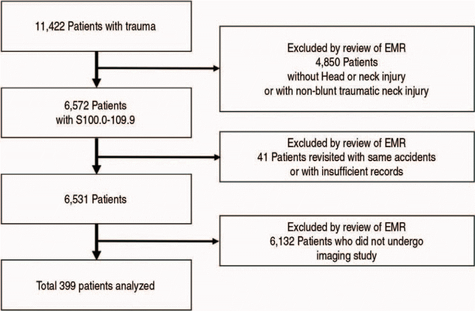
규칙의 각 변수가 개별 환자에 해당하는지 검토한 결과, NEXUS 기준에서“통증으로 초점을 흐리는 손상”이 9.0%로 가장 흔했다(Table 3). CCR에서는 고 위험변수 중“위험한 손상기전(46.1%)”만족 및 저 위험변수 중“지연성 목통증”불만족(17.0%)이 각각 가장 흔했다. “걸을 수 있는 환자”또는“응급실에서 앉을 수 있는 환자”를 만족하지 않은 20명 중 7명은 다리 골절을 동반했고, 4명은 외상성 뇌손상, 1명은 심장정지 및 복합골절 환자였다. PECARN 위험인자를 적용했을 때, “목통증”이 15.8%로 가장 흔했다. 총 291명(72.9%)이 세 규칙에 따라“잠재적으로 영상검사가 필요한 환자”로 분류됐다(Table 4). 상기 규칙에 따라 영상검사가 불필요한 상황임에도, 영상검사를 시행한 환자는 108명(27.1%)이었다.
세 규칙의 경추손상 환자 2명에 대한 민감도 및 음성예측도를 비교한 결과, NEXUS 기준의 민감도가 50%인 것을 제외하면 모두 100%에 가까운 민감도 및 음성예측도를 보였다(Table 5).
컴퓨터단층촬영을 시행한 16명 중 경추손상이 확인된 환자는 2명으로, 모두 경추 골절 및 탈구였다(Appendix 1, https://doi.org/10.22470/pemj.2022.00577). 16명 모두 CCR 및 PECARN 규칙에 따른“잠재적으로 영상검사가 필요한 환자”였다. NEXUS 기준의 저 위험변수를 만족하지 않은 환자는 7명이었다(Table 4, Appendix 1). 16명 중 15명이 PECARN 위험기준의“목통증”을 호소했고 5명은 NEXUS 기준 및 CCR의“경추 정중선 압통”을 보였다. 16명 중 4명이 동반한 중증 외상(간 열상, 신장 열상, 혈흉, 흉추 골절 등)으로 입원했다. 세 규칙의 변수 중 단층촬영의 근거로“목통증”단독으로 있던 환자는 5명이었다.
2명의 경추손상 환자는 모두 낙상으로 인한 경추 골절 및 탈구로 진단됐다. 6세 남자는 집라인을 타다가 떨어지며 뒤통수를 부딪친 후 발생한 목통증으로 방문했다. 당시 “경추 정중선 압통”을 보이지 않아서, NEXUS 기준에 따르면 영상검사가 필요 없었다. 반면, CCR 적용 시“위험한 손상기전(1 m 이상 추락)”에, PECARN 위험인자 적용 시“목통증”에 각각 해당하여, “잠재적으로 영상검사가 필요한 환자”로 분류됐다. 일반영상에서 정상 소견을 보여 귀가했다가 3일 후 목통증이 지속하여 외래를 방문했고, 당시“경추 정중선 압통”이 처음 확인됐으며 컴퓨터단층촬영 결과 경추 4번 골절 및 탈구를 보였다. 14세 남자는 재주넘기를 하다가 머리부터 떨어진 후 목이 앞으로 꺾여 발생한 통증 및 팔의 감각이상으로 방문했고, 세 규칙 모두에 따른“잠재적으로 영상검사가 필요한 환자”였다. NEXUS 기준에서 팔의 감각이상은“국소 신경학적 결손의 부재(absence of focal neurologic deficit)”를 만족하지 않으므로, 영상검사가 필요했다. CCR에서“위험한 손상기전”및“팔다리 감각이상”, PECARN 규칙에서는 “목통증”및“국소 신경학적 검사”의 이상 소견에 각각 해당했다. 일반영상 및 컴퓨터단층촬영에서 경추 5-6번의 탈구가 확인됐고 입원 후 시행한 자기공명영상에서 경추 3번 및 5번 골절을 추가로 진단했다.

고 찰

두경부 무딘 손상에서 불필요한 영상검사를 줄이기 위한 NEXUS 기준 및 CCR은 모두 소아에 적용하기에 적절치 않으며, 소아 대상으로 제시된 PECARN 위험인자는 아직 타당도 평가가 충분히 이뤄지지 않았다. 이에 본 연구는 소아응급환자의 가장 흔한 외상 부위인 두경부에서 경추손상을 배제하기 위해 시행되는 응급진료 현황 및 불필요한 영상검사 시행 빈도를 확인하고, 상기 규칙을 적용하여 그 유용성을 확인하고자 했다.
본 연구에서 경추손상으로 최종 진단된 환자는 2명(0.5%)이었는데, 이전 연구에서도 0.5%-3%였다3,4,12). 이전 연구에서 남자가 60%-66%를 차지했고3,11), 본 연구에서도 남자가 64.2%를 차지했다. 흔한 손상기전은 낙상, 차량승객사고, 구조물과 충돌 순이었는데, 이는 많은 연구에서 낙상 및 차량사고를 소아 경추손상의 가장 흔한 원인으로 보고한 것과 일치했다3,6,13,14).
각 규칙을 연구대상자에게 적용했을 때 많이 해당된 변수를 빈도순으로 2개씩 나열하면, NEXUS 기준은“통증으로 초점을 흐리는 손상”및“경추 정중선 압통”, CCR에서는“위험한 손상기전”만족 및“지연성 목통증”의 불만족(즉 초기에 시작한 통증), PECARN 위험인자에서는 “목통증”및“국소 신경학적 검사”의 이상이었다. 이는 Phillips 등3)이 세 규칙을 호주 소아환자에게 적용한 결과, NEXUS 기준, CCR, PECARN 위험인자에서 각각 “경추 정중선 압통”, “1 m 이상 높이에서 낙상(위험한 손상기전)”,“ 목통증”이 가장 흔했던 점과 대체로 일치했다.
PECARN 위험인자의 민감도 및 특이도는 각각 98% 및 26%로 알려졌고, 이를 적용하면 불필요한 경추 고정 및 영상검사를 25% 이상 줄일 수 있다11). 본 연구에서는 CCR을 적용했을 때보다 영상검사 빈도가 낮았지만, NEXUS 기준과는 유사했다(Table 4). 이는 호주 소아환자에게 세 규칙을 적용한 결과 영상검사가 필요하다고 판단된 빈도가 PECARN 위험인자 적용 시 68.1%로 가장 높고, NEXUS 기준 및 CCR은 각각 44.2% 및 48.4%인 점과 대조된다3). 한편, 10세 이하 환자에서 NEXUS 기준 및 CCR을 적용한 연구에서는 영상검사가 필요하다고 판단된 빈도가 각각 58.3% 및 76.2%였다12).
본 연구에서 컴퓨터단층촬영 시행 빈도가 4.0%에 불과한 점에는 소아환자에서 진정 필요성 또는 방사선위해 가능성이 부담으로 작용한 것 같고, 이는 다른 연구와 일맥 상통한다3,15). 한편, 단층촬영 결과 14명은 경추손상이 없었다. 이들 중 13명이 목통증을 호소했고 이 중 5명은 세 규칙의 변수 중 목통증만 단독으로 해당됐다. Garton과 Hammer16)가 소아환자에 NEXUS 기준 위음성으로 판명된 환자 2명 모두 국소 신경학적 검사에서 정상이었다고 보고한 점을 고려하면, 신경학적으로 정상인 목통증 환자에서 경추손상을 배제하기 어렵다.
NEXUS 기준의 민감도 및 특이도는 각각 99.6% 및 12.9%, CCR은 각각 100% 및 42.5%으로 알려졌다5,6). 하지만 소아환자는 성인과 손상 기전 및 해부학적 구조 면에서 차이가 있으므로, 이 규칙을 소아에게 적용하기 적절치 않을 수 있다12,13,17-21). 본 연구에서 경추손상 환자가 2명에 불과하여 평가하기 어렵지만, NEXUS 기준의 민감도가 CCR 및 PECARN 위험인자의 민감도보다 낮았다(Table 5). Ehrlich 등12)은 NEXUS 기준 및 CCR의 민감도를 각각 43% 및 86%로 보고하여, 본 연구와 비슷했다. NEXUS 기준의 위음성에 관한 기존 연구 결과는 다음과 같다. Viccellio 등5)은 NEXUS 기준을 소아에 적용한 결과, 2명의 어린 경추손상 환자를 놓쳤다고 보고했다. 미식축구 경기 중 태클을 당해 목 손상을 입은 16세 남자 환자가 NEXUS 기준에 따르면 영상검사가 필요 없었지만, 결과적으로 경추골절이 진단된 증례도 있다20). NEXUS 기준의 민감도가 8-19세에서는 100%지만 8-10세 미만에서는 43%-94%였는데12,16), 본 연구에서도 6세 경추손상 환자가 NEXUS 기준에 따른 위음성이었다. 이 환자는 응급실 퇴원 후 경추 정중선 압통이 발견됐다. 이는 첫 응급실 방문 당시, 압통을 정확히 확인하지 못했을 수 있다는 뜻이다. 세 규칙에서 목통증, 압통, 감각이상, 의식저하 등 일부 변수는 영유아에서 확인하기 어려워, 정확도가 떨어질 수 있다.
두경부 외상 소아환자에서 불필요한 영상검사를 줄이기 위한 규칙을 엄격히 적용하기가 현실적으로 어렵다. 이 때문에 의사의 개별적 판단에 따라 영상검사를 더 자주 시행함으로써, 불필요한 방사선위해, 진정, 의료비 상승을 초래할 수 있다. 실제로, 응급실에서 상기 규칙에 따라 영상 검사가 필요없다고 판단된 경우에도 영상검사를 시행하는 경향을 보인다3,10,12,13). 본 연구에서도 규칙에 따르면 영상검사가 필요 없는 상황임에도 영상검사를 시행한 빈도가 27%에 달했다. 적어도 이러한 경우에는 영상검사를 자제해야 한다.
본 연구는 후향적 단일기관 연구로, 경추손상이 드물어 이 손상의 특성을 분석하기 어려웠고, 진단이 누락되거나 기록이 불충분하여 손상 기전 또는 영상검사를 시행한 근거를 판단하기 애매한 경우가 있었다. 이에 따라 각 변수의 해당 여부를 잘못 판단했다면, 이 잘못된 판단이 규칙의 경추손상에 대한 예측성적(예: 민감도)에 영향을 미쳤을 수 있다.
응급실에서 경추손상 관련 영상검사 시행을 결정하기 위한 세 규칙에 따라 공통으로 검사가 필요 없다면, 불필요한 검사를 자제해야 한다. 또한 추가 연구를 통해 어린 소아의 특성을 반영하는 방향으로 규칙을 개선해야 할 것이다.

Notes

모든 저자는 이 논문과 관련된 이해관계가 없음.

재정지원

모든 저자는 이 논문과 관련된 재정지원을 받지 않았음.

References

1. Korea Centers for Disease Control and Prevention. 2020 injury types and cause statistics [Internet]. Cheongju (Korea): Korea Centers for Disease Control and Prevention; c2019 [cited 2023 Jan 1]. Available from: https://www.kdca.go.kr/injury/biz/injury/recsroom/statsS mMain.do. Korean.
2. Shavelle RM, Devivo MJ, Paculdo DR, Vogel LC, Strauss DJ. Long-term survival after childhood spinal cord injury. J Spinal Cord Med. 2007; 30 Suppl 1:S48–54.
3. Phillips N, Rasmussen K, McGuire S, Abel KA, Acworth J, Askin G, et al. Projected paediatric cervical spine imaging rates with application of NEXUS, Canadian C-Spine and PECARN clinical decision rules in a prospective Australian cohort. Emerg Med J. 2021; 38:330–7.
4. Mohseni S, Talving P, Branco BC, Chan LS, Lustenberger T, Inaba K, et al. Effect of age on cervical spine injury in pediatric population: a National Trauma Data Bank review. J Pediatr Surg. 2011; 46:1771–6.
5. Viccellio P, Simon H, Pressman BD, Shah MN, Mower WR, Hoffman JR. A prospective multicenter study of cervical spine injury in children. Pediatrics. 2001; 108:E20.
6. Stiell IG, Wells GA, Vandemheen KL, Clement CM, Lesiuk H, De Maio VJ, et al. The Canadian C-spine rule for radiography in alert and stable trauma patients. JAMA. 2001; 286:1841–8.
7. Hoffman JR, Mower WR, Wolfson AB, Todd KH, Zucker MI; National Emergency X-Radiography Utilization Study Group. Validity of a set of clinical criteria to rule out injury to the cervical spine in patients with blunt trauma. N Engl J Med. 2000; 343:94–9.
8. Pieretti-Vanmarcke R, Velmahos GC, Nance ML, Islam S, Falcone RA Jr, Wales PW, et al. Clinical clearance of the cervical spine in blunt trauma patients younger than 3 years: a multi-center study of the american association for the surgery of trauma. J Trauma. 2009; 67:543–50.
9. Burns EC, Yanchar NL. Using cervical spine clearance guidelines in a pediatric population: a survey of physician practices and opinions. CJEM. 2011; 13:1–6.
10. Yoon KW. Failure of initial neurologic assessment for SCIWORA with brain injury in 22-month-old patient. J Korean Soc Emerg Med. 2019; 30:598–602. Korean.
11. Leonard JC, Kuppermann N, Olsen C, Babcock-Cimpello L, Brown K, Mahajan P, et al. Factors associated with cervical spine injury in children after blunt trauma. Ann Emerg Med. 2011; 58:145–55.
12. Ehrlich PF, Wee C, Drongowski R, Rana AR. Canadian Cspine Rule and the National Emergency X-Radiography Utilization low-risk criteria for C-spine radiography in young trauma patients. J Pediatr Surg. 2009; 44:987–91.
13. Leonard JR, Jaffe DM, Kuppermann N, Olsen CS, Leonard JC; Pediatric Emergency Care Applied Research Network (PECARN) Cervical Spine Study Group. Cervical spine injury patterns in children. Pediatrics. 2014; 133:e1179–88.
14. Leonard JC, Browne LR, Ahmad FA, Schwartz H, Wallendorf M, Leonard JR, et al. Cervical spine injury risk factors in children with blunt trauma. Pediatrics. 2019; 144:e20183221.
15. Kim W, Ahn N, Ata A, Adamo MA, Entezami P, Edwards M. Pediatric cervical spine injury in the United States: defining the burden of injury, need for operative intervention, and disparities in imaging across trauma centers. J Pediatr Surg. 2021; 56:293–6.
16. Garton HJ, Hammer MR. Detection of pediatric cervical spine injury. Neurosurgery. 2008; 62:700–8.
17. Cui LW, Probst MA, Hoffman JR, Mower WR. Sensitivity of plain radiography for pediatric cervical spine injury. Emerg Radiol. 2016; 23:443–8.
18. Slaar A, Fockens MM, Wang J, Maas M, Wilson DJ, Goslings JC, et al. Triage tools for detecting cervical spine injury in pediatric trauma patients. Cochrane Database Syst Rev. 2017; 12:CD011686.
19. Leonard JC, Jaffe DM, Olsen CS, Kuppermann N. Age-related differences in factors associated with cervical spine injuries in children. Acad Emerg Med. 2015; 22:441–6.
20. Ferderber JS, Wolfson AB. The dangers of spear tackling: a case report of a NEXUS-negative high school football player. J Emerg Med. 2019; 56:185–90.
21. Gopinathan NR, Viswanathan VK, Crawford AH. Cervical spine evaluation in pediatric trauma: a review and an update of current concepts. Indian J Orthop. 2018; 52:489–500.

Fig. 1.
Flowchart for the selection of study population. EMR: electronic medical record.
pemj-2022-00577f1.tif
Table 1.
Three clinical decision rules 5,6,11
NEXUS* CCR PECARN
Absence of midline cervical tenderness High-risk Altered mental status
Absence of focal neurologic deficit Age ≥ 65 y Focal neurologic examination
Normal level of alertness and consciousness Dangerous mechanism of injury Neck pain
Paresthesia in the extremities Torticollis
No evidence of intoxication Low-risk High-risk motor vehicle crash
Absence of painful distracting injury Simple rear-end motor vehicle crashes Substantial torso injury
Patient able to sit up in the emergency department Predisposing condition associated with cervical spine injury
Patient ambulatory at any time
Delayed onset of neck pain Diving
Absence of midline cervical tenderness
Low-risk
Can rotate neck 45° to the left and to the right?§

* Plain radiography can be safely avoided in patients who meet all 5 criteria.

Plain radiography can be safely avoided in patients who meet low-risk criteria and no high-risk criteria, and can actively rotate their necks.

Having ≥ 1 factors was 98% sensitive and 26% specific for cervical spine injury without specific indications for imaging studies.

§ Regardless of pain.

NEXUS: National Emergency X-Radiography Utilization Study, CCR: Canadian Cervical Spine Rule, PECARN: Pediatric Emergency Care Applied Research Network.

Table 2.
Characteristics of children with blunt neck trauma by age groups (N = 399)
Category Total (N = 399) Infants (N = 49) Preschoolers (N = 160) Schoolers (N = 145) Teenagers (N = 45)
Boys 256 (64.2) 31 (63.3) 96 (60.0) 93 (64.1) 36 (80.0)
Injury mechanism
Fall 146 (36.6) 31 (63.3) 72 (45.0) 37 (25.5) 6 (13.3)
Passenger car accident 98 (24.6) 12 (24.5) 48 (30.0) 31 (21.4) 7 (15.6)
Collision with structures 73 (18.3) 3 (6.1) 18 (11.3) 36 (24.8) 16 (35.6)
Pedestrian/bicycle versus car 42 (10.5) 1 (2.0) 7 (4.4) 25 (17.2) 9 (20.0)
Slip down 30 (7.5) 1 (2.0) 8 (5.0) 15 (10.3) 6 (13.3)
Rolling 10 (2.5) 1 (2.0) 7 (4.4) 1 (0.7) 1 (2.2)
Computed tomoraphy 16 (4.0) 0 (0) 6 (3.8) 5 (3.4) 5 (11.1)
Hospitalization 19 (4.8) 2 (4.1) 5 (3.1) 9 (6.2) 3 (6.7)
Cervical spine injury 2 (0.5) 0 (0) 0 (0) 1 (0.7) 1 (2.2)

Values are expressed as numbers (%).

Table 3.
Application of 3 clinical decision rules to select children with a potential need for imaging studies*
Criteria Variable Total (N = 399)
NEXUS No absence of painful distracting injury 36 (9.0)
No absence of midline cervical tenderness 26 (6.5)
No absence of focal neurologic deficit 6 (1.5)
No normal level of alertness and consciousness 6 (1.5)
No absence of evidence of intoxication 2 (0.5)
Having at least 1 variable 72 (18.0)
CCR, high-risk (+) Dangerous mechanism of injury 184 (46.1)
Paresthesia in the extremities 2 (0.5)
Having at least 1 variable 198 (49.6)
 Low-risk (-) No delayed onset of neck pain 68 (17.0)
No simple rear-end motor vehicle crashes 62 (15.5)
No absence of midline cervical tenderness 26 (6.5)
No patient ambulatory at any time 12 (3.0)
No patient able to sit up in the emergency department 8 (2.0)
 Low-risk (-) Unable to rotate neck 45。to the left and to the right? 13 (3.3)
Having at least 1 variable 289 (72.4)
PECARN Neck pain 63 (15.8)
Focal neurologic examination 6 (1.5)
Altered mental status 5 (1.3)
Substantial torso injury 5 (1.3)
High-risk motor vehicle crash 2 (0.5)
Torticollis 0 (0)
Predisposing condition associated with cervical spine injury 0 (0)
Diving 0 (0)
Having at least 1 variable 74 (18.5)

Values are expressed as numbers (%).

* The table was intended to be used for selecting children with a potential need for imaging studies. For this purpose, we listed numbers (%) of the absence of low-risk variables (e.g., “delayed onset of neck pain” of CCR) and the presence of high-risk variables (e.g., “dangerous mechanism of injury” of CCR).

Of high-risk variables, “age ≥ 65 years” was omitted due to inapplicability to this study.

NEXUS: National Emergency X-Radiography Utilization Study, CCR: Canadian Cervical Spine Rule, PECARN: Pediatric Emergency Care Applied Research Network.

Table 4.
Summary of application of 3 clinical decision rules
Rules Positivity* No. (%) CT (N = 16) CSI (N = 2)
NEXUS + 72 (18.0) 7 1
- 327 (82.0) 9 1
CCR + 289 (72.4) 16 2
- 110 (27.6) 0
PECARN + 74 (18.5) 16 2
- 325 (81.5) 0
At least 1 rule + 291 (72.9) 16 1
All rules - 108 (27.1) 0 0

* Absence of low-risk variables or presence of high-risk variables.

With a potential need for imaging studies.

Without a need for imaging studies.

CT: computed tomography, CSI: cervical spine injury, NEXUS: National Emergency X-Radiography Utilization Study, CCR: Canadian Cervical Spine Rule, PECARN: Pediatric Emergency Care Applied Research Network.

Table 5.
Accuracy of 3 clinical decision rules
Variable NEXUS CCR PECARN
Sensitivity 50.0 100 100
Specificity 82.1 27.7 81.9
Negative predicted value 99.7 100 100
Positive predicted value 1.4 0.7 2.7
Accuracy 82.0 28.1 82.0

Values are expressed as %.

NEXUS: National Emergency X-Radiography Utilization Study, CCR: Canadian Cervical Spine Rule, PECARN: Pediatric Emergency Care Applied Research Network.

Appendix

Appendix 1

Details of 16 patients who underwent computed tomography

Patient No. Age, y Injury mechanism NEXUS CCR PECARN
1 3 Contusion NA No delayed onset of neck pain Neck pain
2 4 Fall NA No delayed onset of neck pain Neck pain
Dangerous mechanism of injury Substantial torso injury
3 4 Fall NA No delayed onset of neck pain Neck pain
4 5 Passenger car accident No absence of midline cervical tenderness No delayed onset of neck pain Neck pain
No absence of painful distracting injury No absence of midline cervical tenderness Substantial torso injury
No patient ambulatory at any time
No patient able to sit up in the emergency department
5 5 Collision with structures No absence of midline cervical tenderness No delayed onset of neck pain Neck pain
No absence of midline cervical tenderness
6 5 Fall NA No delayed onset of neck pain Neck pain
7* 6 Fall NA Neck pain Neck pain
Dangerous mechanism of injury
8 7 Passenger car accident NA No delayed onset of neck pain Neck pain
Dangerous mechanism of injury Substantial torso injury
High risk motor vehicle crash
9 8 Pedestrian No absence of midline cervical tenderness No delayed onset of neck pain Neck pain
No absence of midline cervical tenderness
10 11 Collision with structures No absence of midline cervical tenderness No delayed onset of neck pain Neck pain
No absence of midline cervical tenderness
Dangerous mechanism of injury
11 12 Fall No normal level of alertness and consciousness No patient able to sit up in the emergency department Altered mental status,
No absence of focal neurologic deficit No patient ambulatory at any time Substantial torso injury
No absence of painful distracting injury Dangerous mechanism of injury Focal neurologic examination
12 13 Fall No absence of midline cervical tenderness No delayed onset of neck pain Neck pain
No absence of midline cervical tenderness
13 13 Collision with structures NA No delayed onset of neck pain Neck pain
14 13 Bicycle-car accident NA No delayed onset of neck pain Neck pain
Dangerous mechanism of injury High risk motor vehicle crash
15 14 Bicycle-car accident NA No delayed onset of neck pain Neck pain
Dangerous mechanism of injury High risk motor vehicle crash
16* 14 Fall No absence of focal neurologic deficit No delayed onset of neck pain Neck pain
No absence of painful distracting injury Paresthesia in the extremities Focal neurologic examination

* Patients with cervical spine injury.

NEXUS: National Emergency X-Radiography Utilization Study, CCR: Canadian Cervical Spine Rule, PECARN: Pediatric Emergency Care Applied Research Network.

TOOLS
Similar articles