Abstract
Subarachnoid hemorrhage (SAH) due to ruptured posterior cerebral artery (PCA) intracranial arterial dolichoectasia (IADE) is very rare. As these lesions are difficult to treat microsurgically, neurointervention is preferred because the dolichoectatic artery does not have a clear neck, and the surgical field of view was deep seated with the SAH. However, in some cases, neurointervention is difficult due to anatomical variation of the blood vessel to access the lesion. In this case, a 30-year-old male patient presented with a ruptured PCA IADE and an aortic arch anomaly. Aortic arch anomalies render it difficult to reach the ruptured PCA IADE via endovascular treatment. The orifice of the vertebral artery (VA) was different from the usual cases, so it was difficult to find the entrance. After only finding the VA and arriving at the lesion along the VA, trapping was performed. Herein, we report the PCA IADE with aortic arch anomaly endovascular treatment methods and results.
Intracranial arterial dolichoectasia (IADE) refers to enlargement of the length, diameter, and curvature of the blood vessels in the brain. In patients with dolichoectasia, the tunica media of the arterial wall expands due to the disruption of the internal elastic lamina, atrophy of the muscle layer, and hyalinization of the connective tissue leading to abnormal dilatation of the affected vessel [6,13,14]. IADE is mainly associated with the posterior circulation and affects the basilar artery in 80% of the cases. IADE is usually asymptomatic, but it can sometimes manifest as cerebral infarction and compression of the cranial nerve or brainstem [6,11,12,14,18-20]. Occasionally, in cases of arterial rupture, it may present as cerebral hemorrhage and subarachnoid hemorrhage (SAH). SAH due to ruptured IADE does not occur commonly and is not well known; however, emergency treatment is required because of the possibility of rebleeding. However, the standard IADE treatment plan is controversial (trapping, resection with reanastomosis, transposition, and wrapping) [17,20]. In addition, IADE may be accompanied by extracranial vascular anomaly such as abdominal aortic aneurysm, coronary artery ectasia, aortic arch anomalies [13-15]. Especially in aortic arch anomaly as the arteries supplying blood to the brain develop during embryogenesis, anatomical deformities may occur, and these deformities may affect the development of arterial disease [2,9]. Anatomical deformities in the aorta may further make endovascular treatment difficult, since endovascular access to the lesion is not easy. During the neuro-intervention procedure, aortic arch anomalies make it difficult to access IADE [1,7,8]. The authors report treatment for intracranial artery dolichoectasia with a literature review (Table 1). Here, we report trapping of SAH with IADE on the left P2 segment and an aortic arch anomaly.
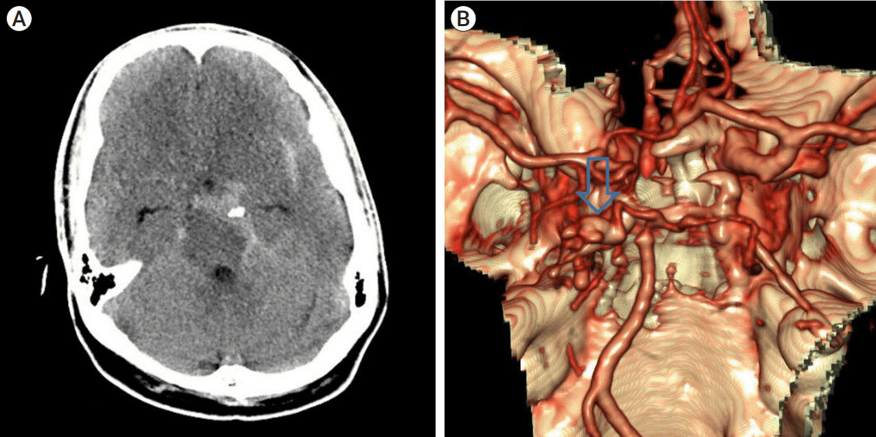
A 30-year-old male patient, who had no history of an underlying disease, presented with a severe headache before admission. Emergency brain multi-phase computed tomography angiography was performed, which revealed SAH. In the non-contrast image, a lot of SAH was found on the left crural cistern and the left ambient cistern (Fig. 1A). In the three-dimensional (3-D) image, we found a vascular abnormality on the left P2 segment that was elongated and tortuous. It was found to be an IADE, rather than the more commonly detected saccular aneurysm, dissection (Fig. 1B). Therefore, we believe that the IADE on the left P2 was likely ruptured. The dolichoectatic artery of the P2 segment is difficult to treat surgically, since it does not have a clear neck so it is difficult to clipping. And trapping and bypass surgery was not planned because it was technically very difficult in acute phase with SAH, and there was a high probability of technical failure and high risk of rebleeding. Therefore, we decided to treat the patient using an endovascular approach.
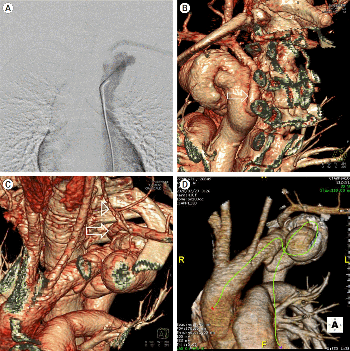
Initially, we tried to access the left vertebral artery (VA) initially in the anteroposterior view, but we could not locate it (Fig. 2A). We inferred that the aorta of the patient might be unusual. After re-evaluating the 3-D image of the aortic arch, we found that the left subclavian artery originated posteriorly from the descending aorta at the T4 level. From the left subclavian artery orifice to the left subclavian artery at T1, it points to the right. After passing through the left first rib, it turned to the left. The left VA came off superiorly from the left subclavian artery at T2 (Fig. 2B, C, D).
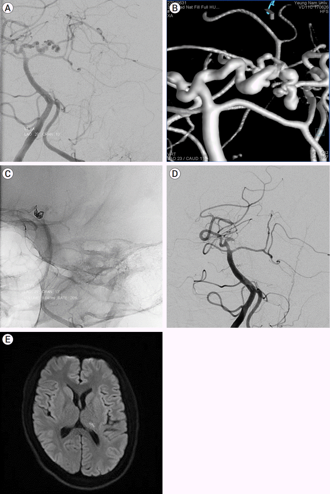
Under the guidance of left VA, IADE was located at the left P2. The IADE on the left P2 was tortuous and elongated (Fig. 3A, B). If access to the left VA was fail, we considered the right VA guiding through the right radial artery because the right VA guiding through femoral artery was judged to be difficult to access due to aortic arch anomaly. It was difficult to access the microcatheter inside the IADE on the left P2. Therefore, we decided to perform endovascular trapping on the left P2 segment before the location of the IADE. To make this access, we used a distal access intermediate catheter (SOFIA 6F; MicroVention Terumo, Tustin, California, United States), which was positioned at the left VA on the C2 level. Next, a microwire (Synchro-14; Stryker, Fremont, California, United States) and microcatheter (Excelsior SL-10; Stryker, Fremont, California, United States) were attached to the left P2, prior to the location of the IADE. Five detachable coils were deposited to the left P2 prior to the location of the IADE, the details of which are as follows: one 4 mm×8 cm, one 1 mm×3 cm, and one 1.5 mm×2 cm Axium Prime (EV3 endovascular, Irvine, California, United States) and one 2 mm×3 cm and one 1 mm×2 cm HydroCoil 10 HydroSoft 3D (MicroVention Terumo, Tustin, California, United States)(Fig. 3C). An angiogram revealed that there was no distal flow to the left posterior cerebral artery after the coil embolization procedure (Fig. 3D). The headache of the patient improved after the procedure. Magnetic resonance imaging for stroke revealed a focal acute infarction in the left lateral thalamus (Fig. 3E). The patient was was discharged without any neurological deficits. Thoracic surgeons were consulted regarding the aortic anomaly for the patient, and they diagnosed the possibility of congenital anomalies or aortic aneurysms that could rupture. Hence, the patient was advised an open thoracic surgery.
An established and appropriate treatment method does not exist currently for patients with SAH and IADE of the left P2 segment [21]. SAH that is associated with IADE in the posterior circulation is fatal [4,16]. In our reported case of SAH associated with IADE, emergency treatment was necessary because of the possibility of rebleeding. We considered two strategies to treat SAH with IADE — microsurgical or endovascular. In addition, both strategies may be considered together as a treatment option [2,3,5,10,20]. Regardless of the treatment method, it is important to ensure that no further bleeding occurs. This case was difficult to treat surgically because the dolichoectatic artery of the P2 segment did not have a clear neck, and the surgical field of view was deep seated with the SAH. However, in the endovascular treatment strategy, it may be difficult to preserve the patent artery while accurately obliterating the pathological lesion only. Since the IADE of the left P2 segment was too tortuous and elongated in our patient, the exact lesion that had ruptured could not be identified. Therefore, we treated the patient with endovascular trapping. However, complications from trapping of the left P2 segment may occur secondary to the occlusion of the perforator or from distal hemodynamic instability [18]. As the purpose was to prevent rebleeding, trapping was performed despite the possibility that some cerebral infarction may occur after trapping. But some studies, proximal occlsuion of posterior cerebral artery (PCA) was well tolerated. It may be no perforators from dolichoectasia [3].
However, from the very beginning, we considered the presence of a vascular abnormality in the patient. In some patients with IADE, arterial disorders such as coronary artery ectasia, abdominal aortic aneurysm, saccular aneurysm, and enlarged thoracic aorta have been reported [13,15]. However, it has also been reported that there was no significant association between aortic arch anomalies and ascending aortic aneurysms [3]. Dolichoectasia has been associated with old age, male sex, hypertension, and connective tissue disorders such as Elher-Danlos syndrome, Marfan’s disease, and Fabry’s disease. Thus, IADE may be associated with damage to various components of the tunica media [1]. Therefore, when planning an endovascular procedure for a patient with dolichoectasia, it should be considered that the endovascular procedure may be difficult to perform due to vascular malformations such as aortic anomaly, aortic aneurysm, and coronary artery disease.
Endovascular therapy may be effective in some patients with PCA IADE. But it should be considered that there may also be malformations in extracranial vessels such as the aorta in endovascular treatment. So, when treating PCA IADE, the treatment method should be decided in consideration of the anatomical variation.
REFERENCES
1. Al-Atassi T, Khoynezhad A. Indications for aortic arch intervention. Semin Cardiothorac Vasc Anesth. 2016; Dec. 20(4):259–64.
2. Baccin CE, Krings T, Alvarez H, Ozanne A, Lasjaunias PL. A report of two cases with dolichosegmental intracranial arteries as a new feature of PHACES syndrome. Child Nerv Syst. 2007; May. 23(5):559–67.
3. Chao KH, Riina HA, Heier L, Steig PE, Gobin YP. Endovascular management of dolichoectasia of the posterior cerebral artery report. AJNR Am J Neuroradiol. 2004; Nov-Dec. 25(10):1790–1.
4. Coert BA, Chang SD, Do HM, Marks MP, Steinberg GK. Surgical and endovascular management of symptomatic posterior circulation fusiform aneurysms. J Neurosurg. 2007; May. 106(5):855–65.
5. Dziewasa R, Freund M, Ludemann P, Muller M, Ritter M, Droste DW, et al. Treatment options in vertebrobasilar dolichoectasia–case report and review of the literature. Eur Neurol. 2003; 49(4):245–7.
6. Gutierrez J, Sacco RL, Wright CB. Dolichoectasia-an evolving arterial disease. Nat Rev Neurol. 2011; Jan. 7(1):41–50.
7. Hanneman K, Newman B, Chan F. Congenital variants and anomalies of the aortic arch. Radiographics. 2017; Jan-Feb. 37(1):32–51.
8. Mantri SS, Raju B, Jumah F, Rallo MS, Nagaraj A, Khandelwal P, et al. Aortic arch anomalies, embryology and their relevance in neuro-interventional surgery and stroke: a review. Interv Neuroradiol. 2022; Aug. 28(4):489–98.
9. Menshawi K, Mohr JP, Gutierrez J. A functional perspective on the embryology and anatomy of the cerebral blood supply. J Stroke. 2015; May. 17(2):144–58.
10. O’Shaughnessy BA, Getch CC, Bendok BR, Parkinson RJ, Batjer HH. Progressive growth of a giant dolichoectatic vertebrobasilar artery aneurysm after complete Hunterian occlusion of the posterior circulation: case report. Neurosurgery. 2004; Nov. 55(5):1223.
11. Passero SG, Calchetti B, Bartalini S. Intracranial bleeding in patients with vertebrobasilar dolichoectasia. Stroke. 2005; Jul. 36(7):1421–5.
12. Pereira-Filho A, Faria M, Bleil C, Kraemer JL. Brainstem compression syndrome caused by vertebrobasilar dolichoectasia: microvascular repositioning technique. Arq Neuropsiquiatr. 2008; Jun. 66(2B):408–11.
13. Ubogu EE, Zaidat OO. Intracranial arterial dolichoectasia is associated with enlarged descending thoracic aorta. Neurology. 2005; Nov. 65(10):1681–2.
14. Pico F, Labreuche J, Amarenco P. Pathophysiology, presentation, prognosis, and management of intracranial arterial dolichoectasia. Lancet Neurol. 2015; Aug. 14(8):833–45.
15. Pico F, Labreuche J, Hauw JJ, Seilhean D, Duyckaerts C, Amarenco P. Coronary and basilar artery ectasia are associated: results from an autopsy case-control study. Stroke. 2016; Jan. 47(1):224–7.
16. Rabb CH, Barnwell SL. Catastrophic subarachnoid hemorrhage resulting from ruptured vertebrobasilar dolichoectasia: case report. Neurosurgery. 1998; Feb. 42(2):379–82.
17. Rathore L, Yamada Y, Kawase T, Kato Y, Senapati SB. A 5-year follow-up of intracranial arterial dolichoectasia: a case report and review of literature. Asian J Neurosurg. 2019; Nov. 14(4):1302–7.
18. Siddiqui A, Chew NS, Miszkiel K. Vertebrobasilar dolichoectasia: a rare cause of obstructive hydrocephalus: case report. Br J Radiol. 2008; Apr. 81(964):e123–6.
19. Sokolov AA, Husain S, Sztajzel R, Croquelois A, Lobrinus JA, Thaler D, et al. Fatal subarachnoid hemorrhage following ischemia in vertebrobasilar dolichoectasia. Medicine (Baltimore). 2016; Jul. 95(27):e4020.
20. Yamaguchi S, Ito O, Maeda Y, Murata H, Imamoto N, Yuhi F, et al. Coil embolization for a ruptured posterior cerebral artery aneurysm with vertebrobasilar dolichoectasia. Neurol Med Chir (Tokyo). 2011; 51(9):657–60.
21. Yuan YJ, Xu K, Luo Q, Yu JL. Research progress on vertebrobasilar dolichoectasia. Int J Med Sci. 2014; Aug. 11(10):1039–48.
Fig. 1.
Brain multi-phased computed tomography angiography is showing subarachnoid hemorrhage in basal on left crural cistern and left ambient cistern (A). Three-dimensional reconstructed angiography showed left P2 segment were elongated and enlarged (arrow) (B).

Fig. 2.
In anteroposterior view of aortogram, the left subclavian artery was identified, but the left vertebral artery (VA) was not seen and the shape of the aortic arch was unusual (A). Aortic arch three-dimensional image, left subclavian artery comes off posteriorly from the descending aorta at the level of T4. Arrow: left subclavian artery (B). From the left subclavian artery orifice to the left subclavian artery at the level of T1, it is pointing to the right. After passing the left first rib, it turns to the left. Left VA comes off superiorly from the left subclavian artery at the level of T2. Arrow: left subclavian artery, Arrowhead: left VA (C). From the aortic arch to the ascending aorta, it is clockwise. Two aortic arch aneurysm is found (D).

Fig. 3.
Left vertebral artery angiogram, working view showing a tortuous and elongated Lt. P2 (A, B). After first, second coil was inserted (C). Followed angiogram, there were no distal flow left posterior cerebral artery after the coil embolization (D). Followed stroke magnetic resonance imaging (MRI), there were focal acute infarction on left lateral thalamus (E).

Table 1.
Treatment for PCA intracranial artery dolichoectasia
| Location | Type | Treatment | Prognosis | |
|---|---|---|---|---|
| Chao KH et al. [3] | Rt. PCA | Incidental | Balloon test occlusion, trapping | Intact |
| This case | Rt. P2 | SAH | Trapping | Intact |


 PDF
PDF Citation
 Print
Print


 XML Download