Journal List > Korean J Anesthesiol > v.76(2) > 1516081921

Lee, Kim, Park, Choi, Kwak, and Shim: Impact of transient decrease in mixed venous oxygen saturation on prognosis in off-pump coronary artery bypass surgery: a retrospective cohort study

Abstract

Background

The prognostic consequences of transient hemodynamic deterioration due to cardiac displacement, which is most severe during left circumflex artery (LCX) grafting in off-pump coronary artery bypass surgery (OPCAB) are unknown. This study aimed to investigate the association between mixed venous oxygen saturation (SvO2) < 60% during LCX grafting and the occurrence of composite of morbidity endpoints.

Methods

Data of patients who underwent elective OPCAB between January 2010 and December 2019 were reviewed. Logistic regression analysis was performed to detect risk factors for the composite of morbidity endpoints, defined as 30-day or in-hospital mortality, postoperative myocardial infarction, prolonged mechanical ventilation > 24 h, cerebrovascular accident, and acute kidney injury.

Results

Among 1,071 patients, the composite of morbidity endpoints occurred in 303 (28%) patients. SvO2 < 60% during LCX grafting was significantly associated with the composite of morbidity (OR: 2.72, 95% CI [1.60, 4.61], P < 0.001) along with advanced age, chronic kidney disease, ratio of early mitral inflow velocity to mitral annular early diastolic velocity, and EuroSCORE II. Other major hemodynamic variables including the cardiac index were not associated with the outcome. Additional regression analysis revealed pre-operative anemia as a predictor of SvO2 < 60% during LCX grafting (OR: 2.09, 95% CI [1.33, 3.29], P = 0.001).

Conclusions

A decrease in SvO2 < 60%, albeit confined to the period of cardiac displacement, was associated with a 2.7-fold increased risk of detrimental outcomes after OPCAB, implying the prognostic importance of this transient deterioration in oxygen supply-demand balance.

Introduction

Evidence suggests that the theoretical advantage of evading cardiopulmonary bypass in off-pump coronary artery bypass surgery (OPCAB) leads to outcome benefits in terms of attenuated risk of stroke, renal failure, and bleeding complications when compared with on-pump coronary artery bypass graft surgery (CABG) [13].
Nonetheless, OPCAB requires mechanical cardiac displacement for exposure of target vessels, which results in alterations to cardiac geometry and motion [4]. Manifested as impaired filling and diastolic dysfunction of the ventricles, these modifications inevitably induce hemodynamic instability, which may complicate the perioperative course [57].
Although hemodynamic instability induced by mechanical cardiac displacement is transient and well tolerated in most patients, previous studies have indicated its potential adverse influence on post-operative outcomes [8,9]. As it is essential to provide sufficient cardiac output against mechanical constraints during OPCAB, it is reasonable to hypothesize that the resultant hemodynamic instability, when severe enough to jeopardize global oxygen supply-demand balance (manifested as severe decline in mixed venous oxygen saturation [SvO2]), could adversely affect outcomes. This is of particular clinical importance in patients receiving multivessel OPCAB, including grafting at the lateral wall when hemodynamic instability is most severe [4]; however, this has not yet been comprehensively scrutinized. A hemodynamic management goal of SvO2 ≥ 60% has been advocated in cardiothoracic and vascular surgeries, including on-pump CABG [1012], without any pertinent evidence related to OPCAB.
Therefore, this study aimed to examine the association between significant cardiac displacement-induced hemodynamic instability, reflected by SvO2 < 60% during left circumflex artery (LCX) grafting, and the clinical outcomes of patients who underwent isolated multivessel OPCAB.  

Materials and Methods

The present study was a retrospective review of a cohort of patients who underwent elective, isolated OPCAB between January 2010 and December 2019 in Severance Hospital, Seoul, Republic of Korea. The study was approved by the Institutional Review Board (IRB no. 4-2021-1412) of Yonsei University Health System (Seoul, Republic of Korea) in November 2021 and the requirement for written informed consent was waived. The study adhered to the Strengthening the Reporting of Observational Studies in Epidemiology (STROBE) guidelines, and was conducted in accordance with the Ethical Principles for Medical Research Involving Human Subjects as outlined in the Helsinki Declaration of 1975 (revised 2013).

Intraoperative management

All patients received standardized anesthetic and surgical management, which was as follows: upon arrival in the operating room, standard monitoring devices were set up, including a pulmonary artery catheter (Swan-Ganz CCOmbo CCO/SvO2TM, Edwards Lifesciences LLC, USA) to monitor cardiac index and SvO2. Anesthesia was maintained with sevoflurane and sufentanil, and rocuronium was used for neuromuscular blockade. Mechanical ventilation was adjusted to maintain normocapnia and partial pressure of arterial oxygen ≥ 100 mmHg, with a tidal volume of 8 ml/kg, an I : E ratio of 1 : 2, and 40% oxygen with air at a positive end-expiratory pressure of 5 cmH2O.
All surgical procedures were performed through a median sternotomy, and the heart was displaced using a posterior pericardial stitch, large gauze (12 × 70 cm) swabs, and tissue stabilizer (Octopus Tissue Stabilization Systems®, Medtronic Inc., USA). During the cardiac displacement period, a crystalloid solution was infused at a fixed rate of 6 ml/kg, and an additional colloid solution was infused to compensate for blood loss. Intraoperative blood loss was collected by a cell salvage device (Cell Saver® EliteTM, Haemonetics, USA), which was re-infused into the patient after grafting was completed. Hemodynamic management during the period of cardiac displacement and grafting was as follows: (1) maintenance of a mean systemic arterial pressure (MAP) above 70 mmHg either with infusion of norepinephrine or vasopressin (up to 0.3 μg/kg/h and 4 IU/h, respectively) and (2) infusion of milrinone in patients with SvO2 < 60% for 10 min or development of mitral regurgitation grade ≥ 3. Transfusion was performed when the hematocrit level was < 25%.

Study protocol

The assessed pre-operative variables included age, sex, body mass index, presence of anemia, hypertension (HTN), diabetes mellitus (DM), chronic obstructive pulmonary disease (COPD), chronic kidney disease (CKD), cerebrovascular accident (CVA), myocardial infarction (MI) within one month, congestive heart failure (CHF, defined as New York Heart Association function classification ≥ 3), EuroSCORE II, and data derived from transthoracic echocardiography. Transthoracic echocardiography was performed 1–3 days before surgery by cardiologists, and the parameters included in the analysis were left ventricular ejection fraction (LVEF), ratio of early mitral inflow velocity to mitral annular early diastolic velocity (E/e’), and left ventricular end-diastolic diameter (LVEDD). Anemia was defined as hemoglobin concentration < 12.0 g/dl in women and < 13.0 g/dl in men [13].
All intraoperative variables were prospectively recorded according to the standardized prearranged format of our institution. Hemodynamic variables including heart rate (HR), MAP, central venous pressure (CVP), mean pulmonary arterial pressure (mPAP), cardiac index, and SvO2 were recorded at the following five time points: after the induction of anesthesia; 10 min after stabilizer application for grafting on the left anterior descending artery (LAD), LCX, and right coronary artery (RCA); and 10 min after sternal closure, with additional recordings of the lowest cardiac index and SvO2 between the time points. In addition, the exact duration of each grafting procedure was recorded.
The retrieved post-operative variables included length of stay in the intensive care unit (ICU) and hospital along with a composite of major morbidity endpoints, including 30-day or in-hospital mortality, post-operative MI, prolonged mechanical ventilation (> 24 h), CVA, and acute kidney injury (AKI). Post-operative MI was defined as an elevation of creatinine kinase-MB level ≥ 50 ng/ml (10-fold more than the upper reference limit) during the first 48 h after surgery and at least one of the following: symptoms of MI, new ischemic changes on electrocardiogram, development of pathological Q waves, or imaging evidence of new loss of viable myocardium or new regional wall motion abnormality [14]. AKI was defined as acute post-operative renal insufficiency resulting in one or more of the following: increase in serum creatinine by 50% within 7 days, increase in serum creatinine by 0.3 mg/dl (26.5 μmol/L) within 2 days, or oliguria [15]. A composite of morbidity endpoints was defined as the presence of at least one of the major morbidity endpoints described above.

Study endpoints

The primary endpoint of this study was to investigate the association between SvO2 < 60% during LCX grafting and the occurrence of the composite of morbidity endpoints. The secondary endpoints were to assess the association of SvO2 as a continuous variable with the composite of morbidity endpoints and identify the risk factors for SvO2 < 60% during LCX grafting when adjusted for well-known, conventional risk factors.

Statistical analysis

Statistical analyses were performed using SPSS 23.0 (IBM Corp., USA). The results are expressed as the median (Q1, Q3) or number of patients (percentage). Patients were allocated to either the SvO2 ≥ 60% group or SvO2 < 60% group according to their nadir SvO2 values measured in the LCX grafting period. Continuous variables were first assessed for normality using the Kolmogorov-Smirnov test, and the Wilcoxon signed-rank test was used to analyze those variables that did not meet normality. The Chi-square or Fisher’s exact tests were used to compare categorical variables between the groups. Additionally, comparisons of hemodynamic variables with their corresponding baseline values were performed using repeated-measures analysis of variance with post-hoc tests using Bonferroni’s correction. To investigate the association between SvO2 < 60% and the composite of morbidity endpoints, variables that showed significant differences between the groups, along with variables regarding SvO2, were included in the logistic regression analysis, and multicollinearity was checked by using the variance inflation factors. The optimal cutoff values for continuous variables were determined using receiver operating characteristic (ROC) curve analysis.
Additionally, a comparison of the intraoperative hemodynamic variables between patients without any morbidity complications (non-morbidity group) and patients with at least one of the major morbidity endpoints (morbidity group) was performed in the same manner to further illustrate the hemodynamic trends of patients who exhibited morbidities afterwards. To identify risk factors for SvO2 < 60%, the following variables were introduced a priori to the logistic regression analysis: age, HTN, DM, COPD, CKD, old CVA, MI within one month, CHF, pre-operative anemia, LVEF, E/e’, and LVEDD. Statistical significance was set at P < 0.05.  

Results

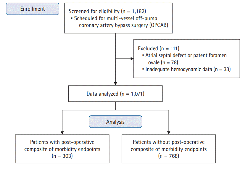
We retrospectively reviewed the electronic medical records of 1,182 patients who underwent elective multivessel OPCAB between January 2010 and December 2019. Patients with atrial septal defects or patent foramen ovale inducing left-to-right shunts (n = 78) or inadequate hemodynamic data due to failure of pulmonary artery catheter insertion were excluded (n = 33). Data from the remaining patients (n = 1,071) were analyzed (Fig. 1).
Among the assessed 1,071 patients, the composite of morbidity endpoints occurred in 303 (28.3%) patients (Supplementary Table 1). Intergroup comparisons of pre-operative variables between the SvO2 ≥ 60% group and SvO2 < 60% group are listed in Table 1. The SvO2 < 60% group exhibited advanced age, higher E/e’ and EuroSCORE II, and higher incidences of CKD and anemia.
The intraoperative hemodynamic data of the SvO2 ≥ 60% group and SvO2 < 60% group are presented in Table 2. The SvO2 < 60% group exhibited lower cardiac index and SvO2 values at all assessed time points when compared to those of the SvO2 ≥ 60% group. Decline in SvO2 below 60% was most frequently observed at the period of LCX grafting (93 patients), followed by periods of RCA grafting (23 patients) and LAD grafting (11 patients). The majority of patients who exhibited nadir SvO2 < 60% during LAD and RCA grafting also exhibited SvO2 < 60% during LCX grafting, with the exception of four patients whose incidence of SvO2 < 60% was confined to the LAD (3 patients) or RCA grafting period (1 patient). None of the patients exhibited SvO2 < 60% at baseline or after sternal closure. Supplementary Table 2 shows the hemodynamic data of the patients grouped into either the morbidity or non-morbidity group. The morbidity group exhibited lower SvO2 values at all assessed time points than the non-morbidity group, while no significant differences in HR, MAP, CVP, mPAP, or cardiac index were observed. The incidence of SvO2 < 60% during the three grafting periods was significantly higher in the morbidity group.
In the multivariable logistic regression analysis, SvO2 < 60% during LCX grafting was identified as an independent risk factor for the composite of morbidity (odds ratio [OR]: 2.72, 95% CI [1.60, 4.61], P < 0.001), along with age, CKD, E/e’, and EuroSCORE II (Table 3). When introduced as a continuous variable, nadir SvO2 during LCX grafting remained an independent risk factor without changes in the statistical significance of the other risk factors mentioned above (OR: 0.94, 95% CI [0.92, 0.96], P < 0.001; Supplementary Table 3). The nadir SvO2 of other grafting periods or cardiac index was not associated with adverse outcomes, and the ROC curve analysis revealed an optimal cut-off value for SvO2 at LCX grafting of 70.5%, with a sensitivity of 58.1% and a specificity of 63.5% (area under the ROC: 0.66, 95% CI [0.62, 0.69], P < 0.001).
Comparisons of post-operative outcomes between the SvO2 ≥ 60% group and SvO2 < 60% group are displayed in Table 4. Patients with SvO2 < 60% at LCX grafting were associated with significantly longer lengths of ICU and hospital stays and higher incidences of AKI and 30-day or in-hospital mortality.
In the multivariable analysis to identify risk factors of nadir SvO2 < 60% during LCX grafting among selected variables, only pre-operative anemia remained as an independent risk factor (OR: 2.09, 95% CI [1.33, 3.29], P = 0.001; Table 5).

Discussion

In this retrospective study, transient decline of SvO2 below 60% induced by mechanical cardiac displacement for LCX grafting was significantly associated with the composite of morbidity endpoints after OPCAB, along with previously well-known risk factors such as advanced age, CKD, E/e’, and EuroSCORE II. In addition, among the known risk factors that may influence the myocardial performance governing the global oxygen supply-demand balance, only the presence of pre-operative anemia was identified as an independent risk factor for SvO2 < 60% during LCX grafting.
SvO2 is perceived as a suitable index for monitoring hemodynamic alterations, reflecting acute changes in the balance between oxygen delivery and consumption [1618]. In general, the oxygen extraction ratio of major organs (except the myocardium) usually resides within the vicinity of 20% [19] and oxygen extraction ratio over 50% has been suggested as a critical value indicating a shock state reaching the maximum oxygen extraction ratio beyond compensation [20]. Thus, an SvO2 between 50% and 60% is typically considered marginal and requires close follow-up [21]. Accordingly, the hemodynamic management goal of SvO2 ≥ 60% has been advocated for cardiothoracic and vascular surgeries [1012]. Likewise, maintaining SvO2 above 60% has been advocated for OPCAB as well, without relevant evidence specific to this procedure [4]. We, therefore, selected the reference value of SvO2 as 60% for an early and safe warning of tissue O2 debt and prediction of inadequate tissue oxygenation.
In the current study, SvO2 < 60% during LAD, LCX, and RCA was associated with an increased risk of composite of morbidity in the univariable analysis. However, when accounting for confounders in the multivariable analysis, only the prognostic importance of SvO2 < 60% during LCX grafting was pronounced, whereas the significance of SvO2 < 60% during LAD and RCA grafting disappeared. We presume that the extremely low incidence of isolated SvO2 < 60% during LAD or RCA grafting (while most of the patients exhibiting SvO2 < 60% during LAD or RCA grafting also exhibited SvO2 < 60% during LCX grafting) contributed to diminishing the prognostic significance of SvO2 during these periods. In addition, our results confirmed that hemodynamic deterioration during lateral wall exposure (LCX grafting) was the most severe, as was previously known [4]. Similar results were observed when SvO2 at each time point were analyzed as a continuous variable; only SvO2 during LCX grafting retained its close association with poor prognosis, with an OR indicating that every 1% decrease in SvO2 during LCX grafting was responsible for a 6.3% increased risk of adverse outcomes. Additionally, the optimal cut-off value of SvO2 to predict outcome was 70.5%, which corresponds to an approximately 10% decrease from baseline. However, the area under the ROC curve as well as sensitivity and specificity of SvO2 as continuous variables were low, possibly because of the overlap of SvO2 ranges among patients. Nonetheless, the OR of SvO2 < 60% as a dichotomous variable on outcome was 2.72, which was even higher than that of CKD and showed the prognostic importance of SvO2 considering that the composite of morbidity was mostly driven by AKI. While the clinical consequences of transient hemodynamic deterioration related to cardiac displacement have not yet been comprehensively investigated, and the advocacy of maintaining SvO2 above 60% in OPCAB was without pertinent evidence [4], the current study provides primary evidence in that regard.
Interestingly, no difference was observed in the serially assessed cardiac index between the morbidity and non-morbidity groups (Supplementary Table 2). Although the trend of cardiac index was similar to that of SvO2, cardiac index during LCX grafting (or at any other grafting period) showed no association with the outcome in the logistic regression analysis. Likewise, serially assessed MAP, CVP, and mPAP were all similar between the morbidity and non-morbidity groups, which may be attributable to our standardized anesthetic protocol to stabilize hemodynamic parameters during the period of cardiac displacement. Nonetheless, SvO2 significantly differed between the two groups; this underlines the prognostic importance of SvO2 reflecting net circulatory menace, which could not be detected by other commonly targeted major hemodynamic variables. Among the other revealed risk factors, high E/e’, reflecting increased left ventricular filling pressure, also showed a close association with poor outcome, which corroborates our previous finding in a similar subset of patients implicating the prognostic significance of diastolic dysfunction [22].
Additionally, we investigated the risk factors for SvO2 < 60% during mechanical cardiac displacement, including variables that affect cardiac function and oxygen delivery. No significant association was observed between SvO2 and parameters derived from pre-operative transthoracic echocardiography, such as LVEF, E/e’, and LVEDD, while pre-operative anemia was identified as the only predictor yielding a 2.1-fold increased risk of developing SvO2 < 60%. This result seems logical because the influence of anemia would be critical in providing adequate oxygen-carrying capacity, especially during periods of hemodynamic instability, and adds background rationale to the importance of pre-operative anemia correction to improve patient outcomes [23].
Efforts to increase the cardiac index may be considered to attenuate the decrease in SvO2 during mechanical cardiac displacement. However, increasing cardiac index inevitably accompanies increased myocardial oxygen consumption, which may incur ischemia and add technical burden to surgeons due to increased HR. Moreover, our results indicated that a decrease in cardiac index during grafting was not associated with detrimental outcomes. On the other hand, previous evidence has indicated that hyperoxia enhances oxygen supply by redistributing blood flow to relatively hypoperfused renal tissues and reduces systemic oxygen consumption without a significant effect on cardiac index [24,25]. Despite the potential drawbacks of hyperoxia [26], it remains to be proven whether a transient increase in the inspired oxygen fraction during grafting may actually yield improved outcomes by attenuating the decrease in SvO2 without jeopardizing the coronary reserve.
This study has inherent limitations owing to its retrospective nature allowing for temporal bias over a 10-year period. In addition, the duration of the SvO2 decline would also be as important as the magnitude of the drop, but this could not be clearly addressed in the current study. However, the median duration of LCX grafting in cases with SvO2 < 60% was 11 (8–15) min, while the SvO2 did not remain consistently below 60% during that period. Despite being a retrospective study, hemodynamic data including SvO2 were serially gathered at the predefined five time points with additional recordings of the lowest cardiac index and SvO2 between the time points, as well as the exact duration of each grafting. Thus, we can assure that the duration of SvO2 < 60% was only limited to the period of grafting. Lastly, an analysis incorporating SvO2 values during the immediate post-operative period would have been more comprehensive.
In conclusion, transient hemodynamic deterioration induced by mechanical cardiac displacement during LCX grafting, when severe enough as reflected by SvO2 < 60%, was associated with a 2.7-fold increased risk of adverse outcomes in patients undergoing OPCAB. Among the pre-operative risk factors including echocardiography, anemia alone was associated with the occurrence of SvO2 < 60% during LCX grafting. The results of the current study should arouse awareness on the importance of transient hemodynamic deterioration during cardiac displacement and SvO2 as a reliable prognostic factor in this context.

Notes

Funding

None.

Conflicts of Interest

Young-Lan Kwak has been an editor in chief for the Korean Journal of Anesthesiology since 2016. However, she was not involved in any process of review for this article, including peer reviewer selection, evaluation, or decision-making. There were no other potential conflicts of interest relevant to this article.

Data Availability

The datasets generated during and/or analyzed during the current study are available from the corresponding author on reasonable request.

Author Contributions

Kyuho Lee (Data curation; Investigation; Writing – original draft)

Kwang-Sub Kim (Investigation; Methodology; Visualization)

Jong-Kwang Park (Conceptualization; Validation; Visualization)

Jun Hyug Choi (Data curation; Investigation)

Young-Lan Kwak (Conceptualization; Project administration; Validation; Writing – review & editing)

Jae-Kwang Shim (Conceptualization; Investigation; Methodology; Validation; Writing – review & editing)

Supplementary Materials

Supplementary Table 1.
Incidence of morbidity endpoints.
kja-22277-suppl1.pdf
Supplementary Table 2.
Intraoperative hemodynamic data of patients classified by the composite of morbidity endpoints.
kja-22277-suppl2.pdf
Supplementary Table 3.
Predictive power of selected variables for composite morbidity according to logistic regression analysis.
kja-22277-suppl3.pdf

References

1. Zhao DF, Edelman JJ, Seco M, Bannon PG, Wilson MK, Byrom MJ, et al. Coronary artery bypass grafting with and without manipulation of the ascending aorta: a network meta-analysis. J Am Coll Cardiol. 2017; 69:924–36.
2. Puskas JD, Martin J, Cheng DC, Benussi S, Bonatti JO, Diegeler A, et al. ISMICS consensus conference and statements of randomized controlled trials of off-pump versus conventional coronary artery bypass surgery. Innovations (Phila). 2015; 10:219–29.
3. Lamy A, Devereaux PJ, Prabhakaran D, Taggart DP, Hu S, Paolasso E, et al. Off-pump or on-pump coronary-artery bypass grafting at 30 days. N Engl J Med. 2012; 366:1489–97.
4. Chassot PG, van der Linden P, Zaugg M, Mueller XM, Spahn DR. Off-pump coronary artery bypass surgery: physiology and anaesthetic management. Br J Anaesth. 2004; 92:400–13.
5. Ngaage DL. Off-pump coronary artery bypass grafting: the myth, the logic and the science. Eur J Cardiothorac Surg. 2003; 24:557–70.
6. Mueller XM, Chassot PG, Zhou J, Eisa KM, Chappuis C, Tevaearai HT, et al. Hemodynamics optimization during off-pump coronary artery bypass: the 'no compression' technique. Eur J Cardiothorac Surg. 2002; 22:249–54.
7. Mathison M, Edgerton JR, Horswell JL, Akin JJ, Mack MJ. Analysis of hemodynamic changes during beating heart surgical procedures. Ann Thorac Surg. 2000; 70:1355–60.
8. Oh SY, Shim JK, Song JW, Kim JC, You KJ, Kwak YL. Cardiac displacement-induced hemodynamic instability during off-pump coronary artery bypass surgery and its predictors. Acta Anaesthesiol Scand. 2011; 55:870–7.
9. Shim JK, Choi YS, Chun DH, Hong SW, Kim DH, Kwak YL. Relationship between echocardiographic index of ventricular filling pressure and intraoperative haemodynamic changes during off-pump coronary bypass surgery. Br J Anaesth. 2009; 102:316–21.
10. Svedjeholm R, Hakanson E, Szabo Z. Routine SvO2 measurement after CABG surgery with a surgically introduced pulmonary artery catheter. Eur J Cardiothorac Surg. 1999; 16:450–7.
11. Holm J, Hakanson RE, Vanky F, Svedjeholm R. Mixed venous oxygen saturation is a prognostic marker after surgery for aortic stenosis. Acta Anaesthesiol Scand. 2010; 54:589–95.
12. Holm J, Hakanson E, Vanky F, Svedjeholm R. Mixed venous oxygen saturation predicts short- and long-term outcome after coronary artery bypass grafting surgery: a retrospective cohort analysis. Br J Anaesth. 2011; 107:344–50.
13. Nutritional anaemias. Report of a WHO scientific group. World Health Organ Tech Rep Ser. 1968; 405:5–37.
14. Thygesen K, Alpert JS, Jaffe AS, Chaitman BR, Bax JJ, Morrow DA, et al. Fourth universal definition of myocardial infarction (2018). Circulation. 2018; 138:e618–51.
15. Levin A, Stevens PE. Summary of KDIGO 2012 CKD guideline: behind the scenes, need for guidance, and a framework for moving forward. Kidney Int. 2014; 85:49–61.
16. Robin E, Costecalde M, Lebuffe G, Vallet B. Clinical relevance of data from the pulmonary artery catheter. Crit Care. 2006; 10 Suppl 3(Suppl 3):S3.
17. Shepherd SJ, Pearse RM. Role of central and mixed venous oxygen saturation measurement in perioperative care. Anesthesiology. 2009; 111:649–56.
18. Lazor MA, Pierce ET, Stanley GD, Cass JL, Halpern EF, Bode RH Jr. Evaluation of the accuracy and response time of STAT-mode continuous cardiac output. J Cardiothorac Vasc Anesth. 1997; 11:432–6.
19. Rotondi S, Tartaglione L, De Martini N, Bagordo D, Caissutti S, Pasquali M, et al. Oxygen extraction ratio to identify patients at increased risk of intradialytic hypotension. Sci Rep. 2021; 11:4801.
20. Kasnitz P, Druger GL, Yorra F, Simmons DH. Mixed venous oxygen tension and hyperlactatemia. Survival in severe cardiopulmonary disease. JAMA. 1976; 236:570–4.
21. Kaplan JA. Kaplan's cardiac anesthesia. 7th ed. Philadelphia: Elsevier Saunders;2016. p. 1295.
22. Jun NH, Shim JK, Kim JC, Kwak YL. Prognostic value of a tissue Doppler-derived index of left ventricular filling pressure on composite morbidity after off-pump coronary artery bypass surgery. Br J Anaesth. 2011; 107:519–24.
23. Raphael J, Mazer CD, Subramani S, Schroeder A, Abdalla M, Ferreira R, et al. Society of Cardiovascular Anesthesiologists Clinical Practice Improvement Advisory for management of perioperative bleeding and hemostasis in cardiac surgery patients. Anesth Analg. 2019; 129:1209–21.
24. Reinhart K, Bloos F, Konig F, Bredle D, Hannemann L. Reversible decrease of oxygen consumption by hyperoxia. Chest. 1991; 99:690–4.
25. Calzia E, Radermacher P, Matejovic M. Splanchnic resuscitation revisited: combining hyperoxia and hypertonic saline during early goal-directed treatment. Crit Care Med. 2006; 34:2858–60.
26. Ottolenghi S, Sabbatini G, Brizzolari A, Samaja M, Chiumello D. Hyperoxia and oxidative stress in anesthesia and critical care medicine. Minerva Anestesiol. 2020; 86:64–75.

Fig. 1.
Flowchart of patient enrollment.
kja-22277f1.tif
Table 1.
Pre-operative Data of Patients Classified by Nadir SvO2 during Grafting of LCX
Variable SvO2 ≥ 60% (n = 978) SvO2 < 60% (n = 93) P value
Age (yr) 66.1 (59.2, 72.0) 69.4 (64.6, 73.2)* 0.021
Sex (M/F) 763 (78.0) / 215 (22.0) 67 (72.0) / 26 (28.0) 0.187
BMI (kg/m2) 24.3 (22.3, 26.4) 24.0 (22.3, 25.5) 0.215
HTN 698 (71.4) 66 (71.0) 0.935
DM 514 (52.6) 53 (57.0) 0.413
COPD 43 (4.4) 1 (1.1) 0.123
CKD 105 (10.7) 17 (18.3)* 0.029
CVA 134 (13.7) 11 (11.8) 0.614
MI within one month 259 (26.5) 28 (30.1) 0.451
CHF 104 (10.6) 13 (14.0) 0.323
Anemia 405 (41.4) 58 (62.4)* < 0.001
Ejection fraction (%) 58 (46, 67) 57 (46, 68) 0.999
E/e’ 12.0 (9.8, 16.0) 13.0 (11.0, 16.5)* 0.026
LVEDD (mm) 51 (47, 54) 51 (47, 54) 0.413
EuroSCORE II 1.15 (0.79, 1.92) 1.51 (0.97, 2.88)* < 0.001

Values are presented as median (Q1, Q3) or number of patients (%). SvO2: mixed venous oxygen saturation, LCX: left circumflex artery, SvO2 ≥ 60% group: patients whose nadir SvO2 during LCX grafting was more than or equal to 60%, SvO2 < 60% group: patients whose nadir SvO2 during LCX grafting was less than 60%, BMI: body mass index, HTN: hypertension, DM: diabetes mellitus, COPD: chronic obstructive pulmonary disease, CKD: chronic kidney disease, CVA: cerebrovascular accident, MI: myocardial infarction, CHF: chronic heart failure, E/e’: early mitral inflow velocity / mitral annular early diastolic velocity, LVEDD: left ventricular end-diastolic diameter.

* P < 0.05, compared with the SvO2 ≥ 60% group.

Table 2.
Intraoperative Hemodynamic Data of Patients Classified by Nadir SvO2 during Grafting of LCX
Variable Baseline LAD grafting LCX grafting RCA grafting Sternal closure Pgroup × time
HR (beats/min) 0.997
 SvO2 ≥ 60% group 59 (53, 65) 66 (60, 72)* 70 (63, 76)* 71 (63, 77)* 77 (71, 82)*
 SvO2 < 60% group 59 (53, 66) 68 (61, 74)* 70 (63, 78)* 71 (65, 79)* 77 (71, 83)*
MAP (mmHg) 0.196
 SvO2 ≥ 60% group 75 (69, 81) 77 (72, 82)* 78 (72, 84)* 77 (72, 82)* 77 (70, 84)
 SvO2 < 60% group 76 (69, 87) 76 (71, 81) 75 (68, 82) 76 (71, 82) 77 (72, 83)
CVP (mmHg) 0.223
 SvO2 ≥ 60% group 9 (8, 11) 12 (10, 14)* 17 (13, 19)* 16 (14, 18)* 10 (8, 11)
 SvO2 < 60% group 9 (7, 11) 11 (10, 13)* 16 (12, 19)* 16 (13, 18)* 9 (8, 11)
mPAP (mmHg) 0.148
 SvO2 ≥ 60% group 17 (15, 20) 21 (19, 23)* 24 (21, 27)* 25 (22, 27)* 18 (16, 20)
 SvO2 < 60% group 17 (15, 20) 20 (18, 22)* 22 (19, 26)* 23 (20, 27)* 18 (16, 21)
Cardiac index (L/min/m2) 0.031
 SvO2 ≥ 60% group 2.3 (1.9, 2.6) 2.0 (1.7, 2.3)* 1.8 (1.6, 2.1)* 1.8 (1.6, 2.0)* 2.2 (2.0, 2.6)
 SvO2 < 60% group 2.1 (1.7, 2.5) 1.7 (1.5, 2.1)* 1.6 (1.4, 1.8)* 1.5 (1.3, 1.7)* 1.9 (1.6, 2.2)*
SvO2 (%) < 0.001
 SvO2 ≥ 60% group 80 (77, 84) 77 (73, 81)* 73 (68, 78)* 74 (69, 78)* 78 (74, 82)*
 SvO2 < 60% group 75 (71, 80) 67 (62, 70)* 57 (54, 58)* 61 (58, 66)* 70 (65, 75)*
Incidence of SvO2 < 60% (n) N/A
 SvO2 ≥ 60% group 0 (0%) 3 (0.3%) 0 (0%) 1 (0%) 0 (0%)
 SvO2 < 60% group 0 (0%) 8 (8.6%) 93 (100%) 22 (23.7%) 0 (0%)

Values are presented as median (Q1, Q3) or number of patients (%). SvO2: mixed venous oxygen saturation, LCX: left circumflex artery, LAD: left anterior descending artery, RCA: right coronary artery, SvO2 ≥ 60% group: patients whose nadir SvO2 during LCX grafting was more than or equal to 60%, SvO2 < 60% group: patients whose nadir SvO2 during LCX grafting was less than 60%, HR: heart rate, MAP: mean systemic arterial pressure, CVP: central venous pressure, mPAP: mean pulmonary arterial pressure.

* P < 0.05, compared with baseline within the SvO2 ≥ 60% group and SvO2 < 60% group, respectively.

P < 0.01, compared with the SvO2 ≥ 60% group.

Table 3.
Predictive Power of Selected Variables for Composite Morbidity according to Logistic Regression Analysis
Variable Univariable analysis Multivariable analysis
OR (95% CI) P value OR (95% CI) P value
Age 1.05 (1.03, 1.06)* < 0.001 1.03 (1.01, 1.05)* 0.008
CKD 3.28 (2.23, 4.81)* < 0.001 2.15 (1.32, 3.51)* 0.002
Pre-operative anemia 1.96 (1.50, 2.56)* < 0.001 1.45 (1.00, 2.12) 0.053
E/e’ 1.07 (1.05, 1.10)* < 0.001 1.05 (1.01, 1.08)* 0.005
EuroSCORE II 1.26 (1.16, 1.37)* < 0.001 1.12 (1.01, 1.24)* 0.040
SvO2 < 60% during LAD grafting 4.67 (1.36, 16.10)* 0.015 2.61 (0.64, 10.56) 0.180
SvO2 < 60% during LCX grafting 3.33 (2.16, 5.13)* < 0.001 2.72 (1.60, 4.61)* < 0.001
SvO2 < 60% during RCA grafting 2.55 (1.11, 5.86)* 0.028 0.46 (0.15, 1.38) 0.166

OR: odds ratio, CKD: chronic kidney disease, E/e’: early mitral inflow velocity / mitral annular early diastolic velocity, SvO2: mixed venous oxygen saturation, LAD: left anterior descending artery, LCX: left circumflex artery, RCA: right coronary artery.

* P < 0.05.

Table 4.
Post-operative Data of Patients Classified by Nadir SvO2 during Grafting of LCX
Variable SvO2 ≥ 60% (n = 978) SvO2 < 60% (n = 93) P value
ICU days 3 (2, 3) 3 (2, 4)* 0.015
Hospital days 13 (10, 15) 14 (11, 20)* 0.003
30-day or in-hospital mortality 13 (1.3) 6 (6.5)* < 0.001
Post-operative MI 4 (0.4) 0 (0) 0.537
Prolonged mechanical ventilation > 24 h 71 (7.3) 10 (10.8) 0.223
Post-operative CVA 21 (2.1) 2 (2.2) 0.998
AKI 192 (19.6) 41 (44.1)* < 0.001

Values are presented as median (Q1, Q3) or number of patients (%). SvO2: mixed venous oxygen saturation, LCX: left circumflex artery, SvO2 ≥ 60% group: patients whose nadir SvO2 during LCX grafting was more than or equal to 60%, SvO2 < 60% group: patients whose nadir SvO2 during LCX grafting was less than 60%, ICU: intensive care unit, MI: myocardial infarction, CVA: cerebrovascular accident, AKI: acute kidney injury.

* P < 0.05 compared with the SvO2 ≥ 60% group.

Table 5.
Predictive Power of Selected Variables for Nadir SvO2 < 60% during Grafting of LCX
Variable Univariable analysis Multivariable analysis
OR (95% CI) P value OR (95% CI) P value
Age 1.03 (1.00, 1.05)* 0.041 1.01 (0.99, 1.04) 0.250
HTN 0.98 (0.61, 1.57) 0.935
DM 1.20 (0.78, 1.84) 0.414
COPD 0.24 (0.32, 1.74) 0.236
CKD 1.86 (1.06, 3.27)* 0.031 1.25 (0.68, 2.30) 0.206
Old CVA 0.85 (0.44, 1.63) 0.614
MI within one month 1.20 (0.75, 1.90) 0.451
CHF 1.37 (0.73, 2.54) 0.325
Pre-operative anemia 2.35 (1.51, 3.64)* < 0.001 2.09 (1.33, 3.29)* 0.001
Ejection fraction 1.00 (0.98, 1.02) 0.953
E/e’ 1.02 (0.98, 1.05) 0.338
LVEDD 1.01 (0.98, 1.04) 0.645

SvO2: mixed venous oxygen saturation, LCX: left circumflex artery, OR: odds ratio, HTN: hypertension, DM: diabetes mellitus, COPD: chronic obstructive pulmonary disease, CKD: chronic kidney disease, CVA: cerebrovascular accident, MI: myocardial infarction, CHF: chronic heart failure, E/e’: early mitral inflow velocity / mitral annular early diastolic velocity, LVEDD: left ventricular end-diastolic diameter.

* P < 0.05.

TOOLS
Similar articles