Journal List > Clin Endosc > v.56(2) > 1516081879

Wasielica-Berger, Rogalski, Pryczynicz, Swidnicka-Siergiejko, and Dabrowski: Methylene blue chromoendoscopy is more useful in detection of intestinal metaplasia in the stomach than mucosal pit pattern or vessel evaluation and predicts advanced Operative Link on Gastric Intestinal Metaplasia stages

Abstract

Background/Aims

Intestinal metaplasia (IM) of the stomach is a precancerous condition that is often not visible during conventional endoscopy. Hence, we evaluated the utility of magnification endoscopy and methylene blue (MB) chromoendoscopy to detect IM.

Methods

We estimated the percentage of gastric mucosa surface staining with MB, mucosal pit pattern, and vessel visibility and correlated it with the presence of IM and the percentage of metaplastic cells in histology, similar to the Operative Link on Gastric Intestinal Metaplasia (OLGIM) stage.

Results

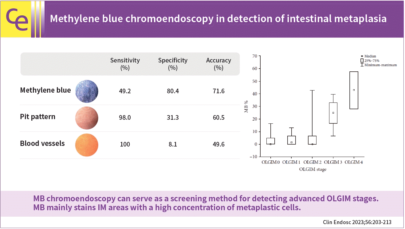
IM was found in 25 of 33 (75.8%) patients and in 61 of 135 biopsies (45.2%). IM correlated with positive MB staining (p<0.001) and other than dot pit patterns (p=0.015). MB staining indicated IM with better accuracy than the pit pattern or vessel evaluation (71.7% vs. 60.5% and 49.6%, respectively). At a cut-off point of 16.5% for the MB-stained gastric surface, the sensitivity, specificity, and accuracy of chromoendoscopy in the detection of advanced OLGIM stages were 88.9%, 91.7%, and 90.9%, respectively. The percentage of metaplastic cells detected on histology was the strongest predictor of positive MB staining.

Conclusions

MB chromoendoscopy can serve as a screening method for detecting advanced OLGIM stages. MB mainly stains IM areas with a high concentration of metaplastic cells.

Graphic abstract

INTRODUCTION

Gastric cancer is one of the most common malignancies worldwide. Clinical symptoms usually manifest only in the advanced stages of the disease, which delays the diagnosis and is associated with a high mortality rate of 75%.1 Intestinal metaplasia (IM) in the stomach is a precancerous condition that increases the risk of gastric cancer by 4-fold.2 Based on the management of epithelial precancerous conditions and lesions in the stomach (MAPS II) guidelines, at least four non-targeted biopsies, along the greater and lesser curvature within the antrum and corpus of the stomach, are recommended to detect IM.3 The severity and extent of IM are determined according to the Operative Link on Gastric Intestinal Metaplasia (OLGIM) classification, in which severe IM at any location or moderate IM in both the corpus and antrum are graded as stages III and IV.4 The presence of these advanced stages requires regular endoscopic surveillance. IM can also be subtyped as complete or incomplete, of which the latter is associated with an 11.3-fold higher risk of developing gastric cancer.5,6
The most important problems associated with the diagnosis and endoscopic surveillance of IM are its focal distribution in the stomach, inconspicuous endoscopic appearance, and the need for histopathological confirmation. Various additional techniques can be used to improve the diagnostic yield of endoscopy. One such method is methylene blue (MB) chromoendoscopy, a vital staining method in which the dye is absorbed by both complete and incomplete IM.7,8 In previous studies, the sensitivity of MB staining in the detection of IM ranged from 71% to 78% and the specificity from 41% to 88%.9,10 It is not yet fully understood what determines the ability of the tissue to absorb MB. Another helpful technique is the evaluation of the gastric surface using optical magnification. Previous studies have suggested that the presence of IM is related to certain types of mucosal surface pit patterns and the architecture of vessels.8,11 However, the diagnostic accuracy of pit pattern and mucosal blood vessel architecture based assessment in the detection of various types of IM is still controversial.
Therefore, we performed a prospective study to identify the factors determining the ability of the gastric IM to take up the MB dye. Our analysis focused on the subtype of IM, its location, extent, and percentage of metaplastic cells in the histopathological sample, and the presence of atrophy, Helicobacter pylori infection, and inflammation. The other study outcomes that were determined were: (1) the relationship between MB staining and OLGIM stage and (2) the comparison of the utility of MB chromoendoscopy, pit pattern, and mucosal blood vessel microarchitecture assessment in detecting IM and differentiating IM subtypes.

METHODS

Thirty-three patients (25 women, 75.8%) were prospectively enrolled in this study. We included patients with a family history of gastric cancer undergoing gastroscopy to exclude precancerous conditions and patients with a history of histologically proven gastric IM, regardless of its extent, referred for gastroscopy for the periodic screening for dysplasia and cancer. The exclusion criteria were as follows: (1) contraindications for biopsies, (2) previous gastric neoplasia, (3) a history of gastric surgery, or (4) intolerance of the endoscopic procedure.
All procedures were performed by an endoscopist experienced in magnification endoscopy and chromoendoscopy (JWB) with an endoscope that allowed an optical magnification of up to 115 times (GIF-Q160Z; Olympus, Tokyo, Japan). A disposable distal attachment (D-201-12402; Olympus) was used to maintain a stable view and focal distance. The patients received topical oropharyngeal anesthesia with 1% lidocaine. Intravenous midazolam was administered depending on the individual needs. Magnified images of the gastric mucosa were analyzed considering the type of surface pit pattern and visibility and the architecture of the venules. The pit patterns of the gastric mucosal surface were classified into the following groups: (1) dot (regularly arranged, pinpoint small orifices of gastric glands), (2) tubular (convex, oval, horizontal rods), (3) villous (resembling simple or branched villi), (4) foveolar (wide orifices of gastric glands, appearing reticular), and (5) cobblestone (convex, round, vertical rods). Examples are shown in Figure 1. The microvessel appearance was classified as (1) regular (clearly visible, regularly arranged collecting venules with branches of uniform length and diameter resembling spider naevi), (2) irregular (visible partially with irregular length or caliber of branches), and (3) obscured (vessels invisible), as shown in Figure 2.12 Subsequently, 20 mL of 1% MB was sprayed through the spray catheter all over the stomach. After one minute, the excess dye was flushed away with water and aspirated. The percentage of the gastric surface positively stained by MB was estimated separately for the antrum (MB% antrum) and corpus (MB% corpus), as it is difficult to visualize the entire surface of the stomach from a single endoscope position. The total stained surface (MB%) was calculated according to the following formula: MB%=1/3×MB% antrum+2/3×MB% corpus. The proportion of the gastric surface comprised of the antrum and corpus was assumed arbitrarily to be 1/3 and 2/3 of the total gastric surface, respectively. Magnification helped us discern whether the stained areas were due to MB absorption by the epithelium or the presence of residual, stained superficial mucus due to inadequate flushing of the same, as shown in Figure 3. Biopsies for histological examination were collected from the antrum and corpus of the stomach, separately from stained and unstained areas, while recording the mucosal pit pattern type and the appearance of microvessels for each sample. Within the areas of both stained and unstained mucosa, the final biopsy site was guided by choice of sites presenting patterns other than dot. Samples from areas presenting a dot pit pattern were collected only when no other type of pit pattern was observed. Each biopsy specimen was placed in a labeled container filled with formalin and was processed separately.
All samples were stained with hematoxylin and eosin. IM severity and extent were graded according to the OLGIM staging system.4 For OLGIM stage evaluation, the pathologist considered all samples irrespective of the endoscopic data on MB staining or pit pattern and chose the most advanced IM changes in the antrum and corpus of the stomach. If IM was found in the histopathological evaluation, it was further differentiated into complete and incomplete by histochemistry (Alcian blue, pH 2.5; PAS; Bio-Optica, Milano, Italy). For each sample, the pathologist reported: (1) the IM subtype: complete, incomplete, or both; (2) the IM extent: focal or diffuse; (3) the percentage of mucosal surface comprised of metaplastic cells at high-power magnification; (4) the activity and grade of inflammation; (5) presence of gastric atrophy; and (6) H. pylori infection.

Statistical analysis

Categorical variables were reported as counts (percentages). Continuous variables were tested for normality using the Shapiro-Wilk test and expressed as mean±standard deviation or median (interquartile range) as appropriate. Continuous variables were compared using the parametric t-test, Mann-Whitney U-test, or Kruskal-Wallis test to detect significant differences between the groups. All tests were two-tailed, and differences were considered significant at p<0.05. To determine the optimal cut-off point for a significant variable as a marker for IM, the receiver operating characteristic curve was prepared using the Youden criterion. The additional analysis included a stepwise multivariate logistic regression, which was used to identify the best combination of parameters predicting IM presence vs. absence of IM and to identify factors that contribute to positive MB staining. Only variables that were significantly different between the groups were included in the regression model as predictors. Statistical analyses were performed using Statistica ver. 13.3 software package (StatSoft Inc.,Tulsa, OK, USA).

Ethical statement

The research protocol followed the guidelines of the Declaration of Helsinki and was approved by Bioethical Committee of Medical University of Bialystok (R-I-002/65/12). The patients provided written informed consent to participate in the study.

RESULTS

Per-patient analysis

IM was found in 25 of the 33 patients (75.8%). Patient characteristics, including age, OLGIM stage, the extent of IM and its subtype, are presented in Table 1.
The mean time of endoscopic examination was 16 minutes 54 seconds (±3 minutes 48 seconds). Positive MB staining was observed in 19 (57.6%) patients. IM was confirmed by histology in 17 (89.5%) of that 19 patients and was excluded in 2 (10.5%) patients. Moreover, IM was found in the histopathology of eight out of 14 patients (57.1%) with negative MB staining. The sensitivity, specificity, accuracy, positive predictive value (PPV), and negative predictive value (NPV) of MB chromoendoscopy for detecting IM in the per-patient analysis were 68.0%, 75.0%, 69.7%, 89.5%, and 42.9%, respectively.
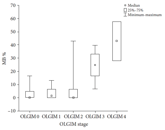
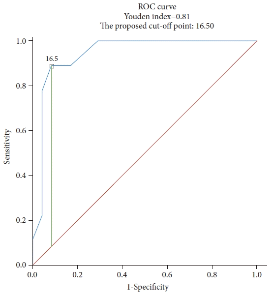
The mean percentage of the MB-stained mucosal surfaces as per the various OLGIM stages was as follows: 3.3%±6.4% for OLGIM 0, 3.3%±4.2% for OLGIM I, 9.9%±18.7% for OLGIM II, 24.2%±12.1% for OLGIM III, and 42.9%±14.8% for OLGIM IV. These values were significantly higher in patients with OLGIM stages III and IV than in those with OLGIM stage 0 (p=0.048 and p=0.039, respectively) as shown in Figure 4. At a cut-off point of 16.5% of the MB-stained surface, the sensitivity, specificity, accuracy, PPV, and NPV of chromoendoscopy in the detection of advanced OLGIM stages (III and IV) were 88.9%, 91.7%, 90.9%, 80.0%, and 95.7%, respectively (Fig. 5).
The percentage of MB-stained gastric surface (25.9%±16.9%) was significantly higher in patients with diffuse IM on histopathology than in patients without IM (3.3%±6.3%, p=0.004), or with focal IM only (5.3%±11.5%, p=0.003). Interestingly, IM was focal in all the eight patients with IM and negative MB staining.

Per-biopsy analysis

1) MB chromoendoscopy

Biopsies were obtained from 135 areas: 37 (27.4%) areas stained with MB, and 97 (71.9%) unstained. In one area (0.7%), MB staining was not documented. IM was detected in 61 samples (45.2%). IM was found more often in the samples obtained from stained areas compared to the unstained ones (30, 81.1% vs. 31, 32.0%; p<0.001).

2) Pit pattern assessment

Pit patterns were recorded in 114 of the 135 sampled locations (84.4%). Twenty-one (18.4%) of these areas presented a dot pit pattern, and 93 (81.6%) presented patterns other than the dot pit pattern. IM was detected in 49 (52.7%) samples taken from areas with pit patterns other than dot and in only one (4.8%) sample obtained from the area with the dot pit pattern (p<0.001). More specifically, IM was significantly less common in areas with a dot pit pattern (n=1, 4.8%) than in the villous (n=20, 48.8%; p=0.047), foveolar (n=9, 60%; p=0.048) and cobblestone pit patterns (n=11, 68.7%, p=0.009). The differences in the frequency of IM between the dot and tubular patterns (n=9, 42.9%; p=0.333) and among the type 2 to 5 pit patterns (p=1.0) were not significant.

3) Appearance of blood vessels

Mucosal blood vessels were assessed in 113 (83.7%) of the 135 sampled areas. Five biopsies (4.4%) were collected from areas with regular blood vessels, 63 (55.8%) from those with obscured blood vessels, and 45 (39.8%) from those with irregular blood vessels. IM was found in 34 (66.7%) areas with obscured vessels, 17 (33.3%) with irregular vessels, and none of the 5 (0%) areas with regular blood vessels (p=0.029).

4) Complex endoscopic evaluation

Of the 31 samples taken from the areas not stained with MB, in which the presence of IM was confirmed by histopathology, 25 (80.6%) samples were obtained from the sites of pit pattern types 2 to 5, one (3.2%) sample was taken from the area of the dot pit pattern. In the remaining 5 (16.1%), the pit pattern was not evaluated. Of the same 31 samples, 27 (87.1%) were collected from sites with abnormal (irregular or obscured) blood vessels, and in four cases (12.9%), blood vessels were not evaluated. The sensitivity, specificity, accuracy, PPV, and NPV of MB chromoendoscopy, pit pattern, and mucosal blood vessel assessment as a single method of IM detection and their combinations are presented in Table 2.

5) Complete and incomplete IM

Complete IM alone was found in four (3.2%) samples, incomplete IM alone was found in 23 (18.2%) samples, and both IM subtypes were found in 31 (24.6%) samples.
The proportion of complete and incomplete IM in histopathology did not differ according to the MB staining result (p=0.41), pit pattern (p=0.224), blood vessel appearance (p=0.444), and localization in the antrum versus corpus of the stomach (p=0.487).

6) Focal and diffuse IM

IM was focal in 32 (52.5%) specimens and diffuse in 29 (47.5%) specimens. Diffuse IM lesions took up the MB stain with a significantly higher frequency than focal IM (21, 72.4% vs. 9, 28.1%; p<0.001). Diffuse IM was significantly associated with a cobblestone pit pattern (2, 40%), while focal IM with a villous pit pattern (8, 66.7%; p=0.031). There was no significant relationship between the extent of IM as determined by histopathology and the appearance of blood vessels (p=0.892), the location from where the sample was obtained (whether from the antrum or corpus of the stomach, p=0.782), or the complete or incomplete subtype of IM (p=0.844).

7) The percentage of the surface of histopathological sample covered by IM

The surface of the histopathological sample comprised of IM was significantly larger in the samples collected from MB-stained areas than in unstained areas (65% [40%–90%] vs. 25% [10%–40%], p<0.001]. It was also significantly more extensive in the samples obtained from a cobblestone pit pattern than in those obtained from a villous pit pattern (68%±26.4% vs. 31.31%±24.4%, p=0.039). No significant differences were observed among the other pit patterns. In areas with obscured vessels, the percentage of sample surface comprised of IM was significantly higher than in areas with irregular vessels (48.5%±32.1% vs. 28.5%±24.4%, p=0.044).

8) H. pylori infection, atrophy and inflammation

Positive MB staining was found less frequently in areas with low-grade inflammation than in areas with moderate and high-grade inflammation (16.5%, 46.3%, and 35.7, respectively, p=0.002). We found no relationship between MB staining and H. pylori infection status (p=0.490), presence of atrophy (p=0.255) and inflammatory activity (p=0.269). Only six (15.4%) patients were H. pylori-positive, which is much less than the average occurrence in the local population. Data on past infections were not obtained.

9) Multivariate logistic regression

Age, positive MB staining, other than dot pit pattern, and abnormal blood vessels (obscured or irregular) were included into the logistic regression model analysis as predictors of IM. Among these features, positive MB staining (p<0.001; odds ratio [OR], 8.36; 95% confidence interval [CI], 2.572–27.176) and other than dot pit pattern (p=0.015; OR, 13.158; 95% CI, 1.659–104.363) were found to be significant.
We also attempted to identify the factors that contributed to positive MB staining. The percentage of the sample surface comprised of IM on histologic examination, the degree of inflammation, and the extent of IM (focal or diffuse) were included in the regression model as potential predictors of positive MB staining. Among these factors, the percentage of metaplastic cells in the histopathological sample was the strongest predictor of positive MB staining (p<0.001; OR, 1.045; 95% CI, 1.028–1.061).

DISCUSSION

IM is an essential transitional step in the progression from gastritis to gastric adenocarcinoma. Conventional endoscopy cannot accurately diagnose IM; therefore, biopsy with histopathological assessment according to the Sydney classification remains mandatory. To allow targeted biopsies, the global consensus from Kyoto recommends using endoscopic techniques developed to reveal IM during gastroscopy.13
We compared three methods for identifying IM: MB chromoendoscopy, evaluation of the pit pattern, and mucosal vessel visibility assessment. IM was found much more often in the samples taken from areas stained with MB than those obtained from unstained areas, and significantly less often in areas with dot pit pattern than villous, foveolar, and cobblestone pit patterns. The assessment of blood vessels is not useful for detecting IM because of its very low specificity. Among these three methods, positive MB staining was the strongest predictor of IM in multivariate logistic regression analysis.
According to some studies, the sensitivity of MB chromoendoscopy for IM diagnosis has reached 76% with 87% specificity.10 In the present study, the sensitivity and specificity of MB staining were 68% and 75% in the per-patient analysis and 49.2% and 90.4% in the per-biopsy analysis, respectively. The low sensitivity of the MB staining in our study can be explained by the study protocol. Samples collected from MB-stained areas were not compared to the random biopsies taken from non-stained places, as in the previous studies, but to the non-stained areas presenting pit patterns other than dot or irregular or obscured vessels. All of the 26 biopsies from unstained areas, in which the presence of IM was confirmed by histopathology, were obtained from the areas with irregular or obscured vessels, while 25 (96.1%) samples were obtained from the sites with pit pattern types 2 to 5. Another possible explanation for the higher sensitivity achieved using the MB chromoendoscopy by other authors may be the pretreatment with the mucolytic agent N-acetylcysteine sprayed over the mucosa before MB staining.10 Omitting this procedure made the examination shorter, cheaper, and easier in routine clinical practice. However, it is difficult to exclude the possibility that mucus may hamper the absorption of MB into the mucosa. Magnification helped us discern whether the visible stained areas were due to uptake of the MB stain by the epithelium or due to residual stained superficial mucus, as shown in Figure 3. Serving a solution of N-acetylcysteine and simethicone to drink before endoscopy may be a perspective.14
To our knowledge, this is the first study to show that the ability of the mucosa to absorb MB correlates with the presence of diffuse IM and the percentage of metaplastic cells on high-power field histological examination. The percentage of the sample surface comprised of the metaplastic cells in the histopathological sample was the strongest predictor of positive MB staining in the multivariate logistic regression analysis. The low concentration of metaplastic cells may explain the false-negative staining results. We also found a correlation between MB staining and inflammation grade, but this was probably due to the coexistence of inflammation and IM. Our results suggest that the ability of the mucosa to absorb MB does not depend on the localization of IM (antrum vs. corpus), type of MB (complete vs. incomplete), presence of atrophy, inflammatory activity, or current H. pylori infection.
The risk of carcinogenesis increases with the extent and severity of IM, which together comprises advanced stages of OLGIM. Our results suggest that the ability of the mucosa to absorb MB correlates with the presence of diffuse IM and the histological percentage of metaplastic cells. Moreover, MB staining has the potential to disclose all foci of IM in the stomach simultaneously, helping to map it. In the present study, the extent of MB staining correlated with the advanced OLGIM stages. The cut-off point of >16.5% of the stained surface indicated advanced OLGIM stages with 88.9% sensitivity and 91.7% specificity. Therefore, our results suggest that MB chromoendoscopy may serve as a screening method for rapid detection of IM and advanced OLGIM stages in clinical practice. Previous concerns about the possible toxic effects of MB on DNA15 have not been justified in recent reports.16,17
Among the other endoscopic methods for detecting advanced OLGIM stages there is the endoscopic grading of gastric intestinal metaplasia method.18 This recently described system of rating the extent of IM using the narrow-band imaging (NBI) method demonstrated 89% sensitivity and 95% specificity in detecting advanced OLGIM stages in a validation study.19 The values obtained in our study using MB chromoendoscopy were comparable. There are many features of IM in NBI to be looked for: whitish green-colored patches, thick borders enclosing a tubulovillous mucosal pattern (the co-called marginal turbid bands), thin fluorescent lines along marginal turbid bands (so-called light blue crests), and white opaque substance obscuring the mucosa surface.20 Absence of standardized training modules for image interpretation is the factor that is potentially impeding a wider distribution of image-enhanced endoscopy.21 Though MB staining is a cumbersome technique compared to techniques that enhance the image by the mere switch of a button on the endoscope, it facilitates an unique ease of image interpretation not possible with other techniques. Areas of IM, after MB staining, are visible as conspicuous blue areas in the white light of the endoscope, and are noticeable without any training. The dye spraying method is easy to master, and endoscopists obtain technical experience quickly.20,22
Another advantage of the MB staining technique is that it does not require any dedicated endoscope so it may be practiced regardless of the type of equipment in the endoscopy unit. This may be of great importance for endoscopists working in areas with limited availability of image-enhanced or high-definition endoscopy equipment.
From our experience, MB chromoendoscopy extends the endoscopy time by only 2 to 3 minutes. This time is needed to spray the dye, wait until it is absorbed, flush, and remove the excess dye. The median 16-minute examination time in our study also included pit pattern and vessel evaluation at high magnification. In most studies on image-enhanced endoscopy, the evaluation is conducted at the beginning of white-light endoscopy and later after image enhancement. This double assessment also increases the duration of the procedure. Unfortunately, most authors have not reported this.18,19,23
In the case of diffuse, widespread positive MB staining, the dark color of the mucosa may transiently hinder the perception of pit patterns, vessels, and minute surface unevenness. This favors the execution of MB staining after standard endoscopic evaluation and sampling of lesions visible in conventional imaging. The staining starts to fade after a few minutes, so if we want to collect samples guided by MB staining, we should do this immediately after flushing away the excess dye. In the present study, the pit pattern evaluation identified IM with a remarkably high sensitivity (98%) but a low specificity (31.3%). We showed that the pit pattern correlates not only with IM but also with the grade of inflammation. Other studies have also shown that inflammation and atrophy change the shape of the mucosal surface.24,25 We were unable to distinguish the pit patterns characteristic of IM from the patterns of inflammation. It is worth noting that most studies are unanimous in concluding that the dot pit pattern does not contain IM.25,26 In the present study, the NPV of the dot pit pattern for IM was 92.9%. There was little agreement on the frequency of the IM in the other pit patterns. The inconsistent results of these studies may result from the variety of patterns causing difficulties in creating easy-to-follow classifications. This is why the availability of standard reference images of each pit pattern is crucial for a better understanding of the authors.
At present, the MAPS II guidelines advise collecting at least four samples during each gastroscopy along the lesser and greater curvature from the antrum and corpus of the stomach.3 In our opinion, because of the 95.2% NPV of the dot pit pattern and 100% NPV of regular collecting venules for IM detection, areas presenting these features may not be sampled. On the other hand, due to the 90.4% specificity of MB for IM diagnosis per sample analysis, the blue-stained areas of the regular pit pattern may be considered metaplastic without biopsy. Therefore, the main benefit of magnification endoscopy and chromoendoscopy is the possibility of reducing the number of biopsies, thus decreasing the cost of the procedure and increasing its safety, especially in patients receiving anticoagulant treatment or with thrombocytopenia. However, it is essential to remember that dysplastic areas may also be stained with MB, so chromoendoscopy can’t replace standard thorough inspection of the stomach.8 Areas with macroscopic abnormalities or obliterated pit patterns should be sampled, regardless of staining.
Incomplete-type IM is associated with an over 11 times higher risk of progression to cancer than complete-type.6 Therefore, the identification of incomplete IM is recommended along with more frequent gastroscopies and more extensive mapping of the gastric mucosa. Currently, the subtypes of IM are differentiated by histopathological examination with Alcian blue staining or evaluation of mucin expression.5,27 The invention of a method allowing complete and incomplete IM discerning during endoscopy is awaited. According to previous studies,8,11 pit pattern analysis enables the prediction of the IM type. In the present study, neither pit pattern evaluation nor MB staining or vessel evaluation was useful for differentiating between complete and incomplete IM.
The current study had some limitations. Because we aimed to compare the utility of MB staining and magnifying endoscopy for IM detection, we used the GIF-Q160Z endoscope, which allows optical magnification of up to 115 times. Unfortunately, this type of endoscope does not support NBI imaging; therefore, we could not compare these techniques simultaneously. Another limitation was the small number of enrolled patients. Nevertheless, even this number allowed us to demonstrate the significant differences between the studied methods.
In summary, IM was found much more often in the samples obtained from areas stained with MB than in the samples from unstained areas and significantly less often in the areas with a dot pit pattern than in villous, foveolar, and cobblestone pit patterns. Assessment of blood vessel visibility was not useful for detecting IM because of its low specificity. MB chromoendoscopy was the strongest predictor of IM in the multivariate logistic regression. The ability of the mucosa to absorb MB correlates with the presence of diffuse MB and the histological percentage of metaplastic cells. The extent of MB staining correlated with the advanced OLGIM stages, and at the cut-off point of >16.5% of the stained surface, the sensitivity and specificity of chromoendoscopy in their detection were 88.9% and 91.7%, respectively. Therefore, our results suggest that MB chromoendoscopy may serve as a screening method for rapid detection of IM and advanced OLGIM stages in clinical practice.

Notes

Conflicts of Interest

The authors have no potential conflicts of interest.

Funding

The study was carried out using funds from the Medical University of Bialystok, grants number 133- 51826 L and 133-14657 P. The funding body did not influence the study design, data collection, analysis, interpretation, or manuscript writing.

Author Contributions

Conceptualization: JWB, AP, PR, ASS, AD; Data curation: JWB, AP; Formal analysis: JWB, PR, ASS, AP, AD; Investigation: JWB, AP; Supervision: AD; Validation: AP, ASS, AD; Visualisation: JWB, AP, ASS;Writing–original draft: JWB, PR, AP; Writing–review & editing: ASS, AD.

REFERENCES

1. Ferlay J, Colombet M, Soerjomataram I, et al. Estimating the global cancer incidence and mortality in 2018: GLOBOCAN sources and methods. Int J Cancer. 2019; 144:1941–1953.
2. Vannella L, Lahner E, Osborn J, et al. Risk factors for progression to gastric neoplastic lesions in patients with atrophic gastritis. Aliment Pharmacol Ther. 2010; 31:1042–1050.
3. Pimentel-Nunes P, Libânio D, Marcos-Pinto R, et al. Management of epithelial precancerous conditions and lesions in the stomach (MAPS II): European Society of Gastrointestinal Endoscopy (ESGE), European Helicobacter and Microbiota Study Group (EHMSG), European Society of Pathology (ESP), and Sociedade Portuguesa de Endoscopia Digestiva (SPED) guideline update 2019. Endoscopy. 2019; 51:365–388.
4. Capelle LG, de Vries AC, Haringsma J, et al. The staging of gastritis with the OLGA system by using intestinal metaplasia as an accurate alternative for atrophic gastritis. Gastrointest Endosc. 2010; 71:1150–1158.
5. Filipe MI, Muñoz N, Matko I, et al. Intestinal metaplasia types and the risk of gastric cancer: a cohort study in Slovenia. Int J Cancer. 1994; 57:324–329.
6. Mera RM, Bravo LE, Camargo MC, et al. Dynamics of Helicobacter pylori infection as a determinant of progression of gastric precancerous lesions: 16-year follow-up of an eradication trial. Gut. 2018; 67:1239–1246.
7. Peitz U, Malfertheiner P. Chromoendoscopy: from a research tool to clinical progress. Dig Dis. 2002; 20:111–119.
8. Dinis-Ribeiro M, da Costa-Pereira A, Lopes C, et al. Magnification chromoendoscopy for the diagnosis of gastric intestinal metaplasia and dysplasia. Gastrointest Endosc. 2003; 57:498–504.
9. Wasielica-Berger J, Baniukiewicz A, Wroblewski E, et al. Magnification endoscopy and chromoendoscopy in evaluation of specialized intestinal metaplasia in Barrett's esophagus. Dig Dis Sci. 2011; 56:1987–1995.
10. Areia M, Amaro P, Dinis-Ribeiro M, et al. External validation of a classification for methylene blue magnification chromoendoscopy in premalignant gastric lesions. Gastrointest Endosc. 2008; 67:1011–1018.
11. Yang JM, Chen L, Fan YL, et al. Endoscopic patterns of gastric mucosa and its clinicopathological significance. World J Gastroenterol. 2003; 9:2552–2556.
12. Nakagawa S, Kato M, Shimizu Y, et al. Relationship between histopathologic gastritis and mucosal microvascularity: observations with magnifying endoscopy. Gastrointest Endosc. 2003; 58:71–75.
13. Sugano K, Tack J, Kuipers EJ, et al. Kyoto global consensus report on Helicobacter pylori gastritis. Gut. 2015; 64:1353–1367.
14. Mahawongkajit P, Kanlerd A. A prospective randomized controlled trial comparing simethicone, N-acetylcysteine, sodium bicarbonate and peppermint for visualization in upper gastrointestinal endoscopy. Surg Endosc. 2021; 35:303–308.
15. Davies J, Burke D, Olliver JR, et al. Methylene blue but not indigo carmine causes DNA damage to colonocytes in vitro and in vivo at concentrations used in clinical chromoendoscopy. Gut. 2007; 56:155–156.
16. Dinis-Ribeiro M, Moreira-Dias L. There is no clinical evidence of consequences after methylene blue chromoendoscopy. Gastrointest Endosc. 2008; 67:1209.
17. Repici A, Ciscato C, Wallace M, et al. Evaluation of genotoxicity related to oral methylene blue chromoendoscopy. Endoscopy. 2018; 50:1027–1032.
18. Pimentel-Nunes P, Libânio D, Lage J, et al. A multicenter prospective study of the real-time use of narrow-band imaging in the diagnosis of premalignant gastric conditions and lesions. Endoscopy. 2016; 48:723–730.
19. Esposito G, Pimentel-Nunes P, Angeletti S, et al. Endoscopic grading of gastric intestinal metaplasia (EGGIM): a multicenter validation study. Endoscopy. 2019; 51:515–521.
20. Cho JH, Jeon SR, Jin SY. Clinical applicability of gastroscopy with narrow-band imaging for the diagnosis of Helicobacter pylori gastritis, precancerous gastric lesion, and neoplasia. World J Clin Cases. 2020; 8:2902–2916.
21. Jang JY. The past, present, and future of image-enhanced endoscopy. Clin Endosc. 2015; 48:466–475.
22. Kiesslich R, Jung M, DiSario JA, et al. Perspectives of chromo and magnifying endoscopy: how, how much, when, and whom should we stain? J Clin Gastroenterol. 2004; 38:7–13.
23. Sobrino-Cossío S, Teramoto-Matsubara O, Emura F, et al. Usefulness of optical enhancement endoscopy combined with magnification to improve detection of intestinal metaplasia in the stomach. Endosc Int Open. 2022; 10:E441–E447.
24. Yagi K, Honda H, Yang JM, et al. Magnifying endoscopy in gastritis of the corpus. Endoscopy. 2005; 37:660–666.
25. Anagnostopoulos GK, Yao K, Kaye P, et al. High-resolution magnification endoscopy can reliably identify normal gastric mucosa, Helicobacter pylori-associated gastritis, and gastric atrophy. Endoscopy. 2007; 39:202–207.
26. Tahara T, Tahara S, Tuskamoto T, et al. Magnifying NBI patterns of gastric mucosa after Helicobacter pylori eradication and its potential link to the gastric cancer risk. Dig Dis Sci. 2017; 62:2421–2427.
27. Filipe MI, Potet F, Bogomoletz WV, et al. Incomplete sulphomucin-secreting intestinal metaplasia for gastric cancer: preliminary data from a prospective study from three centres. Gut. 1985; 26:1319–1326.
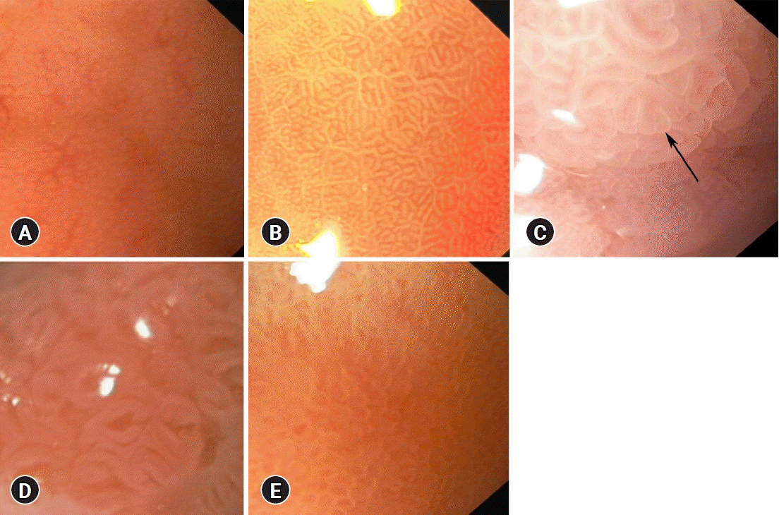
Fig. 1.
Gastric mucosal surface pit pattern types in optical magnification ×105. (A) Dot (regularly arranged, pinpoint small orifices of gastric glands). (B) Tubular (convex, oval, horizontal rods). (C) Villous (resembling simple or branched villi, pattern pointed by arrow). (D) Foveolar (wide orifices of gastric glands, appearing as reticular). (E) Cobblestone (convex, round, vertical rods).
ce-2022-087f1.tif
Fig. 2.
Gastric microvessels appearance at an optical magnification ×105. (A) Regular (clearly visible, regularly arranged collecting venules with the branches of uniform length and diameter resembling spider naevi). (B) Irregular (vessels visible partially with irregular length or diameter of branches, central collecting venules invisible). (C) Obscured (vessels invisible).
ce-2022-087f2.tif
Fig. 3.
Evaluation of methylene blue (MB) staining at an optical magnification ×105. (A) Focal positive MB staining (both light and dark blue areas). (B) Diffuse positive MB staining. (C) Negative MB staining (MB inadequately flushed away).
ce-2022-087f3.tif
Fig. 4.
The relationship between the percentage of the gastric mucosa surface stained with methylene blue and the OLGIM stage. OLGIM 0 vs. OLGIM III, p=0.048; OLGIM 0 vs. OLGIM IV, p=0.039. MB%, the percentage of the gastric mucosa surface stained with methylene blue; OLGIM, Operative Link on Gastric Intestinal Metaplasia.
ce-2022-087f4.tif
Fig. 5.
Receiver operating characteristic (ROC) curve for the percentage of methylene blue stained gastric mucosa surface as a marker of advanced intestinal metaplasia (Operative Link on Gastric Intestinal Metaplasia stages III and IV).
ce-2022-087f5.tif
ce-2022-087f6.tif
Table 1.
Characteristics of patients considering age, IM extent and subtype
Characteristic n (%) Age (yr, mean± SD) p-value
Total 33 (100) 63.15±13.29
OLGIM 0 8 (24.2) 54.63±11.81 0.072
OLGIM I 11 (33.3) 62.09±13.47
OLGIM II 5 (15.2) 61.2±13.71
OLGM III 6 (18.2) 73.17±10.53
OLGIM IV 3 (9.1) 73±4.58
OLGIM 0-II 24 (72.7) 59.42±12.89 0.006
OLGIM III-IV 9 (27.3) 73.11±8.64
IM present 25 (75.8) 65.88±12.75 0.035
IM absent 8 (24.2) 54.63±11.82
Focal IM only 14 (42.4) 59.21±11.75 0.002a)
Diffuse IM present 11 (33.3) 74.36±8.32
IM absent 8 (24.2) 54.63±11.82
Complete IM only 1 (3.0) 59 0.121
Incomplete IM present 23 (69.7) 65.65±12.98
IM absent 8 (24.2) 54.63±11.82

IM, intestinal metaplasia; SD, standard deviation; OLGIM, Operative Link on Gastric Intestinal Metaplasia.

a) Post-hoc analysis: IM absent vs. diffuse IM present, p=0.006; focal IM only vs. diffuse IM present, p=0.008.

Table 2.
The sensitivity, specificity, accuracy, PPV, NPV of MB chromoendoscopy, pit pattern and mucosal vessels assessments in detection of intestinal metaplasia in per biopsy analysis
Sensitivity (%) Specificity (%) Accuracy (%) PPV (%) NPV (%)
MB 49.2 90.4 71.6 81.1 68.0
Pit pattern 98.0 31.3 60.5 52.7 95.2
Blood vessels 100 8.1 49.6 47.2 100
MB+pit pattern 90.2 35.6 60.4 53.9 81.3
MB+blood vessels 93.4 16.4 51.5 48.3 75.0
MB+pit pattern+blood vessels 93.4 12.3 49.3 47.1 69.2

PPV, positive predictive value; NPV, negative predictive value; MB, methylene blue.

TOOLS
Similar articles