Abstract
Most cases of congenital diaphragmatic hernia (CDH) can be diagnosed based on symptoms of severe respiratory failure during the neonatal period or fetal ultrasonography. However, some rare cases are diagnosed in late childhood or adolescence. In this case report, I describe an 11-year-old male patient diagnosed with late-onset CDH presenting with acute abdominal pain. The patient had recently experienced anorexia, nausea, and vomiting after eating. However, he reported no abdominal pain or past history of trauma. The abdomen was generally convex. All laboratory data were within normal limits. A chest X-ray revealed elevation of the left diaphragm. Chest computed tomography showed a defect in the left diaphragm. Based on the above radiologic findings, emergency surgery was performed after the diagnosis of diaphragmatic hernia. A surgical incision was performed in the left subcostal area. Finally, late-presenting Bochdalek hernia was diagnosed. The operation was completed and no specific findings on chest X-ray were found after surgery. The patient was discharged on the fourth day after surgery. In conclusion, CDH in late childhood or adolescence is rare and has various clinical manifestations. To avoid complications such as strangulation and bowel perforation, emergency surgery may be required. Thus, it is necessary to suspect CDH in children with recurrent gastrointestinal or respiratory symptoms, based on which an accurate diagnosis can be made and successful surgical treatment can be performed.
Congenital diaphragmatic hernia (CDH) is caused by malrotation of the pleuroperitoneal membrane during embryogenesis. More than 73% of CDH cases can be diagnosed through the use of fetal ultrasound [1-3]. Among various types of CDH, Bochdalek hernia was first described as a posterior congenital defect of the diaphragm in 1848 by Czechoslovakian anatomist Vincent Alexander Bochdalek [4]. Most CDH can be diagnosed by confirming symptoms of severe respiratory failure during neonatal period or fetal ultrasound. However, some are diagnosed in late childhood or adolescence, although such cases are rare. The purpose of this study was to report an emergency surgery and subsequent course of a child who was diagnosed with late-onset CDH in a radiologic examination performed for acute abdominal pain.
Ethical statements: This case report was approved by the Institutional Ethical Committee of the Dong-A University Hospital (approval No. 22-183) and written informed consent was obtained from the parent of the child patient for this report.
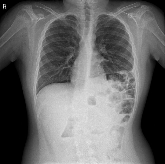
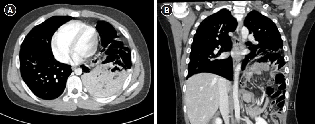
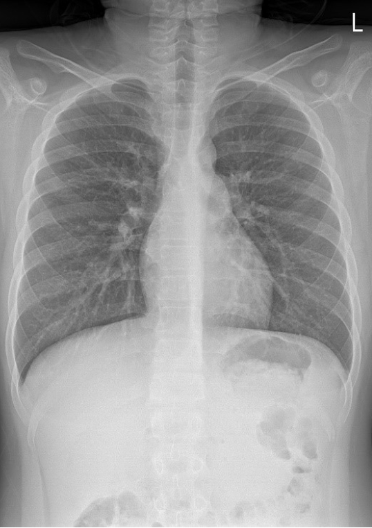
An 11-year-old male patient presented with persistent upper abdominal pain that started on the day of admission. After he underwent a radiologic examination at another hospital, he was transferred to the emergency room of the hospital for surgery. Recently, he experienced anorexia, nausea, and vomiting after eating. However, he had no abdominal pain or past history of trauma. The abdomen was generally convex. All laboratory data were within normal limits. In chest X-ray performed at the hospital, elevation of the left diaphragm was found (Fig. 1). A chest computed tomography (CT) performed in another hospital for an accurate diagnosis showed a defect in the left diaphragm with some small and large bowel herniated into thoracic cavity, based on which emergency surgery was performed after diagnosis of diaphragmatic hernia (Fig. 2). A surgical incision was performed in the left subcostal area. Whole small bowel, transverse colon, and omentum were herniated through the defect in the left posterior site of the diaphragm. The color of the small bowel was slightly dark but peristalsis was confirmed. There were no signs of necrosis or strangulation. The colon was healthy. The oval-shaped diaphragm defect of the 5×3-cm-size was confirmed in the posterolateral area. Hernia sac was not found. Consequentially, late-presenting Bochdalek hernia was diagnosed. First, reduction was conducted in the order of small bowel and colon. Primary repair of a diaphragmatic defect was performed with an interrupted, nonabsorbable suture using black silk 1-0. The operation was completed without placing the chest tube. No specific findings on chest X-ray were found after surgery (Fig. 3). The patient was discharged on the 4th day after surgery. He was clinically followed up for a year without signs of recurrence.
CDH is a rare disease with an incidence of between 1 in 2,000 and 1 in 4,000 live births [5]. The most common type of CDH is a Bochdalek hernia and other types include Morgagni hernia, diaphragm eventration, etc. The majority of this disease is diagnosed within a few hours after birth and 5% to 25% of cases are found even after the neonatal period [6]. But, most cases of Morgagni hernia are diagnosed incidentally in childhood and it can present with acute chest symptoms or can be asymptomatic. Symptoms of a diaphragmatic hernia can include emesis, nausea, abdominal pain, chest pain, dyspnea, wheezing, cough, and absent breath sound [7]. It has been reported that respiratory symptoms are more common in younger patients, while gastrointestinal symptoms are more common in older patients [8]. Delayed presentation of CDH in late childhood or adolescence, in this case, may be confirmed late. As a result, patient's condition might deteriorate.
Haines and Collins [9] have also reported that chest X-rays of asymptomatic diaphragmatic hernia patients are interpreted as left pleural effusion. Hegarty et al. [10] have incorrectly diagnosed a diaphragmatic hernia as pneumothorax, resulting in two cases undergoing thoracentesis. Common misdiagnoses of diaphragmatic hernia include pneumothorax, pneumonia, pleural effusion, infectious etiology, and cystic malformation [11]. Although chest X-rays can be somewhat negative for an initial accurate diagnosis, it can be utilized most commonly for the diagnosis of diaphragmatic hernia [12]. Regarding patients with a diaphragmatic hernia, chest X-rays can also detect bowel gas patterns, air-fluid levels, or cardiac and mediastinal deviations, and lucency within the thoracic cavity, which may complicate or obscure the diagnosis [13]. A thoracic-abdominal contrast CT scan is specific for the diagnosis of diaphragmatic hernia. Currently, routinely using thin-section CT scanning can more accurately predict the prevalence and characteristics of late-presenting Bochdalek hernia. Mullins et al. [14] have reported that 0.17% of a large patient population is diagnosed with incidental Bochdalek hernia through an abdominal CT review. Although CDH is usually asymptomatic for a late-presenting type, immediate surgical treatment should be performed when its diagnosed to prevent complications such as strangulation and bowel perforation. By doing this, potentially fatal consequences in the future can be avoided. If late-presenting CDH can be accurately diagnosed, an excellent survival rate of about 97% to 100% can be ensured compared with neonatal CDH [13].
Late-onset CDH can be operated by approaching the chest or abdomen. Surgery consists of repositioning abdominal contents into the abdominal cavity and closing the diaphragmatic defect. The traditional abdominal approach is often preferred. However, the evidence that the chest approach is not inferior to the abdominal approach in terms of efficiency has been growing [15]. Complications of CDH surgery in older children tend to occur more frequently in the gastrointestinal tract because of associated bowel malrotation and inadequate bowel fixation.
In conclusion, CDH in late childhood or adolescence is observed to be rare. Its clinical manifestations can appear in various symptoms. To avoid complications such as strangulation and bowel perforation in many patients, emergency surgery may be required. Thus, it is necessary to suspect CDH considering children with recurrent gastrointestinal or respiratory symptoms, based on which, accurate diagnosis and successful surgical treatment can be performed.
References
1. Kim DJ, Chung JH. Late-presenting congenital diaphragmatic hernia in children: the experience of single institution in Korea. Yonsei Med J. 2013; 54:1143–8.
2. Testini M, Girardi A, Isernia RM, De Palma A, Catalano G, Pezzolla A, et al. Emergency surgery due to diaphragmatic hernia: case series and review. World J Emerg Surg. 2017; 12:23.
3. Close O. More than just a cough: late presentation of congenital diaphragmatic hernia. J Paediatr Child Health. 2020; 56:1302–4.
4. Puri P, Wester T. Historical aspects of congenital diaphragmatic hernia. Pediatr Surg Int. 1997; 12:95–100.
5. Gallot D, Boda C, Ughetto S, Perthus I, Robert-Gnansia E, Francannet C, et al. Prenatal detection and outcome of congenital diaphragmatic hernia: a French registry-based study. Ultrasound Obstet Gynecol. 2007; 29:276–83.
6. Mishalany H, Gordo J. Congenital diaphragmatic hernia in monozygotic twins. J Pediatr Surg. 1986; 21:372–4.
7. Waseem M, Quee F. A wheezing child: breath sounds or bowel sounds? Pediatr Emerg Care. 2008; 24:304–6.
8. Kitano Y, Lally KP, Lally PA; Congenital Diaphragmatic Hernia Study Group. Late-presenting congenital diaphragmatic hernia. J Pediatr Surg. 2005; 40:1839–43.
9. Haines JO, Collins RB. Bochdalek hernia in an adult simulating a pleural effusion. Radiology. 1970; 95:277–8.
10. Hegarty MM, Bryer JV, Angorn IB, Baker LW. Delayed presentation of traumatic diaphragmatic hernia. Ann Surg. 1978; 188:229–33.
11. Kadian YS, Rattan KN, Verma M, Kajal P. Congenital diaphragmatic hernia: misdiagnosis in adolescence. J Indian Assoc Pediatr Surg. 2009; 14:31–3.
12. Berman L, Stringer D, Ein SH, Shandling B. The late-presenting pediatric Bochdalek hernia: a 20-year review. J Pediatr Surg. 1988; 23:735–9.
13. Blackstone MM, Mistry RD. Late-presenting congenital diaphragmatic hernia mimicking bronchiolitis. Pediatr Emerg Care. 2007; 23:653–6.
14. Mullins ME, Stein J, Saini SS, Mueller PR. Prevalence of incidental Bochdalek's hernia in a large adult population. AJR Am J Roentgenol. 2001; 177:363–6.
15. Obata S, Souzaki R, Fukuta A, Esumi G, Nagata K, Matsuura T, et al. Which is the better approach for late-presenting congenital diaphragmatic hernia: laparoscopic or thoracoscopic? A single institution's experience of more than 10 years. J Laparoendosc Adv Surg Tech A. 2020; 30:1029–35.


 PDF
PDF Citation
 Print
Print


 XML Download