Abstract
Patients with Apert syndrome require repeated limb surgery due to their complex deformities. In this study, a full-thickness isograft was performed for the division of Upton type III hands in identical twins with Apert syndrome. Nine-month-old identical twins presented with Apert syndrome characterized by craniosynostosis, severe syndactyly of the hands and feet, and dysmorphic facial features. Division and full-thickness skin grafting were performed. The siblings were operated consecutively on the same day. Following surgery for the younger sibling, there was an excess of graft left unused. In contrast, the older sibling required an additional skin graft of 1 × 1 cm. Full-thickness skin was successfully transferred between the twins without any rejection as of a 2-month follow-up. Thus, full-thickness skin isografting between monozygotic twins with Apert syndrome was successfully implemented.
Apert syndrome is a form of acrocephalodactyly, characterized by craniosynostosis, severe syndactyly of hands and feet, and dysmorphic facial features [1,2]. The hands in Apert syndrome are classified into types I, II, and III, based on increasing thumb involvement and complexity of pan-syndactyly. The condition is bilateral and involves both hands and feet, requiring multiple divisions and corrective surgeries [3]. For patients requiring multiple divisions, securing donor skin becomes crucial.
It could be assumed that there would be no immune rejection between monozygotic twins. However, there are some differences in phenotype, which require immunosuppression for solid organ transplantation [4]. In 1927, Bauer first reported the successful epidermal grafting between homozygotic twins [5]. The procedure has since been successfully replicated in select identical twin patients (involving severe burn cases) worldwide, without tissue rejection or immunosuppression [6-8]. Herein, we describe the first case of full-thickness skin isografting between homozygotic twins in the division of syndactyly in Apert syndrome.
The review of patients’ medical records in this report was approved by the Institutional Review Board of Seoul National University Hospital (No. H-2208-136-1347).
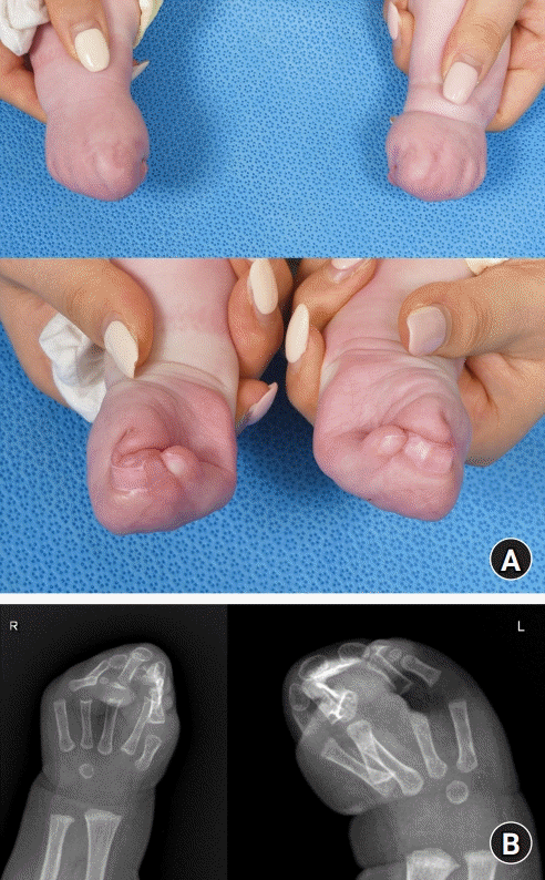
Nine-month-old female twins presented at the outpatient clinic with syndactyly in bilateral hands and feet. On physical examination, all the web spaces of hands and feet were involved. Based on complex syndactyly with distal synostosis of the phalangeal segments and a broad conjoined nail, the hand was categorized as Upton type III (Fig. 1). Previously, both twins were confirmed to have FGFR2 mutation and were clinically diagnosed with Apert syndrome. The twins also had identically coronal synostosis, hypertelorism with face hypoplasia, midgut malrotation, atrial septal defect (ASD), and patent ductus arteriosus (PDA). For the Upton type III hand, the right 1st and 3rd webspace division and full-thickness skin graft were planned.
Surgeries for twin sisters were performed consecutively on the same day, with a slight time difference. For the hand, a zigzag incision was made and a dorsal rectangular flap and a volar triangular flap were elevated. The bony fusion part of the acral side was separated using an oscillating saw. After the division of the first and third webspace, dorsal and volar triangular flaps were interposed to recreate the web. Subsequently, the raw surface was covered with full-thickness skin harvested from the left inguinal crease.
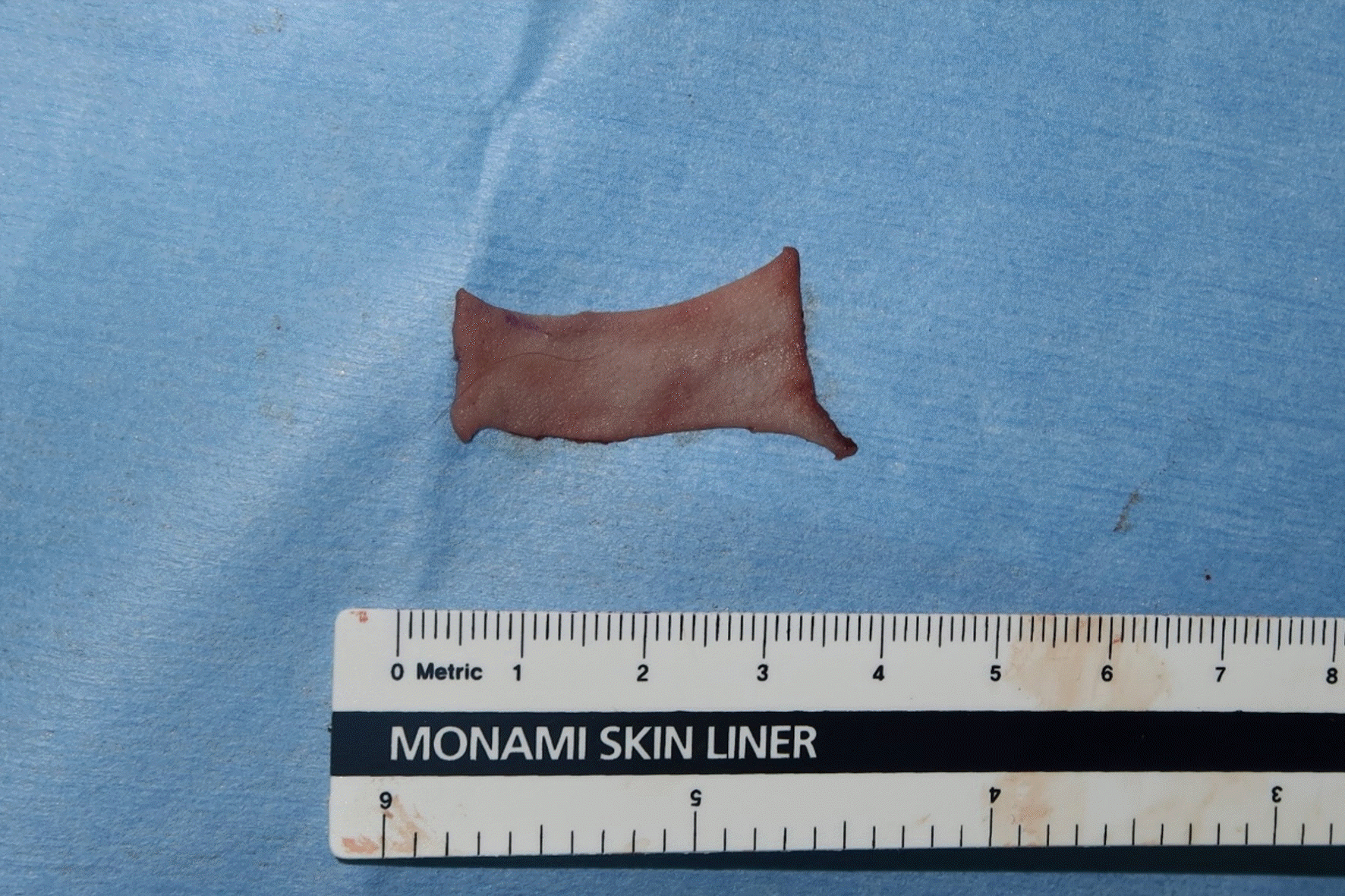
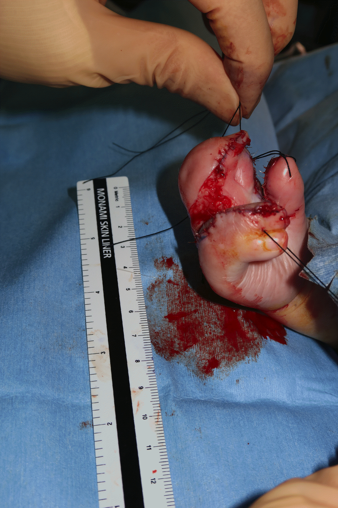
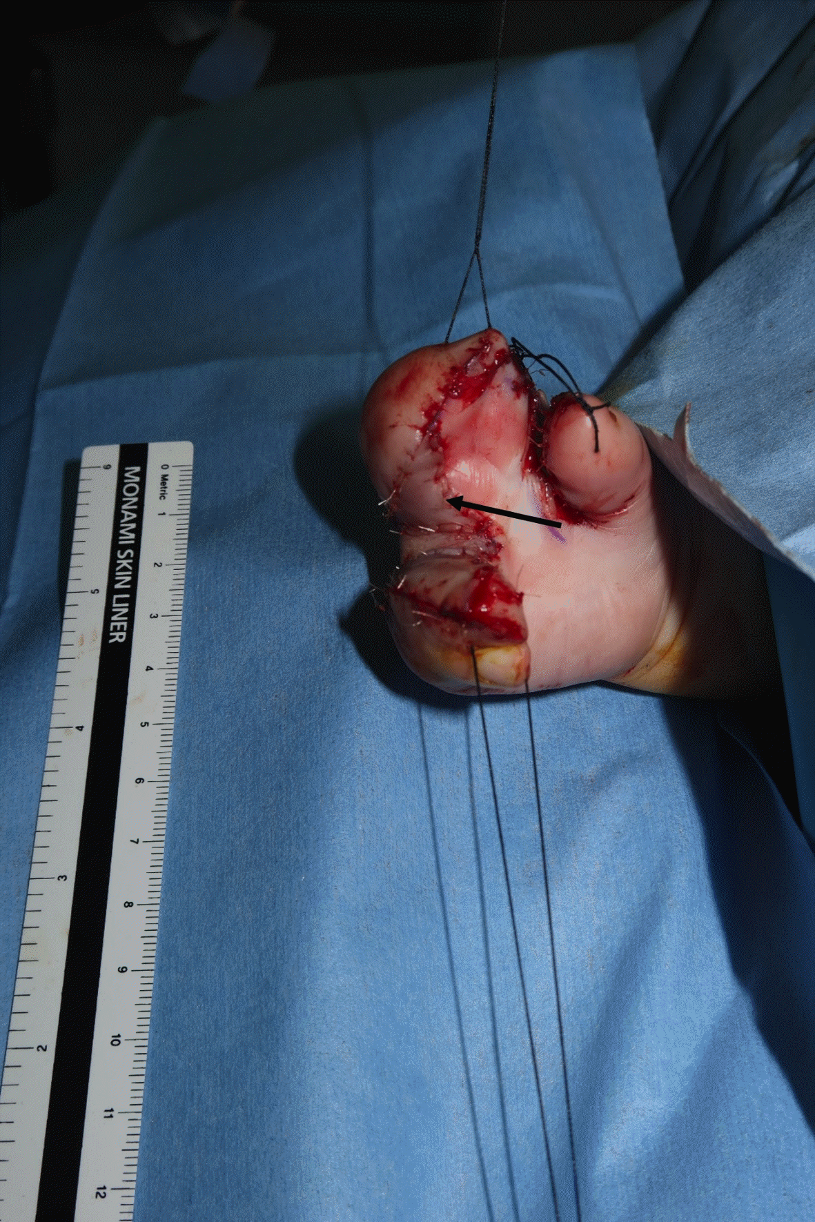
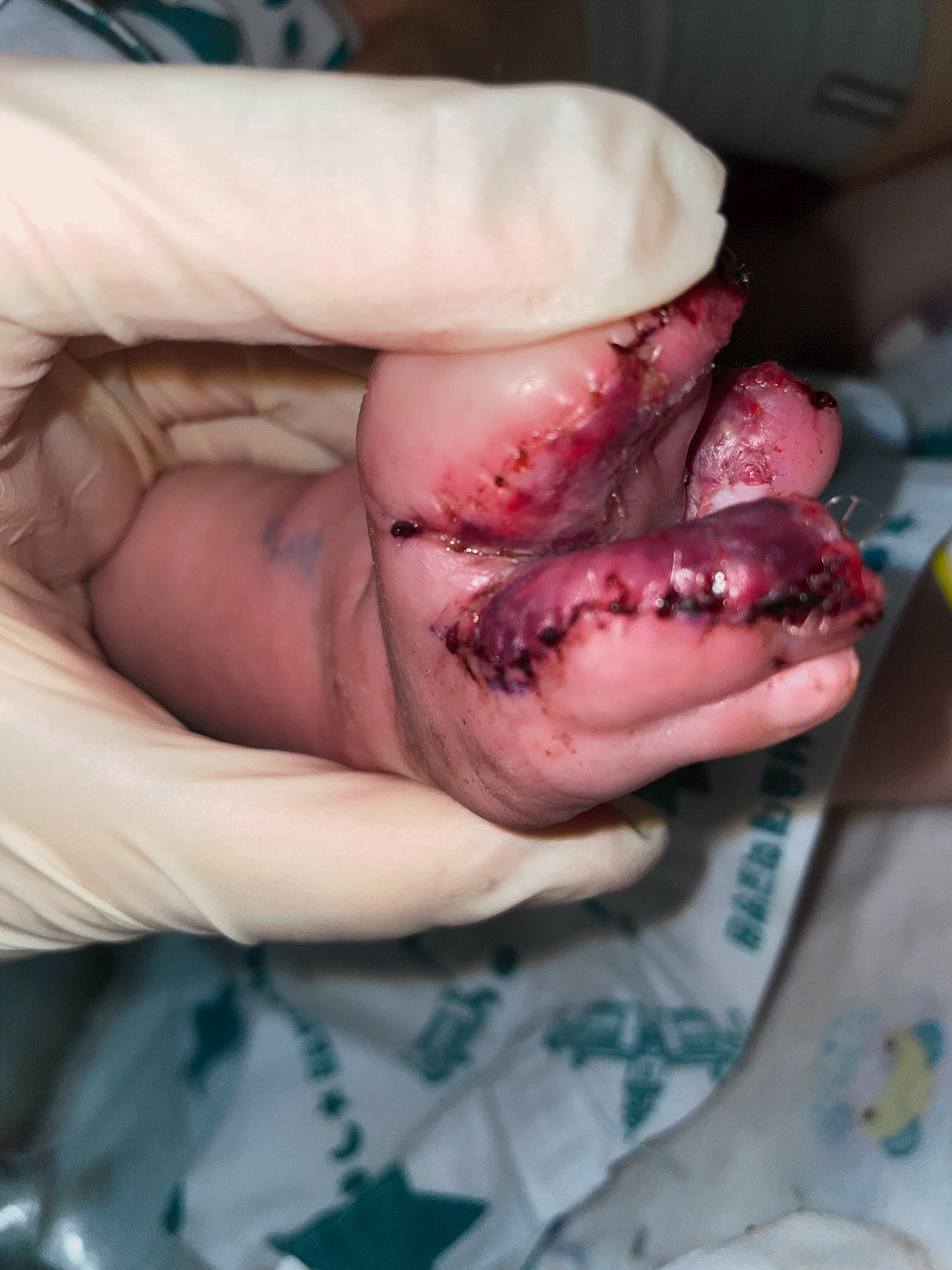
The younger sibling’s surgery was terminated first, and there was a residual skin graft left unused (Fig. 2). However, in the case of the older sibling, after utilizing the skin harvested for the first time, there was a need for an additional 1 × 1-cm size skin graft (Fig. 3). Consequently, the residual skin graft from the younger sibling was utilized for the older sibling (Fig. 4). On the 5th day after surgery, after confirming that skin graft taking was in progress without any specific complications, the patients were discharged from the hospital (Fig. 5). The patients were followed up at the outpatient clinic two months after the operation. The skin was well engrafted without rejection, and the reconstructed web space was maintained without recurrence (Fig. 6).
Despite being genetically identical, variations in next-generation sequencing and discordance in phenotype have been reported in identical twins. Depending on the type of organ to be transplanted, immunosuppression may be performed before or after surgery. The skin graft in an identical twin was first applied in syndactyly by Bauer [5] in 1927 and has been applied ever since when extensive skin grafting is required in patients with trauma, especially burns [6-8]. Bauer [5] reported successful reciprocal epidermis grafting for the correction of syndactyly. In case of skin defect due to trauma, the split-thickness skin graft has been performed without additional immunosuppression [6-8]. There was no report of rejection [4]. Due to the rarity of Apert syndrome, there have been no reports of skin isografting or transplantation between monozygotic twins with Apert syndrome. In this study, the 9-month-old genetically proven identical twins, with no significant difference in phenotype (coronal synostosis, both hand/foot syndactyly, hypertelorism with face hypoplasia and midgut malrotation, ASD, PDA). The full-thickness skin grafting was performed without any rejection reaction or additional immunosuppression.
In Apert syndrome, when syndactyly of the hands and feet is accompanied, multiple surgeries are required owing to the involvement of multiple phalanges [3]. In the cases of repetition of grafting, where the donor skin was insufficient, there have been attempts to stretch the skin of the hand using an expander [9,10]. Consequently, the patient will need multiple surgeries for pan-syndactyly of hand and foot, compromising the donor skin. In contrast, the availability of skin grafts from identical twins prevented additional skin harvests, and there was no wastage of the skin graft. Considering that skin can be shared during syndactyly surgery for identical twins, it can be accommodated in the operation schedule.
In conclusion, full-thickness skin isografting between identical twins can be successfully implemented with effective skin preservation in the treatment of syndactyly in Apert syndrome.
References
1. Fearon JA, Podner C. Apert syndrome: evaluation of a treatment algorithm. Plast Reconstr Surg. 2013; 131:132–42.
2. Raposo-Amaral CE, Raposo-Amaral CA, Garcia Neto JJ, Farias DB, Somensi RS. Apert syndrome: quality of life and challenges of a management protocol in Brazil. J Craniofac Surg. 2012; 23:1104–8.
3. Upton J, McNamara CT, Ali B, Nuzzi LC, Taghinia AH, Labow BI. Distraction lengthening of the Apert thumb. Plast Reconstr Surg. 2022; 149:691e–699e.
4. Day E, Kearns PK, Taylor CJ, Bradley JA. Transplantation between monozygotic twins: how identical are they? Transplantation. 2014; 98:485–9.
5. Niederhuber J, Feller I. Permanent skin homografting in identical twins. Arch Sur. 1970; 100:126–8.
6. Herron PW, Marion LF. Homografting in treatment of severe burns using an identical twin as a skin donor. Pac Med Surg. 1967; 75:4–10.
7. Turk E, Karagulle E, Turan H, et al. Successful skin homografting from an identical twin in a severely burned patient. J Burn Care Res. 2014; 35:e177–9.
8. Ahmed R, Giwa L, Jordan N, Dheansa B. The helpful twin: Skin graft donation in a challenging burn case. JPRAS Open. 2020; 27:58–62.
9. Coombs CJ, Mutimer KL. Tissue expansion for the treatment of complete syndactyly of the first web. J Hand Surg Am. 1994; 19:968–72.
10. Işik S, Selmanpakoğlu N, Aşikel C. The use of tissue expanders in syndactyly repair. Eur J Plast Surg. 1996; 19:225–8.


 PDF
PDF Citation
 Print
Print



 XML Download