Journal List > Brain Tumor Res Treat > v.11(1) > 1516081741

Park and Lee: The Role of Hypoxia in Brain Tumor Immune Responses

Abstract

Oxygen is a vital component of living cells. Low levels of oxygen in body tissues, known as hypoxia, can affect multiple cellular functions across a variety of cell types and are a hallmark of brain tumors. In the tumor microenvironment, abnormal vasculature and enhanced oxygen consumption by tumor cells induce broad hypoxia that affects not only tumor cell characteristics but also the antitumor immune system. Although some immune reactions require hypoxia, hypoxia generally negatively affects immunity. Hypoxia induces tumor cell invasion, cellular adaptations to hypoxia, and tumor cell radioresistance. In addition, hypoxia limits the efficacy of immunotherapy and hinders antitumor responses. Therefore, understanding the role of hypoxia in the brain tumor, which usually does not respond to immunotherapy alone is important for the development of effective anti-tumor therapies. In this review, we discuss recent evidence supporting the role of hypoxia in the context of brain tumors.

INTRODUCTION

Oxygen is indispensable for cellular functions of living organisms. Cells utilize oxygen to generate energy through the conversion of nutrients to adenosine triphosphate (ATP) via an oxygen-dependent pathway known as cellular respiration [1]. An excessive level of oxygen is referred to as hyperoxia, whereas an insufficient level is referred to as hypoxia; an oxygen level within the normal range is termed normoxia. Following the seminal observation by Louis Pasteur that living organisms consume oxygen [2], discoveries by Otto Warburg and Corneille Heymans further elucidated the mechanism by which the protein complex hemoglobin interacts with oxygen [3] and how the central nervous system (CNS) responds to oxygen [45]. Three Nobel Prize recipients proceeded to demonstrate the mechanism by which living organisms sense oxygen levels in the body. In 2019, William G. Kaelin Jr., Sir Peter J. Ratcliffe, and Gregg L. Semenza won the Nobel Prize for their discovery of hypoxia-inducible factor 1-alpha (HIF1A) and its role in oxygen sensing mechanisms [678910]. We now have a better understanding of how cells sense and respond to variations in oxygen levels.
Although the brain comprises only 2% of total body weight, it utilizes 20% of the total oxygen used within the body. Within the brain, the consumption and distribution of oxygen are region-dependent; oxygen levels within the midbrain comprise only 0.5% of total oxygen content, whereas pial oxygen levels comprise approximately 8% [11]. Because oxygen is important for cellular metabolism, tumor cells consume high levels of oxygen. Therefore, brain tumor regions are typically hypoxic, with tumoral and peritumoral regions containing oxygen concentrations of approximately 1.25% and 2.5%, respectively [12]. Hypoxia is a clinical hallmark of many cancers, including brain tumors, and is typically associated with negative outcomes in patients [13]. Multiple factors can contribute to brain tumor hypoxia. The most unique characteristics of tumor cells are their high proliferation rate and metabolic demands. Due to the fast rate of proliferation, tumor tissues can expand a long distance from blood vessels, limiting oxygen diffusion to the center of tumor tissues. In addition, abnormal angiogenesis induced by the tumor microenvironment (TME) results in constriction of blood vessels, further inducing hypoxia [1415].
Hypoxic conditions can affect not only tumor cells, but also blood vessels, stromal cells, and immune cells. Although hypoxia is beneficial for certain processes, including the germinal center (GC) reaction, hematopoiesis, and intestinal barrier maintenance [16], it hinders antitumor immune responses. For example, hypoxia induces the accumulation and immune-suppressing actions of myeloid-derived suppressor cells (MDSCs) and regulatory T cells (Tregs), thereby reducing the functionality of antitumor immune cells such as CD8 T cells [17]. Therefore, hypoxia induces a vicious cycle of antitumor immune responses and it is important to better understand how hypoxia regulates the TME.

MOLECULAR MECHANISM OF SENSING HYPOXIA

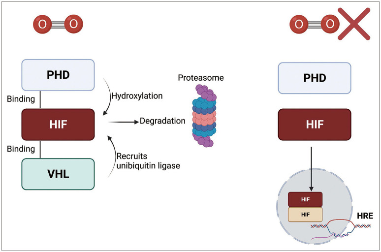
Hypoxia-inducible factor (HIF) is the most well-characterized sensor for oxygen tension [18]. HIF is a transcription factor that is constituted by two distinct subunits, HIFα and HIFβ. Humans express three isoforms of the HIFα subunit (HIF1α, HIF2α, and HIF3α). HIF1α is expressed ubiquitously and is overexpressed in tumor cells, whereas HIF2α expression is restricted to certain cell types such as subsets of tumor-associated macrophages (TAMs). HIF3α expression is also cell-specific, and immune cell expression has not been confirmed [1920]. HIF1α and HIF2α act as transcription factors that target both unique and overlapping sets of target genes. In addition, HIF1α subunits can dimerize with HIF1β, also known as the aryl hydrocarbon receptor nuclear translocator (ARNT), that is ubiquitously expressed. Although HIF3α is a negative regulator of HIF1, it can also function as a transcriptional activator of distinct genes [21].
Under normoxic conditions, HIFα is bound to prolyl hydroxylase domain (PHD) proteins 1–3 through an oxygen-dependent degradation domain (ODDD). PHD is sensitive to oxygen levels due to its 2-oxoglutarate and iron-dependent dioxygenase domains, and is therefore affected by changes in oxygen tension [22]. PHD hydroxylates the prolyl residues of HIFα, which induce ubiquitination through E3 ubiquitin ligase interactions with the von Hippel-Lindau tumor suppressor protein (VHL), thereby promoting the proteasomal degradation of HIFα [23]. However, decreases in oxygen tension result in decreased PHD activity and therefore lead to stabilization of HIFα. Stable HIFα proteins translocate to the nucleus, bind to HIF1β and other coactivators, and influence downstream transcription (Fig. 1) [24].
Downstream genes targeted by HIF-mediated transcription are known as hypoxia-response elements (HREs) that are involved in metabolism and proliferation among other cellular functions. For example, hypoxia stabilizes epidermal growth factor receptor variant iii (EGFRviii) by enhancing its interaction with integrin β1 within brain tumor cells. This interaction facilitates the transport of other integrins to the cell surface leading to activation of focal adhesion kinase (FAK) and tumor cell invasion [25]. Hypoxia also represses cap-dependent translation, resulting in the downregulation of global translation and an increase in the selective translation of stress-related proteins [26]. For example, hypoxia-induced activation of inositol-requiring transmembrane kinase/endoribonuclease 1α (IRE1α) is responsible for activation of the stress-related protein X-box binding protein 1 (XBP1) [2728]. In addition, hypoxia mediates metabolic changes that can lead to mitochondrial dysfunction. Severe hypoxia below an oxygen level of 0.3% disrupts electron transport complex activity [29]. Hypoxia also attenuates the tricarboxylic acid (TCA) cycle, induces mitochondrial fission, and activates mitophagy in both HIF-dependent and HIF-independent manners [303132]. A characteristic feature of hypoxic cells is enhanced glycolysis that is mediated through HIF-induced upregulation in the expression of the glucose transporter, pyruvate kinase M2, lactate dehydrogenase, and phosphoinositide-dependent kinase 1 [3334] and in turn, increased lactate production and low pH levels. In addition, hypoxia regulates the expression of several angiogenesis-related genes, most notably vascular endothelial growth factor (VEGF) as well as placenta growth factor (PGF), angiopoietin, C-X-C motif chemokine ligand 12, and platelet-derived growth factor B [35]. VEGF signaling induces endothelial proliferation, and both VEGF and PGF induce extracellular matrix degradation [3637]. Hypoxia can also regulate the radiosensitivity of brain tumor cells, although the mechanisms are unclear [38].

HYPOXIA THE THE IMMUNE SYSTEM

Like other cells, immune cells need an appropriate level of oxygen that increases with greater activity. Although normoxic air contains 21% oxygen, some tissues may have hypoxic oxygen concentrations despite normal physiological conditions [18], known as “physiological hypoxia” [16]. These low or hypoxic oxygen levels are sometimes beneficial for maintaining the functions of certain tissues. For example, hypoxia is necessary to maintain hematopoietic stem cell (HSC) homeostasis in the bone marrow [39]. Although HIF2α is dispensable, HIF1β is required for multiple HSC functions [4041]. The GC is also hypoxic due to the expansion of B cells. Moreover, hypoxia is known to affect the function of GC B cells [4243]. Female reproductive organs such as the vagina and placenta are hypoxic [4445], which is required for protection of the fetus from maternal immunity. For example, HIF1α induces the expression of trophoblast-regulating nonclassical class I histocompatibility antigens that prevent damage from natural killer (NK) cells [46].

ANTITUMOR CYCLE

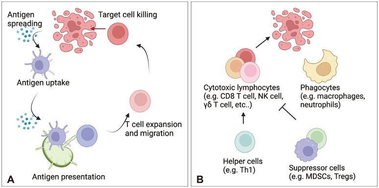
Multiple types of immune cells participate in antitumor responses. Following tumor cell death, antigen presenting cells (APCs), such as dendritic cells (DCs), mediate cellular immune responses by migrating to lymph nodes (LN) and presenting tumor antigens to T cells. Soluble antigens also drain into LNs and can be taken up by LN-resident DCs. CD8 T cells are considered the most important antitumor immune cell population. CD8a+/XCR1+/CD103+ DC1 cells can cross-present antigens to CD8 T cells via the major histocompatibility complex (MHC) class I, whereas CD4+/CD11b+/signal regulatory protein alpha (SIRPα)+ DC2 cells contain higher expression of MHC class II molecules and present antigens to CD4 T cells [47]. However, DC1 cells also prime CD4 T cells via MHC class II and CD40 [48]. Moreover, antigen-specific T cells can be primed by chemokines to migrate to the tumor site, recognize tumor cells via MHC molecules, and kill target cells. Tumor site APCs, such as macrophages, can stimulate the production of cytotoxicity and cytokine production by T cells or can prime them for exhaustion [49]. However, tumor cells can escape T cell responses via multiple mechanisms. For example, tumor cells downregulate MHC molecules and prime T cells for exhaustion via immune checkpoint molecules such as programmed cell death (PD) 1 and PD-1 ligand (PD-L1) [5051]. T cell responses can also be suppressed by anti-inflammatory cytokines such as IL-10 [52] and immune cells such as Tregs and MDSCs [53]. Therefore, researchers are investigating strategies to block inhibitory immune mediators. In addition to T cells, phagocytes including macrophages, microglia, and neutrophils can participate in the antitumor response via phagocytosis [545556]. Unconventional T cells such as NK cells, natural killer T (NKT) cells, mucosal-associated invariant T cells (MAIT cells), and γδ T cells are also involved in antitumor responses (Fig. 2) [5758].

ANTITUMOR RESPONSES IN THE BRAIN TUMORS

The brain is characterized as an “immune-privileged” organ. In 1921, a Japanese scientist attempted to transplant heterologous rat sarcoma tissue to the rat brain parenchyma [59], revealing that the tumor was not rejected. These observations were confirmed by James Murphy and Ernest Sturm using mouse sarcoma transplantation in the rat brain parenchyma [60]. Furthermore, the successful transplantation of skin autografts in the CNS has been demonstrated [61]. Based on these observations, the brain was thought to lack the ability to mount an immune response [62]. The idea of immune privilege was further supported by the notion that there is no lymphatic drainage from the CNS [6364]. However, the re-discovery of the dorsal and basal meningeal lymphatics led to the realization that immune system surveillance can occur around the CNS [656667]. For example, cerebrospinal fluid (CSF) produced from the choroid plexus can circulate through the CNS and the glymphatic system may allow an exchange between CSF and interstitial fluid (ISF). Due to the glymphatic exchange, parenchymal molecules can be drained by meningeal lymphatics [68]. CSF can reach the meninges, skull, and vertebral bone marrow [6669]. Additional drainage routes such as the cribriform plate also support drainage of brain-derived molecules [70]. As a result, antigen presentation primarily occurs at the dura and deep cervical lymph node (dcLN) [71]. Despite immune-surveillance by multiple cell populations, including DCs, immune tolerance is maintained within the CNS to prevent autoimmunity [72]. In addition, immune cell infiltration into the parenchyma of the CNS is restricted by multiple barriers including blood-brain barrier [73].
In addition to the barrier system, the brain tumor site is a unique microenvironment containing many immunosuppressive cells. Brain tumors are known as “cold tumors” due to the lack of neoantigens, low lymphocyte infiltration, and a predominant proportion of myeloid cells [74]. The nutrient-deprived TME is another factor contributing to immunosuppression [75]. Glucose deprivation inhibits immune cell activation and limits glycolysis. Due to the Warburg effect, lactate produced from glycolysis by tumor cells leads to enhanced Treg activation, decreased pH, and inhibition of T cell responses. As previously mentioned, brain tumors are also hypoxic [76], which can further induce T cell exhaustion, Treg cell migration, and γδ T cell malfunction [777879]. Thus, it is important to develop a comprehensive understanding of the complicated immunosuppressive brain TME to develop successful antitumor therapies.

THE ROLE OF HYPOXIA IN THE ANTI-BRAIN TUMOR IMMUNE RESPONSE

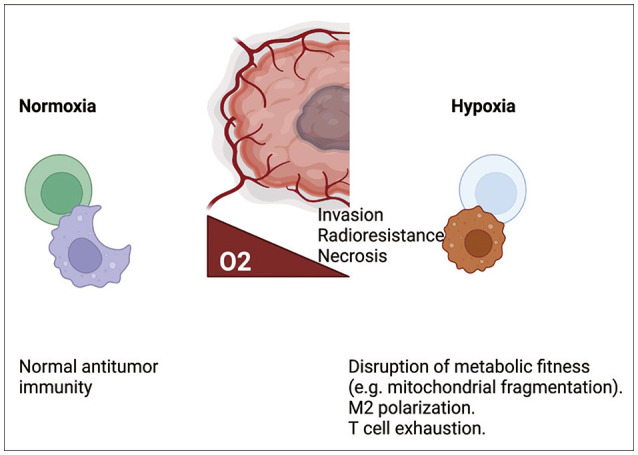
Hypoxia can affect immune cell responses through multiple pathways. Reductions in proliferation, cytokine production, and cytotoxicity, along with an increase in T cell exhaustion, have been observed in hypoxic CD8 T cell cultures [77] and in a murine melanoma model. In addition, exhausted CD8 T cells were more highly enriched in the hypoxic core regions of glioblastoma multiforme (GBM) tissue than in the peripheral regions [80]. Hypoxia also dampens the responses of T helper 1 (Th1) cells, important immune cells that help to mediate CD8 T cell responses and exhibit similar metabolic function, via HIF1α [81]. Several hypoxia-related pathways can promote T cell exhaustion, including PD-L1, a downstream HRE [82]. CD8 T cell exhaustion can also be induced via mitochondrial dysfunction produced by prolonged stimulation with hypoxia [83]; likewise, hypoxia attenuates NK cell responses via mitochondrial fragmentation [84]. A previous study reported that hypoxia is primarily driven by tumor cell oxygen consumption [85], and excessive tumor cell oxygen consumption reduces γδ T cell responses via hypoxia-mediated downregulation of NK group 2 member D (NKG2D) [7986]. Hypoxia can also facilitate the migration of Tregs [78], one of the various subsets of CD4 T cells that are metabolically adapted to hypoxic microenvironments via lactate-mediated stabilization [87]. In addition to T cells, hypoxia can affect macrophage immune responses by promoting M2 macrophage polarization in the glioma [88]. Thus, the effects of hypoxia on the immune system are mostly driven by the attenuation of antitumor immunity (Fig. 3). Therefore, targeting hypoxia may be an attractive option for antitumor therapy. We have shown that metformin, which is known to reduce mitochondrial respiration, reduced oxygen consumption in the glioma cell line GL261 and potentiated γδ T cell responses. Further, combination therapy with metformin or an HIF1α inhibitor administered concomitantly with γδ T cell therapy led to glioma transplant rejection in a murine model [79].

FUTURE DIRECTIONS FOR ANTI-BRAIN TUMOR THERAPIES

The primary therapy options for high-grade brain tumors include surgery, chemotherapy, and radiotherapy [89]. Despite standardized therapy, the average overall survival of patients is only 1–2 years [90]. Although immune checkpoint blockade has produced successful outcomes for certain tumor types, a recent clinical trial of anti-PD-1 had disappointing results [91]. Thus, further trials should be conducted. However, due to the unique immunosuppressive brain TME, it appears hard to be improved. Other studies have attempted to enhance antitumor responses via the abscopal effect using chemotherapy and radiotherapy [92]. However, a recent trial comparing radiotherapy combined with either nivolumab or chemotherapy did not show a beneficial effect with nivolumab [93]. In a murine brain tumor model, VEGF-C synergized with anti-PD-1 therapy to mediate the enhancement of meningeal lymphatics; therefore, it should be evaluated in human trials in the future [9495]. However, a combination of anti-cytotoxic T-lymphocyte associated protein 4 (CTLA4) and anti-PD-1 led to severe immune-related adverse effects (irAE) affecting multiple organs including the pituitary gland [96]. These adverse effects may be explained by the expression of CTLA4 within the pituitary gland [97]. In addition, disrupting immune tolerance within the healthy brain could trigger autoimmune diseases, which should be considered along with the risk for irAEs when developing future clinical trials. In addition to T cell-targeted therapies, targeting macrophages is also an attractive option. In previous studies, blocking M2 polarization via colony stimulating factor 1 (CSF1) and blocking the CD47-SIRPα-induced “don’t eat me” signal produced positive results in the murine model [9899]. However, CSF1 blockade showed disappointing results in another study [100].
Hypoxia induces abnormal vasculature, thereby limiting drug delivery [101]. Hence, acute hypoxia induced by Bevacizumab treatment may be one reason why it did not show favorable efficacy [102]. In addition, hypoxia facilitates M2 polarization of macrophages, limiting the efficacy of immune checkpoint inhibitors [8588], and induces radioresistance leading to dampened efficacy of radiotherapy [103]. Collectively, these findings support hypoxia as a suitable therapeutic target. If tumor cell-specific oxygen consumption can be reduced, we may be able to improve antitumor immune responses during immunotherapy. Reinvigorating the metabolism of tumor-infiltrating immune cells may be another option. A clinical trial evaluating the HIF2α inhibitor, PT2385, is currently ongoing. Other drug compounds, alone or in combination with immunotherapy, should also be evaluated in future trials [104].

CONCLUSION

Hypoxia is a hallmark of brain tumors that affects not only tumor cell characteristics, but also immune cells within the TME. Despite successful results with immunotherapies in multiple types of tumors, recent clinical trials for brain tumors have shown disappointing results. Thus, it is necessary to understand the complicated and unique characteristics of brain tumors. Because tumor cells exhibit high rates of proliferation, their metabolic demands are higher than other normal cells, thereby inducing nutrient deprivation in surrounding cells. The TME promotes hypoxia through the induction of abnormal vasculature and enhanced tumor cell oxygen consumption. Hypoxia may also be a critical factor contributing to the limited efficacy of antitumor therapy. Thus, understanding hypoxia in the brain TME is indispensable for improving strategies for developing antitumor drugs.

Notes

Ethics Statement: Not applicable

Author Contributions:

  • Conceptualization: Jang Hyun Park, Heung Kyu Lee.

  • Data curation: Jang Hyun Park, Heung Kyu Lee.

  • Formal analysis: Jang Hyun Park, Heung Kyu Lee.

  • Funding acquisition: Heung Kyu Lee.

  • Investigation: Jang Hyun Park, Heung Kyu Lee.

  • Methodology: Jang Hyun Park, Heung Kyu Lee.

  • Project administration: Jang Hyun Park, Heung Kyu Lee.

  • Resources: Jang Hyun Park, Heung Kyu Lee.

  • Software: Jang Hyun Park, Heung Kyu Lee.

  • Supervision: Jang Hyun Park, Heung Kyu Lee.

  • Validation: Jang Hyun Park, Heung Kyu Lee.

  • Visualization: Jang Hyun Park, Heung Kyu Lee.

  • Writing—original draft: Jang Hyun Park, Heung Kyu Lee.

  • Writing—review & editing: Jang Hyun Park, Heung Kyu Lee.

Conflicts of Interest: The authors have no potential conflicts of interest to disclose.

Funding Statement: This work was supported by the National Research Foundation of Korea (NRF-2021M3A9D3026428 and NRF-2021M3A9H3015688) funded by the Ministry of Science and ICT of Korea.

Availability of Data and Material

All data generated or analyzed during the study are included in this manuscript.

References

1. Martínez-Reyes I, Chandel NS. Mitochondrial TCA cycle metabolites control physiology and disease. Nat Commun. 2020; 11:102. PMID: 31900386.
crossref
2. Barnett JA. A history of research on yeasts 2: Louis Pasteur and his contemporaries, 1850-1880. Yeast. 2000; 16:755–771. PMID: 10861901.
crossref
3. Warburg O. The chemical constitution of respiration ferment. Science. 1928; 68:437–443. PMID: 17782077.
crossref
4. Heymans C, Ladon A. Recherches physiologiques et pharmacologiques sur la tête isolée et le centre vague du chien. Arch Internat de Pharmacodyn et de Thérap. 1925; 30:415.
5. Heymans JF, Heymans C. Sur les modifications directes et sur la régulation réflexe de l’activité du centre respiratoire de la tête isolée du chien. Arch Int Pharmacodyn Ther. 1927; 33:273–372.
6. Semenza GL, Nejfelt MK, Chi SM, Antonarakis SE. Hypoxia-inducible nuclear factors bind to an enhancer element located 3’ to the human erythropoietin gene. Proc Natl Acad Sci U S A. 1991; 88:5680–5684. PMID: 2062846.
crossref
7. Wang GL, Jiang BH, Rue EA, Semenza GL. Hypoxia-inducible factor 1 is a basic-helix-loop-helix-PAS heterodimer regulated by cellular O2 tension. Proc Natl Acad Sci U S A. 1995; 92:5510–5514. PMID: 7539918.
crossref
8. Maxwell PH, Wiesener MS, Chang GW, Clifford SC, Vaux EC, Cockman ME, et al. The tumour suppressor protein VHL targets hypoxiainducible factors for oxygen-dependent proteolysis. Nature. 1999; 399:271–275. PMID: 10353251.
crossref
9. Ivan M, Kondo K, Yang H, Kim W, Valiando J, Ohh M, et al. HIFα targeted for VHL-mediated destruction by proline hydroxylation: implications for O2 sensing. Science. 2001; 292:464–468. PMID: 11292862.
crossref
10. Jaakkola P, Mole DR, Tian YM, Wilson MI, Gielbert J, Gaskell SJ, et al. Targeting of HIF-α to the von Hippel-Lindau ubiquitylation complex by O2-regulated prolyl hydroxylation. Science. 2001; 292:468–472. PMID: 11292861.
crossref
11. Erecińska M, Silver IA. Tissue oxygen tension and brain sensitivity to hypoxia. Respir Physiol. 2001; 128:263–276. PMID: 11718758.
crossref
12. Beppu T, Kamada K, Yoshida Y, Arai H, Ogasawara K, Ogawa A. Change of oxygen pressure in glioblastoma tissue under various conditions. J Neurooncol. 2002; 58:47–52. PMID: 12160140.
13. Harris AL. Hypoxia--a key regulatory factor in tumour growth. Nat Rev Cancer. 2002; 2:38–47. PMID: 11902584.
14. Thomlinson RH, Gray LH. The histological structure of some human lung cancers and the possible implications for radiotherapy. Br J Cancer. 1955; 9:539–549. PMID: 13304213.
15. Brown JM, Giaccia AJ. The unique physiology of solid tumors: opportunities (and problems) for cancer therapy. Cancer Res. 1998; 58:1408–1416. PMID: 9537241.
16. Taylor CT, Colgan SP. Regulation of immunity and inflammation by hypoxia in immunological niches. Nat Rev Immunol. 2017; 17:774–785. PMID: 28972206.
17. Hendry SA, Farnsworth RH, Solomon B, Achen MG, Stacker SA, Fox SB. The role of the tumor vasculature in the host immune response: implications for therapeutic strategies targeting the tumor microenvironment. Front Immunol. 2016; 7:621. PMID: 28066431.
crossref
18. Park JH, Lee HK. Current understanding of hypoxia in glioblastoma multiforme and its response to immunotherapy. Cancers (Basel). 2022; 14:1176. PMID: 35267480.
crossref
19. Bertout JA, Patel SA, Simon MC. The impact of O2 availability on human cancer. Nat Rev Cancer. 2008; 8:967–975. PMID: 18987634.
crossref
20. Zhong H, De Marzo AM, Laughner E, Lim M, Hilton DA, Zagzag D, et al. Overexpression of hypoxia-inducible factor 1α in common human cancers and their metastases. Cancer Res. 1999; 59:5830–5835. PMID: 10582706.
21. Keith B, Johnson RS, Simon MC. HIF1α and HIF2α: sibling rivalry in hypoxic tumour growth and progression. Nat Rev Cancer. 2011; 12:9–22. PMID: 22169972.
22. Fong GH, Takeda K. Role and regulation of prolyl hydroxylase domain proteins. Cell Death Differ. 2008; 15:635–641. PMID: 18259202.
crossref
23. Cockman ME, Masson N, Mole DR, Jaakkola P, Chang GW, Clifford SC, et al. Hypoxia inducible factor-alpha binding and ubiquitylation by the von Hippel-Lindau tumor suppressor protein. J Biol Chem. 2000; 275:25733–25741. PMID: 10823831.
24. Kallio PJ, Pongratz I, Gradin K, McGuire J, Poellinger L. Activation of hypoxia-inducible factor 1alpha: posttranscriptional regulation and conformational change by recruitment of the Arnt transcription factor. Proc Natl Acad Sci U S A. 1997; 94:5667–5672. PMID: 9159130.
25. Liu Z, Han L, Dong Y, Tan Y, Li Y, Zhao M, et al. EGFRvIII/integrin β3 interaction in hypoxic and vitronectinenriching microenvironment promote GBM progression and metastasis. Oncotarget. 2016; 7:4680–4694. PMID: 26717039.
26. Braunstein S, Karpisheva K, Pola C, Goldberg J, Hochman T, Yee H, et al. A hypoxia-controlled cap-dependent to cap-independent translation switch in breast cancer. Mol Cell. 2007; 28:501–512. PMID: 17996713.
27. Bouchecareilh M, Higa A, Fribourg S, Moenner M, Chevet E. Peptides derived from the bifunctional kinase/RNase enzyme IRE1α modulate IRE1α activity and protect cells from endoplasmic reticulum stress. FASEB J. 2011; 25:3115–3129. PMID: 21680894.
28. Liu CY, Schröder M, Kaufman RJ. Ligand-independent dimerization activates the stress response kinases IRE1 and PERK in the lumen of the endoplasmic reticulum. J Biol Chem. 2000; 275:24881–24885. PMID: 10835430.
crossref
29. Wilson DF, Rumsey WL, Green TJ, Vanderkooi JM. The oxygen dependence of mitochondrial oxidative phosphorylation measured by a new optical method for measuring oxygen concentration. J Biol Chem. 1988; 263:2712–2718. PMID: 2830260.
crossref
30. Hayek I, Fischer F, Schulze-Luehrmann J, Dettmer K, Sobotta K, Schatz V, et al. Limitation of TCA cycle intermediates represents an oxygen-independent nutritional antibacterial effector mechanism of macrophages. Cell Rep. 2019; 26:3502–3510.e6. PMID: 30917307.
31. Fuhrmann DC, Brüne B. Mitochondrial composition and function under the control of hypoxia. Redox Biol. 2017; 12:208–215. PMID: 28259101.
crossref
32. Liu L, Feng D, Chen G, Chen M, Zheng Q, Song P, et al. Mitochondrial outer-membrane protein FUNDC1 mediates hypoxia-induced mitophagy in mammalian cells. Nat Cell Biol. 2012; 14:177–185. PMID: 22267086.
33. Nakazawa MS, Keith B, Simon MC. Oxygen availability and metabolic adaptations. Nat Rev Cancer. 2016; 16:663–673. PMID: 27658636.
34. Lee P, Chandel NS, Simon MC. Cellular adaptation to hypoxia through hypoxia inducible factors and beyond. Nat Rev Mol Cell Biol. 2020; 21:268–283. PMID: 32144406.
35. Abou Khouzam R, Brodaczewska K, Filipiak A, Zeinelabdin NA, Buart S, Szczylik C, et al. Tumor hypoxia regulates immune escape/invasion: influence on angiogenesis and potential impact of hypoxic biomarkers on cancer therapies. Front Immunol. 2021; 11:613114. PMID: 33552076.
36. Zahra FT, Sajib MS, Ichiyama Y, Akwii RG, Tullar PE, Cobos C, et al. Endothelial RhoA GTPase is essential for in vitro endothelial functions but dispensable for physiological in vivo angiogenesis. Sci Rep. 2019; 9:11666. PMID: 31406143.
37. Wang H, Keiser JA. Vascular endothelial growth factor upregulates the expression of matrix metalloproteinases in vascular smooth muscle cells: role of flt-1. Circ Res. 1998; 83:832–840. PMID: 9776730.
38. Marampon F, Gravina GL, Zani BM, Popov VM, Fratticci A, Cerasani M, et al. Hypoxia sustains glioblastoma radioresistance through ERKs/DNA-PKcs/HIF-1α functional interplay. Int J Oncol. 2014; 44:2121–2131. PMID: 24676782.
39. Takubo K, Goda N, Yamada W, Iriuchishima H, Ikeda E, Kubota Y, et al. Regulation of the HIF-1alpha level is essential for hematopoietic stem cells. Cell Stem Cell. 2010; 7:391–402. PMID: 20804974.
40. Guitart AV, Subramani C, Armesilla-Diaz A, Smith G, Sepulveda C, Gezer D, et al. Hif-2α is not essential for cell-autonomous hematopoietic stem cell maintenance. Blood. 2013; 122:1741–1745. PMID: 23894152.
41. Krock BL, Eisinger-Mathason TS, Giannoukos DN, Shay JE, Gohil M, Lee DS, et al. The aryl hydrocarbon receptor nuclear translocator is an essential regulator of murine hematopoietic stem cell viability. Blood. 2015; 125:3263–3272. PMID: 25855602.
42. Cho SH, Raybuck AL, Stengel K, Wei M, Beck TC, Volanakis E, et al. Germinal centre hypoxia and regulation of antibody qualities by a hypoxia response system. Nature. 2016; 537:234–238. PMID: 27501247.
43. Abbott RK, Thayer M, Labuda J, Silva M, Philbrook P, Cain DW, et al. Germinal center hypoxia potentiates immunoglobulin class switch recombination. J Immunol. 2016; 197:4014–4020. PMID: 27798169.
44. Wagner G, Levin R. Oxygen tension of the vaginal surface during sexual stimulation in the human. Fertil Steril. 1978; 30:50–53. PMID: 581075.
45. Kozak KR, Abbott B, Hankinson O. ARNT-deficient mice and placental differentiation. Dev Biol. 1997; 191:297–305. PMID: 9398442.
46. Wakeland AK, Soncin F, Moretto-Zita M, Chang CW, Horii M, Pizzo D, et al. Hypoxia directs human extravillous trophoblast differentiation in a hypoxia-inducible factor–dependent manner. Am J Pathol. 2017; 187:767–780. PMID: 28167044.
47. Chen DS, Mellman I. Oncology meets immunology: the cancer-immunity cycle. Immunity. 2013; 39:1–10. PMID: 23890059.
48. Ferris ST, Durai V, Wu R, Theisen DJ, Ward JP, Bern MD, et al. cDC1 prime and are licensed by CD4+ T cells to induce anti-tumour immunity. Nature. 2020; 584:624–629. PMID: 32788723.
49. Kersten K, Hu KH, Combes AJ, Samad B, Harwin T, Ray A, et al. Spatiotemporal co-dependency between macrophages and exhausted CD8+ T cells in cancer. Cancer Cell. 2022; 40:624–638.e9. PMID: 35623342.
50. Cornel AM, Mimpen IL, Nierkens S. MHC class I downregulation in cancer: underlying mechanisms and potential targets for cancer immunotherapy. Cancers (Basel). 2020; 12:1760. PMID: 32630675.
51. Okazaki T, Honjo T. PD-1 and PD-1 ligands: from discovery to clinical application. Int Immunol. 2007; 19:813–824. PMID: 17606980.
52. Akdis CA, Blaser K. Mechanisms of interleukin-10-mediated immune suppression. Immunology. 2001; 103:131–136. PMID: 11412299.
53. Joyce JA, Fearon DT. T cell exclusion, immune privilege, and the tumor microenvironment. Science. 2015; 348:74–80. PMID: 25838376.
54. Kalafati L, Mitroulis I, Verginis P, Chavakis T, Kourtzelis I. Neutrophils as orchestrators in tumor development and metastasis formation. Front Oncol. 2020; 10:581457. PMID: 33363012.
55. Jaiswal S, Chao MP, Majeti R, Weissman IL. Macrophages as mediators of tumor immunosurveillance. Trends Immunol. 2010; 31:212–219. PMID: 20452821.
56. Kim HJ, Park JH, Kim HC, Kim CW, Kang I, Lee HK. Blood monocyte-derived CD169+ macrophages contribute to antitumor immunity against glioblastoma. Nat Commun. 2022; 13:6211. PMID: 36266311.
57. Godfrey DI, Le Nours J, Andrews DM, Uldrich AP, Rossjohn J. Unconventional T cell targets for cancer immunotherapy. Immunity. 2018; 48:453–473. PMID: 29562195.
58. Park JH, Lee HK. Function of γδ T cells in tumor immunology and their application to cancer therapy. Exp Mol Med. 2021; 53:318–327. PMID: 33707742.
59. Shirai Y. On the transplantation of the rat sarcoma in adult heterogenous animals. Jap Med World. 1921; 1:14–15.
60. Murphy JB, Sturm E. Conditions determining the transplantability of tissues in the brain. J Exp Med. 1923; 38:183–197. PMID: 19868782.
61. Medawar PB. Immunity to homologous grafted skin; the fate of skin homografts transplanted to the brain, to subcutaneous tissue, and to the anterior chamber of the eye. Br J Exp Pathol. 1948; 29:58–69. PMID: 18865105.
62. Carson MJ, Doose JM, Melchior B, Schmid CD, Ploix CC. CNS immune privilege: hiding in plain sight. Immunol Rev. 2006; 213:48–65. PMID: 16972896.
63. Retzius MG, Key A. Studien in der Anatomie des Nervensystems und des Bindegewebes, von Axel Key und Gustaf Retzius. Stockholm: Samson & Wallin;1875.
64. Lukić IK, Gluncić V, Ivkić G, Hubenstorf M, Marusić A. Virtual dissection: a lesson from the 18th century. Lancet. 2003; 362:2110–2113. PMID: 14697818.
65. Mascagni P, Sanctius C. Vasorum lymphaticorum corporis humani historia et ichnographia. Siena: ex Typographia Pazzini Carli;1787.
66. Louveau A, Smirnov I, Keyes TJ, Eccles JD, Rouhani SJ, Peske JD, et al. Structural and functional features of central nervous system lymphatic vessels. Nature. 2015; 523:337–341. PMID: 26030524.
67. Aspelund A, Antila S, Proulx ST, Karlsen TV, Karaman S, Detmar M, et al. A dural lymphatic vascular system that drains brain interstitial fluid and macromolecules. J Exp Med. 2015; 212:991–999. PMID: 26077718.
68. Iliff JJ, Wang M, Liao Y, Plogg BA, Peng W, Gundersen GA, et al. A paravascular pathway facilitates CSF flow through the brain parenchyma and the clearance of interstitial solutes, including amyloid β. Sci Transl Med. 2012; 4:147ra111.
69. Mazzitelli JA, Smyth LCD, Cross KA, Dykstra T, Sun J, Du S, et al. Cerebrospinal fluid regulates skull bone marrow niches via direct access through dural channels. Nat Neurosci. 2022; 25:555–560. PMID: 35301477.
70. Hsu M, Rayasam A, Kijak JA, Choi YH, Harding JS, Marcus SA, et al. Neuroinflammation-induced lymphangiogenesis near the cribriform plate contributes to drainage of CNS-derived antigens and immune cells. Nat Commun. 2019; 10:229. PMID: 30651548.
71. Rustenhoven J, Drieu A, Mamuladze T, de Lima KA, Dykstra T, Wall M, et al. Functional characterization of the dural sinuses as a neuroimmune interface. Cell. 2021; 184:1000–1016.e27. PMID: 33508229.
72. Mohammad MG, Tsai VW, Ruitenberg MJ, Hassanpour M, Li H, Hart PH, et al. Immune cell trafficking from the brain maintains CNS immune tolerance. J Clin Invest. 2014; 124:1228–1241. PMID: 24569378.
73. Furtado D, Björnmalm M, Ayton S, Bush AI, Kempe K, Caruso F. Overcoming the blood–brain barrier: the role of nanomaterials in treating neurological diseases. Adv Mater. 2018; 30:e1801362. PMID: 30066406.
74. Duan Q, Zhang H, Zheng J, Zhang L. Turning cold into hot: firing up the tumor microenvironment. Trends Cancer. 2020; 6:605–618. PMID: 32610070.
75. Leone RD, Powell JD. Metabolism of immune cells in cancer. Nat Rev Cancer. 2020; 20:516–531. PMID: 32632251.
76. Bhandari V, Hoey C, Liu LY, Lalonde E, Ray J, Livingstone J, et al. Molecular landmarks of tumor hypoxia across cancer types. Nat Genet. 2019; 51:308–318. PMID: 30643250.
77. Scharping NE, Menk AV, Whetstone RD, Zeng X, Delgoffe GM. Efficacy of PD-1 blockade is potentiated by metformin-induced reduction of tumor hypoxia. Cancer Immunol Res. 2017; 5:9–16. PMID: 27941003.
78. Miska J, Lee-Chang C, Rashidi A, Muroski ME, Chang AL, Lopez-Rosas A, et al. HIF-1α is a metabolic switch between glycolytic-driven migration and oxidative phosphorylation-driven immunosuppression of Tregs in glioblastoma. Cell Rep. 2019; 27:226–237.e4. PMID: 30943404.
79. Park JH, Kim HJ, Kim CW, Kim HC, Jung Y, Lee HS, et al. Tumor hypoxia represses γδ T cell-mediated antitumor immunity against brain tumors. Nat Immunol. 2021; 22:336–346. PMID: 33574616.
80. Kim AR, Choi SJ, Park J, Kwon M, Chowdhury T, Yu HJ, et al. Spatial immune heterogeneity of hypoxia-induced exhausted features in high-grade glioma. Oncoimmunology. 2022; 11:2026019. PMID: 35036078.
81. Shehade H, Acolty V, Moser M, Oldenhove G. Cutting edge: hypoxia-inducible factor 1 negatively regulates Th1 function. J Immunol. 2015; 195:1372–1376. PMID: 26179900.
82. Noman MZ, Desantis G, Janji B, Hasmim M, Karray S, Dessen P, et al. PD-L1 is a novel direct target of HIF-1α, and its blockade under hypoxia enhanced MDSC-mediated T cell activation. J Exp Med. 2014; 211:781–790. PMID: 24778419.
83. Scharping NE, Rivadeneira DB, Menk AV, Vignali PDA, Ford BR, Rittenhouse NL, et al. Mitochondrial stress induced by continuous stimulation under hypoxia rapidly drives T cell exhaustion. Nat Immunol. 2021; 22:205–215. PMID: 33398183.
84. Zheng X, Qian Y, Fu B, Jiao D, Jiang Y, Chen P, et al. Mitochondrial fragmentation limits NK cell-based tumor immunosurveillance. Nat Immunol. 2019; 20:1656–1667. PMID: 31636463.
85. Najjar YG, Menk AV, Sander C, Rao U, Karunamurthy A, Bhatia R, et al. Tumor cell oxidative metabolism as a barrier to PD-1 blockade immunotherapy in melanoma. JCI Insight. 2019; 4:e124989. PMID: 30721155.
86. Park JH, Kang I, Kim HC, Lee Y, Lee SK, Lee HK. Obesity enhances antiviral immunity in the genital mucosa through a microbiota-mediated effect on γδ T cells. Cell Rep. 2022; 41:111594. PMID: 36351403.
87. Watson MJ, Vignali PDA, Mullett SJ, Overacre-Delgoffe AE, Peralta RM, Grebinoski S, et al. Metabolic support of tumour-infiltrating regulatory T cells by lactic acid. Nature. 2021; 591:645–651. PMID: 33589820.
88. Guo X, Xue H, Shao Q, Wang J, Guo X, Chen X, et al. Hypoxia promotes glioma-associated macrophage infiltration via periostin and subsequent M2 polarization by upregulating TGF-beta and M-CSFR. Oncotarget. 2016; 7:80521–80542. PMID: 27602954.
89. Tan AC, Ashley DM, López GY, Malinzak M, Friedman HS, Khasraw M. Management of glioblastoma: state of the art and future directions. CA Cancer J Clin. 2020; 70:299–312. PMID: 32478924.
90. Minniti G, Niyazi M, Alongi F, Navarria P, Belka C. Current status and recent advances in reirradiation of glioblastoma. Radiat Oncol. 2021; 16:36. PMID: 33602305.
91. Reardon DA, Brandes AA, Omuro A, Mulholland P, Lim M, Wick A, et al. Effect of nivolumab vs bevacizumab in patients with recurrent glioblastoma: the CheckMate 143 phase 3 randomized clinical trial. JAMA Oncol. 2020; 6:1003–1010. PMID: 32437507.
92. Dewan MZ, Galloway AE, Kawashima N, Dewyngaert JK, Babb JS, Formenti SC, et al. Fractionated but not single-dose radiotherapy induces an immune-mediated abscopal effect when combined with anti-CTLA-4 antibody. Clin Cancer Res. 2009; 15:5379–5388. PMID: 19706802.
93. Omuro A, Brandes AA, Carpentier AF, Idbaih A, Reardon DA, Cloughesy T, et al. Radiotherapy combined with nivolumab or temozolomide for newly diagnosed glioblastoma with unmethylated MGMT promoter: an international randomized phase III trial. Neuro Oncol. 2023; 25:123–134. PMID: 35419607.
94. Song E, Mao T, Dong H, Boisserand LSB, Antila S, Bosenberg M, et al. VEGF-C-driven lymphatic drainage enables immunosurveillance of brain tumours. Nature. 2020; 577:689–694. PMID: 31942068.
95. Hu X, Deng Q, Ma L, Li Q, Chen Y, Liao Y, et al. Meningeal lymphatic vessels regulate brain tumor drainage and immunity. Cell Res. 2020; 30:229–243. PMID: 32094452.
96. Wang X, Guo G, Guan H, Yu Y, Lu J, Yu J. Challenges and potential of PD-1/PD-L1 checkpoint blockade immunotherapy for glioblastoma. J Exp Clin Cancer Res. 2019; 38:87. PMID: 30777100.
97. Iwama S, De Remigis A, Callahan MK, Slovin SF, Wolchok JD, Caturegli P. Pituitary expression of CTLA-4 mediates hypophysitis secondary to administration of CTLA-4 blocking antibody. Sci Transl Med. 2014; 6:230ra45.
98. Pyonteck SM, Akkari L, Schuhmacher AJ, Bowman RL, Sevenich L, Quail DF, et al. CSF-1R inhibition alters macrophage polarization and blocks glioma progression. Nat Med. 2013; 19:1264–1272. PMID: 24056773.
99. von Roemeling CA, Wang Y, Qie Y, Yuan H, Zhao H, Liu X, et al. Therapeutic modulation of phagocytosis in glioblastoma can activate both innate and adaptive antitumour immunity. Nat Commun. 2020; 11:1508. PMID: 32198351.
100. Butowski N, Colman H, De Groot JF, Omuro AM, Nayak L, Wen PY, et al. Orally administered colony stimulating factor 1 receptor inhibitor PLX3397 in recurrent glioblastoma: an Ivy Foundation Early Phase Clinical Trials Consortium phase II study. Neuro Oncol. 2016; 18:557–564. PMID: 26449250.
101. Schoch HJ, Fischer S, Marti HH. Hypoxia-induced vascular endothelial growth factor expression causes vascular leakage in the brain. Brain. 2002; 125(Pt 11):2549–2557. PMID: 12390979.
102. Rapisarda A, Melillo G. Role of the hypoxic tumor microenvironment in the resistance to anti-angiogenic therapies. Drug Resist Updat. 2009; 12:74–80. PMID: 19394890.
103. Horsman MR, Overgaard J. The impact of hypoxia and its modification of the outcome of radiotherapy. J Radiat Res. 2016; 57 Suppl 1:i90–i98. PMID: 26983987.
104. Strowd RE, Ellingson BM, Wen PY, Ahluwalia MS, Piotrowski AF, Desai AS, et al. Safety and activity of a first-in-class oral HIF2-alpha inhibitor, PT2385, in patients with first recurrent glioblastoma (GBM). J Clin Oncol. 2019; 37:2027.
Fig. 1

Molecular mechanism of HIF1α degradation and stabilization. Oxygen-dependent PHD activity hydroxylates HIF1α. Hydroxylation of HIF1α allows VHL to bind to HIF1α. VHL facilitates the recruitment of ubiquitin ligases. Ubiquitinated HIF1α is degraded by the proteasome. Hypoxia stabilizes HIF1α. HIF1α translocates to the nucleus and binds to HIF1β. The HIF dimer regulates transcription of HRE genes. HIF1α, hypoxia-inducible factor 1-alpha; PHD, prolyl hydroxylase domain; VHL, von Hippel-Lindau tumor suppressor protein; HRE, hypoxiaresponse element.

btrt-11-39-g001
Fig. 2

Mechanisms of brain tumor immunity. A: Tumor cell death spreads antigens. Antigen presenting cells uptake antigens and migrate to the lymph node. At the lymph node, antigen presenting cells present antigens to the T cells. T cells undergo clonal expansion and activation. Antigen-specific T cells migrate to the tumor site and kill tumor cells. B: Cytotoxic lymphocytes such as CD8 T cells, NK cells, and γδ T cells kill tumor cells. Helper cells, including Th1 promote antitumor immunity. In contrast, suppressor cells such as MDSCs and Tregs suppress antitumor immunity. Phagocytes such as macrophages and neutrophils phagocytose tumor cells or tumor cell debris. MDSC, myeloid-derived suppressor cells; NK, natural killer; Th1, T helper 1; Tregs, regulatory T cells.

btrt-11-39-g002
Fig. 3

Role of hypoxia in antitumor immunity. Oxygen tension decreases close to the tumor core. Under normoxia, antitumor immunity occurs normally. Under hypoxia, oxygen deprivation induces tumor cell invasion, radioresistance, and necrosis. Immune cells also undergo hypoxia. The metabolic fitness of immune cells is disrupted. Immunosuppression resulting from M2 macrophage polarization and T cell exhaustion is mediated by hypoxia.

btrt-11-39-g003
TOOLS
Similar articles