Journal List > Brain Tumor Res Treat > v.11(1) > 1516081737

Byun and Kim: Revisiting the Role of Surgical Resection for Brain Metastasis

Abstract

Brain metastasis (BM) is the most common type of brain tumor in adults. The contemporary management of BM remains challenging. Advancements in systemic cancer treatment have increased the survival of patients with cancer. Although the treatment of BM is still complicated, advances in radiotherapy, including stereotactic radiosurgery and chemotherapy, have improved treatment outcomes. Surgical resection is the traditional treatment for BM and its role in the surgical resection of BM has been well established. However, refinement of the surgical resection technique and strategy for BM is needed. Herein, we discuss the evolving role of surgery in patients with BM and the future of BM treatment.

INTRODUCTION

Brain metastasis (BM) is the most common type of brain tumor in adults. It occurs approximately 10 times more frequently than a primary malignant brain tumor [1]. Population-based studies estimate that 8%–10% of cancer patients develop BM [2]. Lung cancer and breast cancer are the most common primary origin of systemic cancer [34]. As cancer treatment, diagnosis, and surveillance improve, the incidence rates of BM continue to increase. However, the prognosis for BM remains poor, and less than 50% of patients die within 24 months [5].
With the advancement of stereotactic radiosurgery (SRS), it has been widely used to treat small-to-medium-sized metastatic tumors. SRS has a local control rate comparable to surgery [6]. Additionally, minimal invasiveness and a low rate of acute neurological complications are important advantages of SRS [78].
The management of BM remains complex and should be performed after multidisciplinary discussions with neurosurgeons, oncologists, and radiation oncologists. The development of new chemotherapeutic drugs, evolving radiation therapy and SRS techniques, and treatment options for BM are varied. However, surgery remains a unique and essential tool for the treatment of BM [910]. In the SRS era, the role of surgery in the treatment of BMs needs to be refined, and efforts to overcome the drawbacks of surgery are necessary. This review covers the refinement role of surgery for BM and recent innovations in intraoperative considerations.

PREOPERATIVE CONSIDERATION

The preoperative workup of lesions suggestive of BM centers on neuroimaging. CT rules out neurosurgical emergencies and allows superior visualization of bony details, including bony involvement. CT angiography shows the arterial and venous relationships of the tumor, and a detailed evaluation of the adjacent arteries and draining veins may help avoid postoperative complications. Contrast-enhanced MRI offers superior sensitivity to detect metastases, especially when the lesions are small. Other MRI modalities, such as diffusion-weighted imaging, perfusion imaging, susceptibility-weighted imaging, and spectroscopy, can help differentiate metastasis from other malignant tumors and inflammatory and infectious conditions [1112]. In addition, advanced MR imaging modalities may help evaluate posttreatment changes or responses to adjuvant radiation or chemotherapy [1113].
For lesions located in eloquent areas of the brain, functional brain MRI and diffusion tensor imaging help establish a surgical strategy [14].
The general medical condition and performance status of the patient are important to evaluate the prognosis of BM treatment. Expected life expectancy is important factors for decision of surgical resection. Although there is no consensus, in our institution, we have decided surgery when life expectancy expect over 3–6 months.
Disease-specific graded prognostic assessment (DS-GPA) has been widely used to assess prognosis and survival in patients with BM. The DS-GPA score provides a more accurate prediction of prognosis based on the primary origin of the cancer than the original graded prognostic assessment; age, Karnofsky performance status, extracranial metastasis, number of BM, and genetic alterations of each cancer are included in the DS-GPA score [15]. Summary of preoperative work up is presented in Table 1.
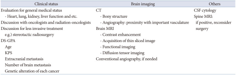
INDICATION OF SURGICAL RESECTION IN BM

Surgical resection is still important treatment of BM. However, decision of surgery should be made considering various factors including patient general condition, alternative noninvasive treatment, and patient preference. Surgical resection is not superior treatment compared to other treatment modalities. The traditional roles of surgery are limited to large mass removal, rapid relief of neurological symptoms, and obtaining tissue for histopathological examination [1016]. However, the treatment option for BM has been varied, and with increasing numbers of treatments by SRS, the role of surgery should be revisited.

Large-mass removal

Fractionated SRS and hypofractionated radiotherapy have shown favorable efficacy for medium-to-large-sized BMs [1718]. However, surgery is undoubtedly the most important treatment for large BM. Surgery can relieve high intracranial pressure and establish coincidental tissue diagnosis. Surgery is the only treatment for emergent situations, such as intracranial hypertension due to large tumors, acute expansion of tumor cysts, or hemorrhage from metastatic tumors [19].

Rapid relief of neurological symptoms

Neurological deficits in the BM can be relieved by surgical removal of the BM. BM usually accompanies a large extent of peritumoral edema, in which both BM and peritumoral edema can lead to neurological deficits [20]. Ambulatory function is the most important function in cancer patients because it is directly related to performance status [21]. SRS can be an option for small-to-medium-sized tumors; however, in case of motor weakness due to peritumoral edema, surgery can relieve peritumoral edema faster than SRS [2022]. From the study by Rahman et al. [23], a mean period of 4.8 months is needed for a 50% volume reduction after SRS for BM. During this latent period, the patient needs long-term steroid usage; sometimes, patients show neurological deterioration. Furthermore, patients may miss the appropriate treatment time for primary origin cancer, which may adversely affect the overall survival of patients with BM. In selected cases, surgical resection of the BM can relieve peritumoral edema more rapidly than SRS or radiation.

Obtaining tissue for histopathological examination

Obtaining tissue through brain biopsy is required for BM from an unknown origin cancer (MUO) patient [2425]. High-grade gliomas can mimic metastatic brain tumors. However, the treatment of glial neoplasms differs from that of metastatic brain tumors; thus, the histopathological diagnosis is essential in cases of BM of unknown origin. Furthermore, SRS carries the risk of radiological misdiagnosis in MUO cases. Surgical resection may be used to diagnose metastatic brain tumors and cancers of primary origin. In addition, patients with only brain recurrence without extracranial disease require brain tumor tissue biopsy for histopathological and genetic tests for systemic treatment.
In some cases, systemic cancer is diagnosed through a brain tumor workup. Primary cancer biopsy, such as lung and breast masses with SRS for BM, is a good option; however, in cases of large BM, simultaneous removal of BM and histopathologic diagnosis through BM may be an alternative for simultaneous treatment and diagnosis.

Treatment of multiple metastatic tumors

Treatment of multiple metastatic tumors is still complicated; whole brain irradiation (WBRT) and SRS may be first considered to treat multiple BM [262728]. Previously, WBRT had been first chosen for multiple metastases; however, WBRT harbors the risk of leukoencephalopathy, which is a cause of severe memory and cognitive impairment [282930]. Numerous studies have shown the efficacy and favorable outcomes of SRS for brain multiple BM, and SRS has been widely accepted as a treatment modality for two to four BM [82627]. However, recently, it has been reported that SRS can be chosen for two to 10 BM and more than 10 BM lesions [27].
In the case of multiple BM that harbors a large mass and is accompanied by other small- or medium-sized tumors, SRS or WBRT alone may be ineffective; it is difficult to decrease the mass effect of large tumors and harbor the risk of adverse radiation effect (ARE) with radiotherapy [831]. Removal of the largest mass combined with additional SRS or WBRT for other lesions may help prolong overall survival and improve quality of life in selected patients [3233]. Resection of multiple large BM tumors located on the same side and same lobe using a single craniotomy may be a good option for patients who show a good performance status or need histological examination [33]. However, surgical treatment may not be recommended for multiple BM combined with leptomeningeal disease or patients with poor performance status. Case-by-case decisions regarding treatment are essential after multidisciplinary discussions.

Rescue surgery for failure of radiosurgery and radiation therapy

Recurrent tumors can be considered salvage options after SRS or radiation therapy, re-irradiation, or surgical resection [343536]. In previous studies, re-irradiation for recurrent BM showed a 2-year local control rate of more than 70% [353637]. Although considering the short survival of patients with BM, long-term ARE may not be a significant problem. However, irradiation carries the risk of ARE, including medically intractable radiation necrosis [2636].
Surgical resection can achieve oncological control and removal of radiation-affected tissue through mass removal during recurrence [34]. When recurrent tumors are located in non-eloquent areas of the brain, surgical resection is a better option than re-irradiation. In high-dose conventional skull base radiation, osteonecrosis, radiation-induced osteomyelitis, or arterial stenosis can be problematic [383940]. In this case, rescue surgery, including surgical debridement and vascularized reconstruction, is needed to manage radiation necrosis of the skull base [40]. Differentiation between radiation necrosis and the recurrent tumor is needed before rescue or surgery after radiotherapy. Positron emission tomography and perfusion MR are helpful for the differential diagnosis between recurrence and radiation necrosis [11].

Skull bone involving metastasis

The cranium is the site of blood-borne metastasis from lung, breast, thyroid, and renal cell carcinoma [414243]. Skull metastasis is classified into cranial vault and skull base metastasis [43]. Clinical syndromes, including orbital syndrome, parasellar syndrome, middle fossa syndrome, jugular foramen syndrome, and occipital condyle, are caused by skull base metastasis [44]. Most skull base metastases are unresectable; therefore, radiation therapy may be chosen as the first choice of local treatment [45]. SRS is a good option for localized small skull bone metastasis, and miliary small bony involvement can be treated with radiation therapy or chemotherapy [4145].
However, surgical resection may be preferred in cases of intracranial cranial vault metastasis involving the dura or brain parenchyma to achieve local control and relieve symptoms [42]. In addition, surgical decompression or resection is first considered for skull metastasis, inducing local pain, exophthalmos, and protrusion of the scalp.

SURGICAL STRATEGY

Surgery plays an important role in the treatment of BM. However, surgical resection of the BM carries the risk of intraoperative tumor cell dissemination and the inherent difficulty of microscopic total resection. Neurosurgeons should attempt to minimize tumor cell dissemination and improve local control.

Improving local control

The extent of resection is an important factor in the local recurrence of BM. Total resection showed better outcomes than subtotal resection [46]. Furthermore, Yoo et al. [47] reported that microscopic total resection, including “5 mm further resection margin of the tumor,” showed a better local control rate and prolonged overall survival than gross total resection in BM surgery. Therefore, total resection plus obtaining a safe resection margin may yield the best outcome in BM surgery. However, care should be taken to minimize postoperative neurological deterioration.
The surgical methodology is also important in BM surgery. En-bloc resection of the tumor showed better local control than piecemeal tumor resection [48], and necessarily accompanied the small thickness of the adjacent normal brain resection. En-bloc resection is also known to prevent intraoperative tumor spreading compared to piecemeal resection [49]. Moreover, Patel et al. [50] reported that en-bloc resection was not associated with a high rate of complications or poor functional outcomes. In addition, according to the study by Suki et al. [4951], en-bloc resection can reduce intraoperative tumor spread. However, en-bloc resection is not always feasible, especially in large tumors or cystic tumors. Caution should be taken to minimize intraoperative spillage of internal content and spread of tumor pieces. In addition, a careful examination of the cavity after mass removal is needed to achieve total resection after piecemeal tumor resection.

REDUCING TUMOR SPREADING AND LEPTOMENINGEAL DISEASE

The spread of tumor cells is an important issue during BM surgery. In a previous study, proximity to the cerebrospinal fluid pathway and piecemeal resection were risk factors for leptomeningeal dissemination (LMS) [1]. Breast cancer histology, younger age, larger tumor volume, and posterior fossa location are known risk factors for LMS [525354]. In a recent meta-analysis, breast cancer primary and multiple BM tumors were meaningful factors for a high risk of LMS [53]. However, there is still debate regarding the risk factors for postoperative LMS.
WBRT has been known to reduce the incidence of LMS after BM surgery [5255]; however, Ahn et al. [1] reported that WBRT did not show efficacy in reducing LMS. It is still debated that minimizing contact of the tumor with the cerebrospinal fluid space, preventing spillage of tumor content, and reducing intraoperative saline irrigation during tumor resection may help to minimize tumor spread during neurosurgical procedures. Shunt surgery may be helpful to improve quality of life in leptomeningeal disease patients.

POSTOPERATIVE COMPLICATIONS

Postoperative complications are an important issue in BM surgery. The development of postoperative neurological deficits is a devastating factor for overall survival and significantly impacts the quality of life of patients with malignant brain tumors [56]. Cancer patients are more vulnerable to medical or surgical complications than the general population. Venous thromboembolism and pneumonia are the most important medical complications [10]. Compression stockings or pneumatic compressors for the lower leg help reduce deep vein thrombosis during the immobilization period. Encouraging sputum expectoration and deep breathing in the immediate postoperative period are required. A detailed evaluation of the patient’s medical condition is needed before surgery.
Postoperative hemorrhage and infarction are the most significant surgical complications after brain tumor surgery [57]. These complications are directly related to the performance status of the patients. Respecting arteries and veins during tumor resection are important to reduce complications. The subpial resection technique helps preserve important vascular and neural structures [58]. Preserving the major arteries is crucial during surgery, and meticulous skeletonization of arteries from the tumor is needed. In addition, preservation of the cortical or deep vein is essential to avoid venous infarction or postoperative swelling [59]. Surgery through multiple cortical openings that preserve the cortical veins and leave a small residual portion of the tumor around the vein could be an alternative surgical strategy. The prolonged use of prophylactic antibiotics did not help reduce surgical site infections [60].

ADJUVANT TREATMENT

Surgery plus radiation therapy, including resection cavity irradiation or WBRT, showed better local control than surgery alone [2961]. Adjuvant resection cavity SRS and WBRT showed comparable efficacy for local recurrence; however, the resection cavity SRS showed superiority for neurotoxicity, including cognitive impairment and AREs, compared to WBRT [6162]. However, postoperative resection cavity SRS has difficulty in identifying the appropriate target. A large surgical cavity after resection and an appropriate radiation dose are also considerable issues in postoperative resection cavity SRS [6163].
Preoperative SRS may be an alternative adjuvant option to postoperative SRS or RT. Preoperative SRS can sterilize tumor cells that spread during surgery. Furthermore, preoperative SRS can show effects similar to adjuvant SRS to eradicate microscopic invasion and avoid neurocognitive decline [6364]. The optimal dose and timing of preoperative SRS should be evaluated in future studies.
If there is another adjuvant treatment option, such as tyrosine kinase inhibitor (TKI) chemotherapy in non-small cell lung cancer, postoperative TKI chemotherapy can replace adjuvant SRS and RT in non-small cell lung cancer [6566].

FUTURE DIRECTION

Innovations in neuronavigation, intraoperative ultrasound, brain mapping, endoscopy, exoscopy, and fluorescence have advanced BM surgery. Minimally invasive surgery has been widely used in brain tumor surgery [67]. Conventional craniotomies typically produce openings larger than the target, while keyhole craniotomies can create small openings smaller than the target with complete exposure achieved by subtending the angles of the approach. Keyhole approaches minimize soft tissue and bony exposure, decrease postoperative complications, and improve cosmetic results [68]. However, minicraniotomies, including the keyhole approach, require a learning curve [69] and should be applied to patients after adequate training and sufficient experience.
Endoscopic surgery can be used in BM surgery, including the endoscopic endonasal approach (EEA) or the endoscopic transorbital approach (ETOA) [7071]. Zacharia et al. [70] reported a case series of EEA for sellar/parasellar metastatic tumors. They reported very low complication rates, such as cerebrospinal fluid leakage; however, the gross total resection rate was below 50%. EEA can be used for tissue confirmation of the unknown etiology of parasellar tumor; however, there are inherent limitations of total or wide resection of the parasellar area.
Tumors in the temporal lobe or insular area can be treated with ETOA. Park et al. [71] reported the feasibility of ETOA for intrinsic brain tumors in the temporal lobe. There has been a lack of reports of ETOA for metastatic tumor surgery; it may be used in selected cases.
Fluorescence-guided surgery using 5-aminolevulinic acid (ALA) has been used in BM surgery [7273]. Ahrens et al. reported the benefits of 5-ALA in BM surgery [1374]. Previous studies reported the benefits of 5-ALA in BM surgery [7374]. However, not all BM tumors showed positivity for 5-ALA fluorescence; only approximately 60% of BM tumors showed 5-ALA fluorescent staining [7475]. The 5-ALA fluorescent is well detected in the aggressive pathology of metastatic tumors [74]. However, 5-ALA staining can be observed in the area of peritumoral edema that is free of tumor cells [76], which is related to false positivity of 5-ALA fluorescent staining in BM. Therefore, tailored usage of 5-ALA is needed in BM surgery.
Brachytherapy involves the implantation of radioactive isotopes into the tumor cavity and has been investigated as both primary and adjuvant therapy for BMs. Radioactive materials, such as 131Cs and 125I, are commonly used for brachytherapy [7778]. Brachytherapy shows local control rates comparable to SRS. In addition, brachytherapy showed improvement in neurocognitive status and self-assessment of quality of life [79]. The use of brachytherapy as an adjuvant treatment after surgical resection of BM should be included in future studies.

CONCLUSION

Neurosurgical advances have altered the treatment of patients with BM, the most common adult brain tumor. The role of surgery in BM has been refined and its use has not been weakened in BM treatment. Total resection and obtaining a safe resection margin are the goals of surgery to improve the local control rate. En-bloc resection may lead to good local control and decreased intraoperative dissemination. Postoperative radiotherapy or chemotherapy can reduce the incidence of local recurrence. Minimally invasive BM surgery can be applied for BM surgery to shorten the recovery period and improve cosmetic results. Preoperative SRS and brachytherapy have highlighted improvements in clinical outcomes.

Notes

Ethics Statement: This retrospective study was approved by the appropriate ethics committee and was performed in accordance with the ethical standards laid down in the 1964 Declaration of Helsinki and its later amendments. For this type of study, formal consent was not required.

Author Contributions:

  • Conceptualization: Joonho Byun.

  • Data curation: Joonho Byun, Jong Hyun Kim.

  • Funding acquisition: Joonho Byun.

  • Investigation: Joonho Byun.

  • Methodology: Joonho Byun, Jong Hyun Kim.

  • Project administration: Joonho Byun.

  • Supervision: Jong Hyun Kim.

  • Validation: Joonho Byun, Jong Hyun Kim.

  • Visualization: Joonho Byun, Jong Hyun Kim.

  • Writing—original draft: Joonho Byun, Jong Hyun Kim.

  • Writing—review & editing: Joonho Byun, Jong Hyun Kim.

Conflicts of Interest: The authors have no potential conflicts of interest to disclose.

Funding Statement: This study was funded in part by the Korea University Guro Hospital and Korea University College of Medicine Research Program (Program number: K2205991).

Availability of Data and Material

The datasets generated or analyzed during the study are available from the corresponding author on reasonable request.

References

1. Ahn JH, Lee SH, Kim S, Joo J, Yoo H, Lee SH, et al. Risk for leptomeningeal seeding after resection for brain metastases: implication of tumor location with mode of resection. J Neurosurg. 2012; 116:984–993. PMID: 22339161.
2. Schouten LJ, Rutten J, Huveneers HA, Twijnstra A. Incidence of brain metastases in a cohort of patients with carcinoma of the breast, colon, kidney, and lung and melanoma. Cancer. 2001; 94:2698–2705.
3. Delattre JY, Krol G, Thaler HT, Posner JB. Distribution of brain metastases. Arch Neurol. 1988; 45:741–744. PMID: 3390029.
4. Gavrilovic IT, Posner JB. Brain metastases: epidemiology and pathophysiology. J Neurooncol. 2005; 75:5–14. PMID: 16215811.
5. Nayak L, Lee EQ, Wen PY. Epidemiology of brain metastases. Curr Oncol Rep. 2012; 14:48–54. PMID: 22012633.
6. O’Beirn M, Benghiat H, Meade S, Heyes G, Sawlani V, Kong A, et al. The expanding role of radiosurgery for brain metastases. Medicines (Basel). 2018; 5:90. PMID: 30110927.
7. Wagner S, Lanfermann H, Wohlgemuth WA, Gufler H. Effects of effective stereotactic radiosurgery for brain metastases on the adjacent brain parenchyma. Br J Cancer. 2020; 123:54–60. PMID: 32362656.
crossref
8. Minniti G, Clarke E, Lanzetta G, Osti MF, Trasimeni G, Bozzao A, et al. Stereotactic radiosurgery for brain metastases: analysis of outcome and risk of brain radionecrosis. Radiat Oncol. 2011; 6:48. PMID: 21575163.
9. Ng PR, Choi BD, Aghi MK, Nahed BV. Surgical advances in the management of brain metastases. Neurooncol Adv. 2021; 3(Suppl 5):v4–v15. PMID: 34859228.
10. Gupta S, Dawood H, Giantini Larsen A, Fandino L, Knelson EH, Smith TR, et al. Surgical and peri-operative considerations for brain metastases. Front Oncol. 2021; 11:662943. PMID: 34026641.
11. Overcast WB, Davis KM, Ho CY, Hutchins GD, Green MA, Graner BD, et al. Advanced imaging techniques for neuro-oncologic tumor diagnosis, with an emphasis on PET-MRI imaging of malignant brain tumors. Curr Oncol Rep. 2021; 23:34. PMID: 33599882.
12. Goo HW, Ra YS. Advanced MRI for pediatric brain tumors with emphasis on clinical benefits. Korean J Radiol. 2017; 18:194–207. PMID: 28096729.
13. Verma N, Cowperthwaite MC, Burnett MG, Markey MK. Differentiating tumor recurrence from treatment necrosis: a review of neuro-oncologic imaging strategies. Neuro Oncol. 2013; 15:515–534. PMID: 23325863.
14. Costabile JD, Alaswad E, D’Souza S, Thompson JA, Ormond DR. Current applications of diffusion tensor imaging and tractography in intracranial tumor resection. Front Oncol. 2019; 9:426. PMID: 31192130.
15. Sperduto PW, Kased N, Roberge D, Xu Z, Shanley R, Luo X, et al. Summary report on the graded prognostic assessment: an accurate and facile diagnosis-specific tool to estimate survival for patients with brain metastases. J Clin Oncol. 2012; 30:419–425. PMID: 22203767.
16. Ene CI, Ferguson SD. Surgical management of brain metastasis: challenges and nuances. Front Oncol. 2022; 12:847110. PMID: 35359380.
17. Masucci GL. Hypofractionated radiation therapy for large brain metastases. Front Oncol. 2018; 8:379. PMID: 30333955.
crossref
18. Kim JW, Park HR, Lee JM, Kim JW, Chung HT, Kim DG, et al. Fractionated stereotactic gamma knife radiosurgery for large brain metastases: a retrospective, single center study. PLoS One. 2016; 11:e0163304. PMID: 27661613.
crossref
19. Sankey EW, Tsvankin V, Grabowski MM, Nayar G, Batich KA, Risman A, et al. Operative and peri-operative considerations in the management of brain metastasis. Cancer Med. 2019; 8:6809–6831. PMID: 31568689.
crossref
20. Shimony N, Shofty B, Harosh CB, Sitt R, Ram Z, Grossman R. Surgical resection of cerebral metastases leads to faster resolution of peritumoral edema than stereotactic radiosurgery: a volumetric analysis. Ann Surg Oncol. 2017; 24:1392–1398. PMID: 27896517.
crossref
21. Amidei C, Kushner DS. Clinical implications of motor deficits related to brain tumors. Neurooncol Pract. 2015; 2:179–184. PMID: 31386054.
crossref
22. Schwartz C, Rautalin I, Niemelä M, Korja M. Symptomatic peritumoral edema is associated with surgical outcome: a consecutive series of 72 supratentorial meningioma patients ≥ 80 years of age. J Neurooncol. 2020; 148:109–116. PMID: 32318913.
crossref
23. Rahman M, Cox JB, Chi YY, Carter JH, Friedman WA. Radiographic response of brain metastasis after linear accelerator radiosurgery. Stereotact Funct Neurosurg. 2012; 90:69–78. PMID: 22286386.
crossref
24. Cirak M, Guclu DG, Akar E, Kazanci MH, Tural D. Retrospective analysis of survival in patients with brain metastases from an unknown primary tumor. Turk Neurosurg. 2020; 30:932–936. PMID: 33216341.
crossref
25. Polyzoidis KS, Miliaras G, Pavlidis N. Brain metastasis of unknown primary: a diagnostic and therapeutic dilemma. Cancer Treat Rev. 2005; 31:247–255. PMID: 15913895.
crossref
26. Sahgal A, Ruschin M, Ma L, Verbakel W, Larson D, Brown PD. Stereotactic radiosurgery alone for multiple brain metastases? A review of clinical and technical issues. Neuro Oncol. 2017; 19(suppl_2):ii2–ii15. PMID: 28380635.
crossref
27. Kraft J, Zindler J, Minniti G, Guckenberger M, Andratschke N. Stereotactic radiosurgery for multiple brain metastases. Curr Treat Options Neurol. 2019; 21:6. PMID: 30758726.
crossref
28. Patil CG, Pricola K, Sarmiento JM, Garg SK, Bryant A, Black KL. Whole brain radiation therapy (WBRT) alone versus WBRT and radiosurgery for the treatment of brain metastases. Cochrane Database Syst Rev. 2017; 9:CD006121. PMID: 28945270.
crossref
29. Kocher M, Soffietti R, Abacioglu U, Villà S, Fauchon F, Baumert BG, et al. Adjuvant whole-brain radiotherapy versus observation after radiosurgery or surgical resection of one to three cerebral metastases: results of the EORTC 22952-26001 study. J Clin Oncol. 2011; 29:134–141. PMID: 21041710.
crossref
30. Mut M. Surgical treatment of brain metastasis: a review. Clin Neurol Neurosurg. 2012; 114:1–8. PMID: 22047649.
crossref
31. Sneed PK, Mendez J, Vemer-van den Hoek JG, Seymour ZA, Ma L, Molinaro AM, et al. Adverse radiation effect after stereotactic radiosurgery for brain metastases: incidence, time course, and risk factors. J Neurosurg. 2015; 123:373–386. PMID: 25978710.
crossref
32. Pollock BE, Brown PD, Foote RL, Stafford SL, Schomberg PJ. Properly selected patients with multiple brain metastases may benefit from aggressive treatment of their intracranial disease. J Neurooncol. 2003; 61:73–80. PMID: 12587798.
33. Salvati M, Tropeano MP, Maiola V, Lavalle L, Brogna C, Colonnese C, et al. Multiple brain metastases: a surgical series and neurosurgical perspective. Neurol Sci. 2018; 39:671–677. PMID: 29383618.
crossref
34. Matsuda R, Morimoto T, Tamamoto T, Inooka N, Ochi T, Miyasaka T, et al. Salvage surgical resection after linac-based stereotactic radiosurgery for newly diagnosed brain metastasis. Curr Oncol. 2021; 28:5255–5265. PMID: 34940078.
crossref
35. Nieder C, Yobuta R, Mannsåker B. Second re-irradiation of brain metastases: a review of studies involving stereotactic radiosurgery. Cureus. 2018; 10:e3712. PMID: 30788201.
crossref
36. Loi M, Caini S, Scoccianti S, Bonomo P, De Vries K, Francolini G, et al. Stereotactic reirradiation for local failure of brain metastases following previous radiosurgery: systematic review and meta-analysis. Crit Rev Oncol Hematol. 2020; 153:103043. PMID: 32650217.
crossref
37. Kowalchuk RO, Niranjan A, Lee CC, Yang HC, Liscak R, Guseynova K, et al. Reirradiation with stereotactic radiosurgery after local or marginal recurrence of brain metastases from previous radiosurgery. Int J Radiat Oncol Biol Phys. 2022; 112:726–734. PMID: 34644606.
crossref
38. Lalani N, Huang SH, Rotstein C, Yu E, Irish J, O’Sullivan B. Skull base or cervical vertebral osteomyelitis following chemoradiotherapy for pharyngeal carcinoma: a serious but treatable complication. Clin Transl Radiat Oncol. 2017; 8:40–44. PMID: 29594240.
crossref
39. Trojanowski P, Sojka M, Trojanowska A, Wolski A, Roman T, Jargiello T. Management of radiation induced carotid stenosis in head and neck cancer. Transl Oncol. 2019; 12:1026–1031. PMID: 31146165.
crossref
40. Habib A, Hanasono MM, DeMonte F, Haider A, Breshears JD, Nader ME, et al. Surgical management of skull base osteoradionecrosis in the cancer population–treatment outcomes and predictors of recurrence: a case series. Oper Neurosurg (Hagerstown). 2020; 19:364–374. PMID: 32324878.
crossref
41. Kotecha R, Angelov L, Barnett GH, Reddy CA, Suh JH, Murphy ES, et al. Calvarial and skull base metastases: expanding the clinical utility of gamma knife surgery. J Neurosurg. 2014; 121 Suppl:91–101.
crossref
42. Park I, Chung DH, Yoo CJ, Shin DB. Skull metastasis of gastric gastrointestinal stromal tumor successfully managed by surgery. J Korean Neurosurg Soc. 2017; 60:94–97. PMID: 28061498.
crossref
43. Chaichana KL, Flores M, Acharya S, Sampognaro P, Bettegowda C, Rigamonti D, et al. Survival and recurrence for patients undergoing surgery of skull base intracranial metastases. J Neurol Surg B Skull Base. 2013; 74:228–235. PMID: 24436917.
crossref
44. Mitsuya K, Nakasu Y, Horiguchi S, Harada H, Nishimura T, Yuen S, et al. Metastatic skull tumors: MRI features and a new conventional classification. J Neurooncol. 2011; 104:239–245. PMID: 21110218.
crossref
45. Laigle-Donadey F, Taillibert S, Martin-Duverneuil N, Hildebrand J, Delattre JY. Skull-base metastases. J Neurooncol. 2005; 75:63–69. PMID: 16215817.
crossref
46. Winther RR, Hjermstad MJ, Skovlund E, Aass N, Helseth E, Kaasa S, et al. Surgery for brain metastases-impact of the extent of resection. Acta Neurochir (Wien). 2022; 164:2773–2780. PMID: 35080651.
crossref
47. Yoo H, Kim YZ, Nam BH, Shin SH, Yang HS, Lee JS, et al. Reduced local recurrence of a single brain metastasis through microscopic total resection. J Neurosurg. 2009; 110:730–736. PMID: 19072310.
crossref
48. Patel AJ, Suki D, Hatiboglu MA, Abouassi H, Shi W, Wildrick DM, et al. Factors influencing the risk of local recurrence after resection of a single brain metastasis. J Neurosurg. 2010; 113:181–189. PMID: 20035574.
crossref
49. Suki D, Hatiboglu MA, Patel AJ, Weinberg JS, Groves MD, Mahajan A, et al. Comparative risk of leptomeningeal dissemination of cancer after surgery or stereotactic radiosurgery for a single supratentorial solid tumor metastasis. Neurosurgery. 2009; 64:664–674. discussion 674-6. PMID: 19197219.
crossref
50. Patel AJ, Suki D, Hatiboglu MA, Rao VY, Fox BD, Sawaya R. Impact of surgical methodology on the complication rate and functional outcome of patients with a single brain metastasis. J Neurosurg. 2015; 122:1132–1143. PMID: 25794344.
crossref
51. Suki D, Abouassi H, Patel AJ, Sawaya R, Weinberg JS, Groves MD. Comparative risk of leptomeningeal disease after resection or stereotactic radiosurgery for solid tumor metastasis to the posterior fossa. J Neurosurg. 2008; 108:248–257. PMID: 18240919.
crossref
52. Ha B, Chung SY, Kim YJ, Gwak HS, Chang JH, Lee SH, et al. Effects of postoperative radiotherapy on leptomeningeal carcinomatosis or dural metastasis after resection of brain metastases in breast cancer patients. Cancer Res Treat. 2017; 49:748–758. PMID: 27809457.
crossref
53. Tewarie IA, Jessurun CAC, Hulsbergen AFC, Smith TR, Mekary RA, Broekman MLD. Leptomeningeal disease in neurosurgical brain metastases patients: a systematic review and meta-analysis. Neurooncol Adv. 2021; 3:vdab162. PMID: 34859226.
crossref
54. van der Ree TC, Dippel DW, Avezaat CJ, Sillevis Smitt PA, Vecht CJ, van den Bent MJ. Leptomeningeal metastasis after surgical resection of brain metastases. J Neurol Neurosurg Psychiatry. 1999; 66:225–227. PMID: 10071105.
crossref
55. Patchell RA, Tibbs PA, Regine WF, Dempsey RJ, Mohiuddin M, Kryscio RJ, et al. Postoperative radiotherapy in the treatment of single metastases to the brain: a randomized trial. JAMA. 1998; 280:1485–1489. PMID: 9809728.
56. De la Garza-Ramos R, Kerezoudis P, Tamargo RJ, Brem H, Huang J, Bydon M. Surgical complications following malignant brain tumor surgery: an analysis of 2002-2011 data. Clin Neurol Neurosurg. 2016; 140:6–10. PMID: 26615463.
crossref
57. Lassen B, Helseth E, Rønning P, Scheie D, Johannesen TB, Mæhlen J, et al. Surgical mortality at 30 days and complications leading to recraniotomy in 2630 consecutive craniotomies for intracranial tumors. Neurosurgery. 2011; 68:1259–1268. discussion 1268-9. PMID: 21273920.
crossref
58. Przybylowski CJ, Whiting AC, Preul MC, Smith KA. Anatomical subpial resection of tumors in the amygdala and hippocampus. World Neurosurg. 2021; 151:e652–e662. PMID: 33940265.
crossref
59. Nakase H, Shin Y, Nakagawa I, Kimura R, Sakaki T. Clinical features of postoperative cerebral venous infarction. Acta Neurochir (Wien). 2005; 147:621–626. PMID: 15770350.
crossref
60. Liu W, Ni M, Zhang Y, Groen RJ. Antibiotic prophylaxis in craniotomy: a review. Neurosurg Rev. 2014; 37:407–414. PMID: 24526365.
crossref
61. Mahajan A, Ahmed S, McAleer MF, Weinberg JS, Li J, Brown P, et al. Post-operative stereotactic radiosurgery versus observation for completely resected brain metastases: a single-centre, randomised, controlled, phase 3 trial. Lancet Oncol. 2017; 18:1040–1048. PMID: 28687375.
crossref
62. Brown PD, Ballman KV, Cerhan JH, Anderson SK, Carrero XW, Whitton AC, et al. Postoperative stereotactic radiosurgery compared with whole brain radiotherapy for resected metastatic brain disease (NCCTG N107C/CEC·3): a multicentre, randomised, controlled, phase 3 trial. Lancet Oncol. 2017; 18:1049–1060. PMID: 28687377.
crossref
63. Prabhu RS, Patel KR, Press RH, Soltys SG, Brown PD, Mehta MP, et al. Preoperative vs postoperative radiosurgery for resected brain metastases: a review. Neurosurgery. 2019; 84:19–29. PMID: 29771381.
crossref
64. Ginzac A, Dupic G, Brun L, Molnar I, Casile M, Durando X, et al. Preoperative stereotactic radiosurgery for brain metastases: the STEP study protocol for a multicentre, prospective, phase-II trial. BMC Cancer. 2021; 21:864. PMID: 34320940.
crossref
65. Lee HH, Chen CH, Chuang HY, Huang YW, Huang MY. Brain surgery in combination with tyrosine kinase inhibitor and whole brain radiotherapy for epidermal growth factor receptor-mutant non-small-cell lung cancer with brain metastases. Sci Rep. 2019; 9:16834. PMID: 31728013.
crossref
66. Magnuson WJ, Lester-Coll NH, Wu AJ, Yang TJ, Lockney NA, Gerber NK, et al. Management of brain metastases in tyrosine kinase inhibitor–naïve epidermal growth factor receptor–mutant non–small-cell lung cancer: a retrospective multi-institutional analysis. J Clin Oncol. 2017; 35:1070–1077. PMID: 28113019.
crossref
67. Phang I, Leach J, Leggate JRS, Karabatsou K, Coope D, D’Urso PI. Minimally invasive resection of brain metastases. World Neurosurg. 2019; 130:e362–e367. PMID: 31233927.
crossref
68. Baker CM, Glenn CA, Briggs RG, Burks JD, Smitherman AD, Conner AK, et al. Simultaneous resection of multiple metastatic brain tumors with multiple keyhole craniotomies. World Neurosurg. 2017; 106:359–367. PMID: 28652117.
crossref
69. Eroglu U, Shah K, Bozkurt M, Kahilogullari G, Yakar F, Dogan İ, et al. Supraorbital keyhole approach: lessons learned from 106 operative cases. World Neurosurg. 2019; 124:e667–e674.
crossref
70. Zacharia BE, Romero FR, Rapoport SK, Raza SM, Anand VK, Schwartz TH. Endoscopic endonasal management of metastatic lesions of the anterior skull base: case series and literature review. World Neurosurg. 2015; 84:1267–1277. PMID: 26079759.
crossref
71. Park HH, Roh TH, Choi S, Yoo J, Kim WH, Jung IH, et al. Endoscopic transorbital approach to mesial temporal lobe for intra-axial lesions: cadaveric study and case series (SevEN-008). Oper Neurosurg (Hagerstown). 2021; 21:E506–E515. PMID: 34528091.
crossref
72. Ferraro N, Barbarite E, Albert TR, Berchmans E, Shah AH, Bregy A, et al. The role of 5-aminolevulinic acid in brain tumor surgery: a systematic review. Neurosurg Rev. 2016; 39:545–555. PMID: 26815631.
crossref
73. Ahrens LC, Krabbenhøft MG, Hansen RW, Mikic N, Pedersen CB, Poulsen FR, et al. Effect of 5-aminolevulinic acid and sodium fluorescein on the extent of resection in high-grade gliomas and brain metastasis. Cancers (Basel). 2022; 14:617. PMID: 35158885.
crossref
74. Kamp MA, Fischer I, Bühner J, Turowski B, Cornelius JF, Steiger HJ, et al. 5-ALA fluorescence of cerebral metastases and its impact for the local-in-brain progression. Oncotarget. 2016; 7:66776–66789. PMID: 27564260.
crossref
75. Kamp MA, Grosser P, Felsberg J, Slotty PJ, Steiger HJ, Reifenberger G, et al. 5-aminolevulinic acid (5-ALA)-induced fluorescence in intracerebral metastases: a retrospective study. Acta Neurochir (Wien). 2012; 154:223–228. PMID: 22080159.
crossref
76. Utsuki S, Miyoshi N, Oka H, Miyajima Y, Shimizu S, Suzuki S, et al. Fluorescence-guided resection of metastatic brain tumors using a 5-aminolevulinic acid-induced protoporphyrin IX: pathological study. Brain Tumor Pathol. 2007; 24:53–55. PMID: 18095131.
crossref
77. Raleigh DR, Seymour ZA, Tomlin B, Theodosopoulos PV, Berger MS, Aghi MK, et al. Resection and brain brachytherapy with permanent iodine-125 sources for brain metastasis. J Neurosurg. 2017; 126:1749–1755. PMID: 27367240.
crossref
78. Huang K, Sneed PK, Kunwar S, Kragten A, Larson DA, Berger MS, et al. Surgical resection and permanent iodine-125 brachytherapy for brain metastases. J Neurooncol. 2009; 91:83–93. PMID: 18719856.
crossref
79. Pham A, Yondorf MZ, Parashar B, Scheff RJ, Pannullo SC, Ramakrishna R, et al. Neurocognitive function and quality of life in patients with newly diagnosed brain metastasis after treatment with intra-operative cesium-131 brachytherapy: a prospective trial. J Neurooncol. 2016; 127:63–71. PMID: 26650067.
crossref
Table 1

Summary of preoperative work-up for brain metastasis

btrt-11-1-i001
Clinical status Brain imaging Others
Evaluation for general medical status CT CSF cytology
- Heart, lung, kidney, liver function and etc. - Bony structure Spine MRI: if positive, reconsider surgery
Discussion with oncologists and radiation-oncologists - Angiography: proximity with important vasculature
Discussion for less invasive treatment Brain MRI
e.g.) stereotactic radiosurgery - Contrast enhancement
DS-GPA - Acquisition of thin sliced image
Age - Functional imaging
KPS - Diffusion tensor imaging
Extracranial metastasis Conventional angiography, if needed
Number of brain metastasis
Genetic alteration of each cancer

DS-GPA, disease-specific graded prognostic assessment; KPS, Karnofsky Performance scale; CSF, cerebrospinal fluid

TOOLS
Similar articles