Journal List > Brain Tumor Res Treat > v.10(1) > 1516081729

Jeong, Lee, and Yang: Intradiploic Encephalocele at the Parietal Bone: A Case Report and Literature Review

Abstract

Intradiploic encephalocele is a rare condition of herniation of the brain parenchyma through the diploic space. A 52-year-old man presented with a parietal intradiploic encephalocele manifesting as an intermittent headache for 7 months. CT revealed an osteolytic lesion involving the right parietal bone. MRI demonstrated brain herniation within the diploic space. Surgery may be unnecessary in the absence of concurrent symptoms or neurological deficits. After 2 years of follow-up, symptoms were improved without neurological deficits and CT findings. We report the X-ray, CT, and MRI findings of an extremely rare case of parietal intradiploic encephalocele in adulthood.

INTRODUCTION

Encephaloceles are protrusions of the brain parenchyma through osseous defects of the skull base or cranial vault [1]. It may develop congenitally (similar to the neural tube defect) or may occur after acquired events, such as infection, trauma, neoplasms, and iatrogenic causes [23]. The incidence of the congenital form has been estimated at 1 in every 3,000–10,000 live births [4]. Many classification systems for encephalocele have been suggested; however, the most accepted is that from Matson [5], which follows the location of the encephalocele: basal, sincipital, convexity, and atretic. These lesions are usually along the midline, ranging from the nasal to the occiput, where three-quarters encephaloceles occur posteriorly [6].
In cases where the defect occupies only the dura and inner table with an intact outer table of the skull, parenchymal herniation occurs in the intradiploic space and are called intradiploic encephaloceles [7]. Especially, off the midline parietal encephaloceles are rare, comprising only 1% of all cerebrospinal malformations and 10% of encephaloceles [28]. Here, we report a case of parietal intradiploic encephalocele during a 2-year follow-up with CT and MR images.

CASE REPORT

A 52-year-old man was referred for further evaluation of a skull osteolytic lesion on CT due to intermittent headache for 7 months. He had no history of seizures, central nervous system infections, stroke, brain tumor, or trauma. His neurological examination was normal, without any palpable mass or pulsation on his scalp. We proceeded with MRI to exclude osteolytic lesion such as vascular lesions or other tumorous conditions. We confirmed a herniation of the brain parenchyma with the surrounding cerebrospinal fluid (CSF) space into the diploe. These confirmed a diagnosis of right parietal intradiploic encephalocele (Fig. 1). The headache was controlled with medicines without neurological symptoms and signs. Two-year follow-up CT findings showed no change in the lesion (Fig. 2) and the patient doing well without symptoms.

DISCUSSION

Intradiploic encephalocele refers to brain parenchyma herniation into a diploic space with a destroyed inner table and an intact outer table [7]. Intradiploic encephalocele has characteristics that distinguish it from classic encephaloceles. In fact, it is debatable whether the nomenclature reflects the true nature of this disease and should be referred to as brain herniation into the calvaria surrounding the CSF [9].
Intradiploic encephaloceles share more common morphological features with expanding skull fractures and intradiploic arachnoid cysts [2]. Causes are unclear, but the mechanism of lesion may be similar with the “growing skull fracture” [27]. Cranial fractures in childhood can lead to a growing skull fracture [1011]. Fracture with a dural tear may cause herniation of the arachnoid tissue through the dural defect, and the fracture is gradually enlarged, and bone margins are separated by the continuous pulsation of the CSF [12]. In some cases, the CSF and brain tissue may herniate into the diploic cavity through a dura and inner table defect without damaging the outer table. The integrity of the outer table can be elucidated by a strong occipital muscle and pericranium that prevent bone erosion from the pulsation of CSF [13].
A similar mechanism can also be developed for low-velocity blunt injury that causes fracture only in the thinner inner table. The cracked inner table depresses and rebounds during initial insult. The depression then tears the dura, and the recoil creates a negative pressure that forces the underlying arachnoid and brain into the diploic space [14]. The lesion then grew with CSF pulsation.
As in the present case, anatomical factors are considered more important because of the lack of history of acquired risk factors. An anatomical and physiological defect of the cranial inner table, foveolae granulare, generate intradiploic encephalocele [15]. These granulations are usually located in the frontoparietal bone and near the superior sagittal sinus. In a situation of head trauma, the dura and arachnoid membranes rupture without skull fracture. CSF pulsation then widens the intradiploic space over time. Communication could also allow the brain parenchyma to escape into the diploic space.
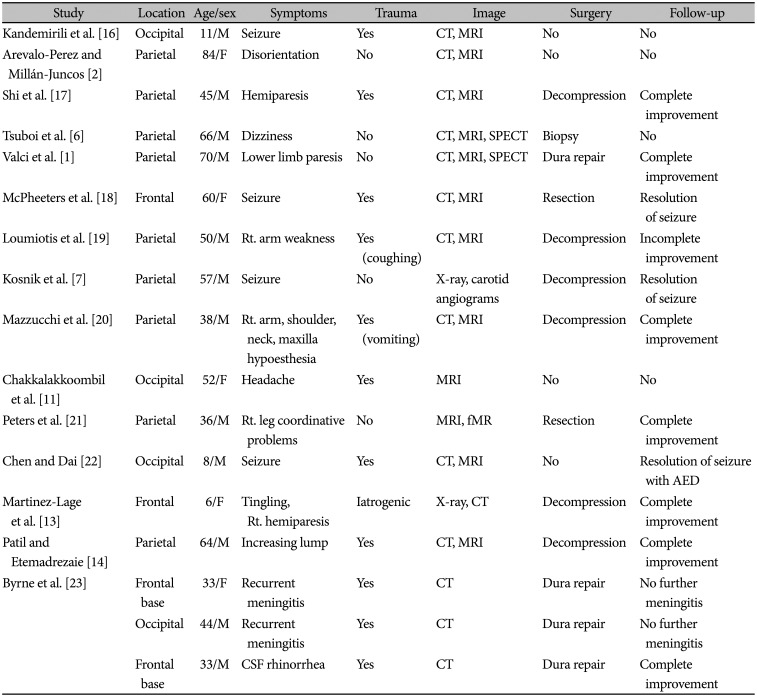
In the literature, 10 patients (10/17 cases) presented with seizure activity or neurologic deficits (Table 1). Others have non-specific symptoms such as headache, dizziness, and cognitive problems. Most intradiploic encephaloceles were found incidentally. Various symptom presentations might be related to the location of involvement; herniation of the eloquent cortex may cause non-specific symptoms. Some patients could present with acute onset of clinical symptoms when a sudden increase in intracranial pressure occurs, such as in an episode of vomiting or coughing. Surgery was performed in 13 cases if the radiologic findings corresponded to the clinical findings. The main purpose of surgery is to repair the dural defect along with the decompression of the herniated cortex.
The differential diagnosis of an osteolytic calvarial lesion in both pediatric and adult populations includes eosinophilic granuloma, plasmacytoma, metastasis, hemangioma, epidermoid or dermoid cyst, and intradiploic arachnoid cyst [1]. The confirmation of a herniated brain parenchyma through the osseous defect into the diploic cavity by MRI is the most important feature that separates intradiploic encephalocele from other possible diagnoses.
Intradiploic encephalocele is extremely rare, and the present case adds to the existing literature. The diagnosis of the lesion can be with typical MRI findings. MRI shows osteolytic lesion involved inner layer of skull and presence of herniated brain tissue within the diploic space. Understanding this condition helps to facilitate diagnosis and avoid confusion with vascular and malignant lesions. Ultimately, it provides adequate management and prevents potential surgery in asymptomatic patients.

Acknowledgments

We would like to thank Editage (www.editage.co.kr) for English language editing.

Notes

Ethics Statement: The Institutional Review Board of National Health Insurance Service Ilsan Hospital exempted the requirement for written informed consent in this case of verbal allowance to publish the retrospective case report with a minimal risk for the patient (NHIMC 2022-01-023).

Author Contributions:

  • Conceptualization: Kookhee Yang.

  • Data curation: Boeun Lee.

  • Formal analysis: Boeun Lee.

  • Funding acquisition: Kookhee Yang.

  • Investigation: Dong Jun Jeong.

  • Methodology: Boeun Lee.

  • Project administration: Boeun Lee.

  • Resources: Kookhee Yang.

  • Software: Boeun Lee.

  • Supervision: Kookhee Yang.

  • Validation: Boeun Lee.

  • Visualization: Dong Jun Jeong.

  • Writing—original draft: Dong Jun Jeong.

  • Writing—review & editing: Kookhee Yang.

Conflicts of Interest: The authors have no potential conflicts of interest to disclose.

Funding Statement: None

Availability of Data and Material

The datasets generated or analyzed during the study are available from the corresponding author on reasonable request.

References

1. Valci L, Dalolio M, Kuhlen D, Pravatà E, Gobbi C, Reinert M. Intradiploic encephalocele of the primary motor cortex in an adult patient: electrophysiological implications during surgery. J Neurosurg. 2018; 128:871–874. PMID: 28452617.
crossref
2. Arevalo-Perez J, Millán-Juncos JM. Parietal intradiploic encephalocele: report of a case and review of the literature. Neuroradiol J. 2015; 28:264–267. PMID: 26246093.
3. Mealey J Jr, Dzenitis AJ, Hockey AA. The prognosis of encephaloceles. J Neurosurg. 1970; 32:209–218. PMID: 5411997.
crossref
4. Martínez-Lage JF, Poza M, Sola J, Soler CL, Montalvo CG, Domingo R, et al. The child with a cephalocele: etiology, neuroimaging, and outcome. Childs Nerv Syst. 1996; 12:540–550. PMID: 8906370.
5. Matson DD. Neurosurgery of infancy and childhood. 2nd ed. Springfield: Charles C Thomas;1969.
6. Tsuboi Y, Hayashi N, Noguchi K, Kurimoto M, Nagai S, Endo S. Parietal intradiploic encephalocele--case report. Neurol Med Chir (Tokyo). 2007; 47:240–241. PMID: 17527054.
7. Kosnik EJ, Meagher JN, Quenemoen LR. Parietal intradiploic encephalocele. Case report. J Neurosurg. 1976; 44:617–619. PMID: 1262923.
8. Naidich TP, Altman NR, Braffman BH, McLone DG, Zimmerman RA. Cephaloceles and related malformations. AJNR Am J Neuroradiol. 1992; 13:655–690. PMID: 1566723.
9. Battal B. Letter to the editor. Brain herniation with surrounding CSF into the skull. J Neurosurg. 2018; 128:949–951. PMID: 29271708.
crossref
10. Roy S, Sarkar C, Tandon PN, Banerji AK. Cranio-cerebral erosion (growing fracture of the skull in children). Part I. Pathology. Acta Neurochir (Wien). 1987; 87:112–118. PMID: 3673689.
11. Chakkalakkoombil SV, Venkatesh M, Nair PP, Sowkar UB. Intradiploic cephalocele: a rare entity at a rare site. BMJ Case Rep. 2018; 2018:bcr2017223262.
crossref
12. Winston K, Beatty RM, Fischer EG. Consequences of dural defects acquired in infancy. J Neurosurg. 1983; 59:839–846. PMID: 6619936.
crossref
13. Martínez-Lage JF, López F, Piqueras C, Poza M. Iatrogenic intradiploic meningoencephalocele. Case report. J Neurosurg. 1997; 87:468–471. PMID: 9285618.
14. Patil AA, Etemadrezaie H. Posttraumatic intradiploic meningoencephalocele. Case report. J Neurosurg. 1996; 84:284–287. PMID: 8592236.
15. Menkü A, Koç RK, Tucer B, Akdemir H. Is skull fracture necessary for developing an intradiploic pseudomeningocele as a complication of head injury in adulthood? Acta Neurochir (Wien). 2004; 146:623–627. discussion 627. PMID: 15168231.
16. Kandemirli SG, Candan S, Bilgin C. Post-traumatic occipital intradiploic encephalocele. World Neurosurg. 2019; 129:9–12. PMID: 31150845.
17. Shi C, Flores B, Fisher S, Barnett SL. Symptomatic parietal intradiploic encephalocele—a case report and literature review. J Neurol Surg Rep. 2017; 78:e40–e45. PMID: 28299248.
18. McPheeters MJ, Kainth DS, Lam CH. Spontaneous frontal intradiploic meningoencephalocele. Surg Neurol Int. 2015; 6:S651–S654. PMID: 26713172.
19. Loumiotis I, Jones L, Diehn F, Lanzino G. Symptomatic left intradiploic encephalocele. Neurology. 2010; 75:1027. PMID: 20837973.
20. Mazzucchi E, Bianchi F, Vigo V, Anile C, Frassanito P. Parietal intradiploic encephalocele in an adult: a delayed complication of pediatric head injury? Childs Nerv Syst. 2017; 33:217–219. PMID: 28005171.
21. Peters J, Raab P, Marquardt G, Zanella FE. Intradiploic meningoencephalocele. Eur Radiol. 2002; 12 Suppl 3:S25–S27.
22. Chen X, Dai H. Intradiploic encephalocele following linear skull fracture: a rare evolution of growing skull fracture. Childs Nerv Syst. 2021; 37:3967–3971. PMID: 33715079.
23. Byrne JV, Britten JA, Kaar G. Chronic post-traumatic erosion of the skull base. Neuroradiology. 1992; 34:528–531. PMID: 1436467.
Fig. 1

Initial MR image. A: T2-weighted axial MR image showing a parietal osteolytic lesion, confirming the presence of a herniated brain cortex within the diploe. B: Magnified the white square of T2-weighted axial image. C: T2-weighted coronal MR image. D: Magnified the white square of T2-weighted coronal image. E: Gadolinium (Gd)-enhanced T1-weighted axial MR image. F: Magnified the white square of Gd-enhanced T1-weighted axial image reveal no enhancement of lesion.

btrt-10-38-g001
Fig. 2

Skull X-ray and CT findings. A: Initial skull X-ray showing an osteolytic lesion (white arrow heads) in the right parietal bone. B: Initial CT scan showing intradiploic herniation of brain parenchyma. C: Initial CT scan (bone window) reveal skull osteolytic lesion confined to inner table. D: Two-year follow up CT scan shows no change of the lesion.

btrt-10-38-g002
Table 1

Review of intradiploic encephalocele cases

btrt-10-38-i001
Study Location Age/sex Symptoms Trauma Image Surgery Follow-up
Kandemirili et al. [16] Occipital 11/M Seizure Yes CT, MRI No No
Arevalo-Perez and Millán-Juncos [2] Parietal 84/F Disorientation No CT, MRI No No
Shi et al. [17] Parietal 45/M Hemiparesis Yes CT, MRI Decompression Complete improvement
Tsuboi et al. [6] Parietal 66/M Dizziness No CT, MRI, SPECT Biopsy No
Valci et al. [1] Parietal 70/M Lower limb paresis No CT, MRI, SPECT Dura repair Complete improvement
McPheeters et al. [18] Frontal 60/F Seizure Yes CT, MRI Resection Resolution of seizure
Loumiotis et al. [19] Parietal 50/M Rt. arm weakness Yes (coughing) CT, MRI Decompression Incomplete improvement
Kosnik et al. [7] Parietal 57/M Seizure No X-ray, carotid angiograms Decompression Resolution of seizure
Mazzucchi et al. [20] Parietal 38/M Rt. arm, shoulder, neck, maxilla hypoesthesia Yes (vomiting) CT, MRI Decompression Complete improvement
Chakkalakkoombil et al. [11] Occipital 52/F Headache Yes MRI No No
Peters et al. [21] Parietal 36/M Rt. leg coordinative problems No MRI, fMR Resection Complete improvement
Chen and Dai [22] Occipital 8/M Seizure Yes CT, MRI No Resolution of seizure with AED
Martinez-Lage et al. [13] Frontal 6/F Tingling, Rt. Hemiparesis Iatrogenic X-ray, CT Decompression Complete improvement
Patil and Etemadrezaie [14] Parietal 64/M Increasing lump Yes CT, MRI Decompression Complete improvement
Byrne et al. [23] Frontal base 33/F Recurrent meningitis Yes CT Dura repair No further meningitis
Occipital 44/M Recurrent meningitis Yes CT Dura repair No further meningitis
Frontal base 33/M CSF rhinorrhea Yes CT Dura repair Complete improvement

CSF, cerebrospinal fluid; SPECT, single photon emission computer tomography; fMR, functional MRI; AED, anti-epileptic drug

TOOLS
Similar articles