Journal List > Brain Tumor Res Treat > v.10(4) > 1516081715

Kim and Lee: Engineered Aurotherapy for the Multimodal Treatment of Glioblastoma

Abstract

Glioblastoma multiforme (GBM) is the most aggressive brain tumor, characterized by fatal prognosis and high rates of recurrence. Although there are various treatment strategies such as surgical resection, radiotherapy, and chemotherapy, these traditional approaches still have not improved the survival rates and prolongation. Therefore, there is a pressing requirement for developing novel technologies to combat GBM. Nanoparticle-based GBM therapy can be considered a promising approach to precisely treat tumors with minimal side effects. Among various nanoparticles, gold nanoparticle (AuNP) has been demonstrated to be effective in treating GBM because of its advantages such as easy functionalization due to self-assembled monolayers of thiols, surface plasmon resonance effect on its surface, and relatively low toxicity issues. By using nanoscale (5–100 nm) and facile functionalization with a targeting ligand, AuNP can overcome the obstacles caused by blood-brain barrier, which selectively inhibits AuNP penetration into the brain tumor mass. AuNPs delivered into brain tissue and targeted with GBM have been mostly explored for photothermal therapy and photodynamic therapy, but also investigated in the development of complex therapies including radiotherapy, chemotherapy, and immunotherapy using AuNP-based nanoplatforms. Therefore, the aim of this mini review is to summarize recent works on the AuNPs-based nanoplatforms for treating GBM with a multimodal approach.

INTRODUCTION

Glioblastoma multiforme (GBM) is the primary grade IV brain malignancy and carries a fatal prognosis. Despite significant advances in molecular understanding and predictive biomarkers identification, only a handful of drugs are currently approved for GBM [1]. The standard treatment for GBM involves combinational therapies including surgical resection, chemotherapy, and radiotherapy [2]. Nevertheless, the survival rate for patients with GBM is still less than one year [34]. From a clinical point of view, effective treatment of GBM is limited by several challenges. First, the anatomical complexity and size of the tumor influence the extent of resection. This is because a fine balance must be struck between maximum removal of malignant tissue and minimum surgical risk [5]. Even with safe operative resection, residual infiltrating GBM cells that cannot be detected by imaging techniques remain in the tumor periphery, leading to disease progression and inevitable death [6]. Second, the central nervous system features distinctive microenvironment that is surrounded by the blood-brain barrier (BBB), restricting systemically delivered drugs from accessing the brain. This reduces the treatment options available for GBM. Additionally, GBM is characterized by high heterogeneity between intra-tumor and inter-tumor at the cellular level, and histologically characterized by the dissimilar presence of subclonal and clonal differentiated tumor cell population, tumor stem cells, and components of the tumor microenvironment. These characteristics of the tissues that make up GBM lead to the failure of targeted therapy due to different susceptibility to therapeutic agents [78]. Therefore, the development of more sophisticated and powerful treatments is one of the most pressing challenges. Herein, we introduce alternative GBM treatment strategies such as photothermal therapy (PTT) and photodynamic therapy (PDT) utilizing gold nanoparticles (AuNPs), and their potential for oral drug delivery platform featuring the most non-invasive drug administration route.

PTT IN GBM TREATMENT

PTT is based on the local application of high temperatures to tissues to induce irreversible cellular damage at the target site and is considered a promising strategy for cancer treatments [9]. It is a non-invasive treatment that requires local irradiation of the tumor using an external near-infrared (NIR) laser. Upon laser irradiation, the photothermal agent (PTA) harvests the light energy, converts, and releases it as heat [10]. Afterwards, temperature rises for a certain period of time causing protein denaturation, cellular membrane disruption, enzyme dysfunction, and mitochondrial corruption, leading to tumor cell death and coagulative necrosis [11]. The advantage of PTT as a cancer treatment is based on the fact that cancerous tissue is more sensitive to heat than normal tissue. This is probably due to their acidic interstitial environment, reduced heat dissipation capacity, and increased metabolic stress, allowing for maximum anti-tumor effects while limiting damage to surrounding healthy tissue [12]. In addition, when the focal tip of a heat generator is applied to the tumor site, it rises the temperature gradient and a lesion is divided into three distinct areas: 1) a central region with maximum temperature rise (over 50℃) that causes irreversible damage, 2) a peripheral region characterized by sub-lethal PTT (41℃–45℃), where cells undergo apoptosis or recover from injury, 3) as a non-affected zone (<37℃) exists, PTA does not cause hyperthermia in normal tissue if accurately localized to the tumor lesion.
PTT is regarded as a paradigm shifting strategy for the treatment of GBM by using heat to destroy tumors as a non-chemical treatment for disease. It possesses a number of advantages that circumvent the limitations of GBM heterogeneity, drug resistance mechanisms, and adverse effects on surrounding healthy tissues [9]. However, there are challenges to develop PTT nanoparticles (NPs), enabling effective PTT on GBM. Upon systemic administration, it must overcome the BBB and reach the GBM site at therapeutic concentrations, and the NIR irradiation must cross multiple barriers (scalp, skull, and normal brain tissue) to reach the tumor site without adverse effects.

PDT IN GBM TREATMENT

PDT involves photoactivation of photosensitizer (PS) that are selectively incorporated into tumor cells. NIR light irradiation initiate PS activation by energy transfer to excite molecular oxygen to a singlet state and converts it to a triplet state by the intersystem relaxation. In the singlet state, the energy is either converted to heat by internal conversion or emitted as fluorescence. In the triplet state, the energy produces the reactive oxygen species (ROS) required to induce cell death. The generated ROS rapidly react with macromolecules such as unsaturated fatty acids, proteins, and cholesterols. These reactions cause disruption on the membranes of intracellular organelles, such as lysosome, mitochondria, and the endoplasmic reticulum [13]. Thereby, it ultimately induces tumor apoptosis, necrosis, local ischemia (due to occlusion of tumor micro-vessels) as well as subsequent host immunological reactions [14]. Unlike radiotherapy and surgical resection, PDT can treat microinvasive region while protecting sensitive brain areas [15]. These advantages over current therapies may improve the efficacy of GBM in patient populations with low survival and high recurrence rates.

PRECLINICAL STUDIES WITH RECENT ADVANCES IN NP-MEDIATED PHOTOTHERAPY FOR THE TREATMENT OF GBM

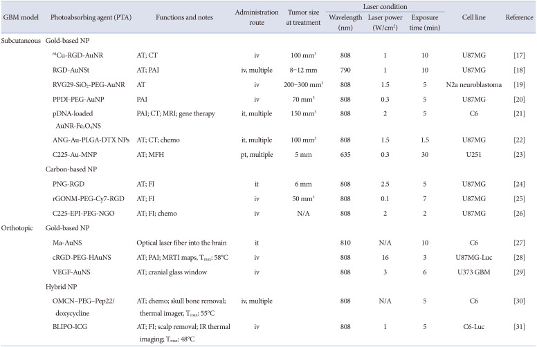
PTT and PDT are promising strategies for the treatment of GBM because they can destroy tumors as non-chemical treatments for diseases that circumvent the limitations of GBM heterogeneity, existing drug resistance mechanisms, and adverse effects on surrounding healthy tissues [16]. However, because they are hampered by the unique properties of the brain, strategies such as active targeting, multimodal imaging, or chemo/gene therapy must be developed to enhance the therapeutic efficacy of phototherapy on GBM. Therefore, to date, NPs with unique optical properties (e.g., Au-based or other semiconducting metal-based NPs, magnetic Fe3O4 NPs, carbon-based nanomaterials) or small photo-absorbable (PTA) molecules (e.g., cyanine or porphyrin derivatives) have been used for phototherapy (Table 1) [171819202122232425262728293031].
Since energy conversion and heat dissipation only occur when the frequency of the incident light overlaps the absorption band of the PTA agent, it should exhibit a strong NIR extinction coefficient. In fact, the NIR region corresponds to the transparent window of biological tissues, and light in this wavelength range (approximately 650–900 nm) can pass through soft tissues with minimal absorption, avoiding energy loss prior to irradiation with PTA preparations. In addition, NPs for phototherapy must be non-toxic without laser exposure and must have physicochemical properties (e.g., size, shape, zeta potential, and stability) to be administered systemically and to accumulate efficiently in the tumor, either passively or actively.
Given the complexity of GBM in most of the studies described above, targeted therapies have been developed that actively target tumor cells to bypass the BBB and achieve maximal NP tumor uptake. These strategies rely on grafting targeting ligands such as proteins, antibodies, and peptides on the NPs surface to selectively recognize overexpressed receptors in a specific cell population. Targeted drug delivery can bypass the blood-brain tumor barrier, the BBB, or directly access the tumor by specifically targeting brain capillary endothelial cells, tumor neovascular endothelial cells, and/or brain tumor cells. The most common targeting moieties for GBM are the transferrin (Tf), arginine-glycine-aspartic acid sequence (RGD), folic acid (FA), interleukin-13 (IL-13) peptides, and angiopep-2 peptide (ANG). Otherwise, local delivery strategies such as direct intracranial administration or convective-enhanced delivery can be used to bypass the BBB and enhance intracerebral NP concentrations by local delivery to the brain.
However, for phototherapy to treat brain tumors in clinical practice, three typical issues need to be resolved: 1) accumulation of PTA into tumors is restricted by the BBB, 2) monitoring the temperature rise and ROS generation inside the brain is not a simple task, it is essential to obtain safe and efficient photo-treatment, 3) local photoreactive responses, including heat dissipation and ROS generation, should be sufficient to achieve tumor cell death even when tumor localization is deep in the brain. How to develop and test systems to overcome these barriers is being continuously researched to achieve a clear vision for advancements in this field. The systems reported to date have not led to complete eradication of GBM due to the large number of invasive cells penetrating the peritumoral region and non-cancerous subpopulations residing within the tumor mass. However, we expect that selective targeting and efficient phototherapy can lead to partial resection of tumors. Moreover, the combination therapy such as PDT and PTT could potentially lead to enhanced anti-tumor consequences.

AuNPs FOR COMBINATIONAL PTT AND PDT IN GBM TREATMENT

Rapid growth of nanotechnology is an emerging tendency in cancer treatment. AuNPs have aroused interest in this field due to their unparalleled light-to-heat energy conversion and their capability to load and deliver numerous of PDT drugs to target site. Therefore, photothermal (PTT) and photodynamic (PDT) combined cancer treatment is possible by the role of AuNPs itself as a heat agonist and a drug carrier for PS transport. In the presence of AuNPs and PS drugs under photoactivating light wavelength, cytotoxic heat dissipation and ROS induces apoptotic and necrotic cancer cell death [32]. In terms of PTT, the collective oscillation of electrons on the AuNP surface causes surface plasmon resonance (SPR) in light of specific wavelength to generate a strong extinction coefficient, thereby dissipate sufficient heat [33]. In our study, the proximate conjugation of PS on AuNPs accelerated its PDT outcome [34]. The phenomenon that enhances the ability of PS to generate ROS was denoted as metal-enhanced ROS generation (MERos) and it is mediated by intersystem crossover between PS and AuNP. Therefore, heat generation from AuNPs by external light irradiation was sufficient, and the ROS generating ability of PS drugs was greatly increased by the role of nanoantennas of AuNPs, thereby the combination therapy of PTT and PDT was promising for GBM treatment.
A number of recent studies demonstrated nanotherapeutics that combine NIR wavelength-responsive PTA, PS drug achieve synergistic PTT, and PDT effects in tumor therapy [35363738]. Nonetheless, additional research is required to improve treatment protocols by regulating ROS generating capacity, laser power density, light irradiation dose, and improved tumor targeting effect to establish clinical feasibility.

ORAL SYSTEMIC ADMINISTRATION FOR THE TREATMENT OF PTT/PDT IN GBM

Oral drug administration is the preferred mode of drug delivery, primarily because of its simplicity. For daily medications, most patients prefer pills over injections. This arises from certain factors close related to patient compliance, including non-invasive and self-administering medications. In this regard, the large surface area (300–400 m2) of the gastrointestinal tract (GI) lined with a viscous mucosa provides an opportunity for long-term residence of the drug and promotes subsequent absorption [39]. In addition, endocrine cells, microfold cells (M cells), mucous secretary goblet cells, and Paneth cells that make up the GI tract may be promising targets for enhancing oral absorption of drugs [40]. However, the problems faced by oral drug delivery, such as low bioavailability and insufficient drug efficacy, remain unresolved. To this end, novel drug formulations have been developed that utilize NP drug delivery systems to overcome their respective limitations [41].
Nanopharmaceuticals being studied in oral drug delivery systems to date contain NPs (particles <100 nm) that are readily absorbed through the intestine and flow into the bloodstream. The typical oral drug delivery NPs include liposomes [424344], polymeric micelles [4546], drug-conjugated polymers [47], and noble metal NPs [48]. Orally delivered NPs are prepared to target specific region of the GI tract, such as duodenum, ileum, and jejunum, or to passively permeate NPs through the spaces between tight junctions at the epithelial barrier [49505152]. Nanopharmaceuticals have many advantages in oral drug delivery. They represent a large surface area, allowing them to interact with the GI tract and can be altered in a variety methods to overcome barriers in oral delivery. Through modifications in size, shape, and surface chemistry, the nanoscale drug delivery carriers described above have improved hydrophobicity, hydrophilicity, and oral bioavailability of biological agents, which can significantly affect cell transport [53].

CONCLUSION

In our study, AuNPs capable of dissipating high heat under light irradiation by SPR effect were used as PTT formulations. Furthermore, chlorin e6 (Ce6), a type of PDT PS, was bonded to the surface of AuNPs through disulfide bonds. As a result, AuNPs acted as nanoantennas, enhancing the ability of Ce6 to generate ROS. Moreover, AuNPs resolved the low water solubility of Ce6 as a carrier and achieved better tumor accumulation. The aim of this study was to develop a GBM-targeted PTT/PDT through oral administration. Therefore, lactoferrin (Lf) was conjugated to the AuNPs surface to improve the low bioavailability and GBM targeting of AuNPs. Lactoferrin is generally delivered orally and is known to be absorbed by interaction with the lactoferrin receptor (LfR) expressed in intestinal endothelial cells, BBB, and GBM. Receptor-mediated endocytosis occurred by utilizing lactoferrin as a ligand to enhance oral absorption in the GI tract and targeting GBM. Additionally, the nanoscale (10–20 nm) of our AuNP-based platforme allowed passive transport into the BBB by the enhanced permeability and retention effect, which are commonly caused by abnormal vascularization and tumor pressure in the GBM microenvironment. As a result, the developed PTT/PDT formulation enabled effective GBM anticancer treatment by irradiating NIR wavelength to generate high-efficiency heat and ROS. Furthermore, oral absorption and GBM targeting were enhanced by lactoferrin. Therefore, our study demonstrates great significance in that it developed an eating-type photothermal/photodynamic brain tumor treatment technology platform and opened up the possibility of application to the development of brain tumor therapeutics by loading other anti cancer drugs in the future.

Notes

Ethics Statement: Not applicable

Author Contributions:

  • Conceptualization: Hyung Shik Kim, Dong Yun Lee.

  • Data curation: Hyung Shik Kim, Dong Yun Lee.

  • Formal analysis: Hyung Shik Kim, Dong Yun Lee.

  • Investigation: Hyung Shik Kim, Dong Yun Lee.

  • Methodology: Hyung Shik Kim, Dong Yun Lee.

  • Project administration: Dong Yun Lee.

  • Supervision: Dong Yun Lee.

  • Visualization: Hyung Shik Kim, Dong Yun Lee.

  • Writing—original draft: Hyung Shik Kim.

  • Writing—review & editing: Dong Yun Lee.

Conflicts of Interest: The authors have no potential conflicts of interest to disclose.

Funding Statement: This work was supported by grant NRF-2020R1A2C3005834, NRF-2022R1A4A1030421, and NRF-2022R1A6A3A01085835.

Availability of Data and Material

Data sharing not applicable to this article as no datasets were generated or analyzed during the study.

References

1. Bleeker FE, Molenaar RJ, Leenstra S. Recent advances in the molecular understanding of glioblastoma. J Neurooncol. 2012; 108:11–27. PMID: 22270850.
crossref
2. Stupp R, Hegi ME, Mason WP, van den Bent MJ, Taphoorn MJ, Janzer RC, et al. Effects of radiotherapy with concomitant and adjuvant temozolomide versus radiotherapy alone on survival in glioblastoma in a randomised phase III study: 5-year analysis of the EORTC-NCIC trial. Lancet Oncol. 2009; 10:459–466. PMID: 19269895.
crossref
3. Krex D, Klink B, Hartmann C, von Deimling A, Pietsch T, Simon M, et al. Long-term survival with glioblastoma multiforme. Brain. 2007; 130(Pt 10):2596–2606. PMID: 17785346.
crossref
4. Burzynski S, Burzynski G. Long-term progression-free survival of recurrent glioblastoma multiforme treated with a combination of targeted agents: a case report. Neuro Oncol. 2014; 16(Suppl 5):v11.
5. Yong RL, Lonser RR. Surgery for glioblastoma multiforme: striking a balance. World Neurosurg. 2011; 76:528–530. PMID: 22251498.
crossref
6. Bergmann N, Delbridge C, Gempt J, Feuchtinger A, Walch A, Schirmer L, et al. The intratumoral heterogeneity reflects the intertumoral subtypes of glioblastoma multiforme: a regional immunohistochemistry analysis. Front Oncol. 2020; 10:494. PMID: 32391260.
crossref
7. Denicolaï E, Tabouret E, Colin C, Metellus P, Nanni I, Boucard C, et al. Molecular heterogeneity of glioblastomas: does location matter? Oncotarget. 2016; 7:902–913. PMID: 26637806.
crossref
8. Becker AP, Sells BE, Haque SJ, Chakravarti A. Tumor heterogeneity in glioblastomas: from light microscopy to molecular pathology. Cancers (Basel). 2021; 13:761. PMID: 33673104.
crossref
9. Bastiancich C, Da Silva A, Estève MA. Photothermal therapy for the treatment of glioblastoma: potential and preclinical challenges. Front Oncol. 2021; 10:610356. PMID: 33520720.
crossref
10. Doughty ACV, Hoover AR, Layton E, Murray CK, Howard EW, Chen WR. Nanomaterial applications in photothermal therapy for cancer. Materials (Basel). 2019; 12:779.
crossref
11. Liu Y, Bhattarai P, Dai Z, Chen X. Photothermal therapy and photoacoustic imaging via nanotheranostics in fighting cancer. Chem Soc Rev. 2019; 48:2053–2108. PMID: 30259015.
crossref
12. Chu KF, Dupuy DE. Thermal ablation of tumours: biological mechanisms and advances in therapy. Nat Rev Cancer. 2014; 14:199–208. PMID: 24561446.
crossref
13. Hirschberg H, Berg K, Peng Q. Photodynamic therapy mediated immune therapy of brain tumors. Neuroimmunol Neuroinflamm. 2018; 5:27. PMID: 30221185.
crossref
14. Kaneko S, Fujimoto S, Yamaguchi H, Yamauchi T, Yoshimoto T, Tokuda K. Photodynamic therapy of malignant gliomas. Prog Neurol Surg. 2018; 32:1–13. PMID: 29990969.
crossref
15. Akimoto J, Haraoka J, Aizawa K. Preliminary clinical report on safety and efficacy of photodynamic therapy using talaporfin sodium for malignant gliomas. Photodiagnosis Photodyn Ther. 2012; 9:91–99. PMID: 22594978.
crossref
16. Hussein EA, Zagho MM, Nasrallah GK, Elzatahry AA. Recent advances in functional nanostructures as cancer photothermal therapy. Int J Nanomedicine. 2018; 13:2897–2906. PMID: 29844672.
crossref
17. Sun X, Huang X, Yan X, Wang Y, Guo J, Jacobson O, et al. Chelator-free (64)Cu-integrated gold nanomaterials for positron emission tomography imaging guided photothermal cancer therapy. ACS Nano. 2014; 8:8438–8446. PMID: 25019252.
crossref
18. Nie L, Wang S, Wang X, Rong P, Ma Y, Liu G, et al. In vivo volumetric photoacoustic molecular angiography and therapeutic monitoring with targeted plasmonic nanostars. Small. 2014; 10:1585–1593. PMID: 24150920.
crossref
19. Lee C, Hwang HS, Lee S, Kim B, Kim JO, Oh KT, et al. Rabies virus-inspired silica-coated gold nanorods as a photothermal therapeutic platform for treating brain tumors. Adv Mater. 2017; 29:1605563.
crossref
20. Yang Z, Song J, Dai Y, Chen J, Wang F, Lin L, et al. Self-assembly of semiconducting-plasmonic gold nanoparticles with enhanced optical property for photoacoustic imaging and photothermal therapy. Theranostics. 2017; 7:2177–2185. PMID: 28740543.
crossref
21. Hu Y, Zhou Y, Zhao N, Liu F, Xu FJ. Multifunctional pDNA-conjugated polycationic Au nanorod-coated Fe3O4 hierarchical nanocomposites for trimodal imaging and combined photothermal/gene therapy. Small. 2016; 12:2459–2468. PMID: 26996155.
crossref
22. Wang L, Hu Y, Hao Y, Li L, Zheng C, Zhao H, et al. Tumor-targeting core-shell structured nanoparticles for drug procedural controlled release and cancer sonodynamic combined therapy. J Control Release. 2018; 286:74–84. PMID: 30026078.
crossref
23. Lu Q, Dai X, Zhang P, Tan X, Zhong Y, Yao C, et al. Fe3O4@Au composite magnetic nanoparticles modified with cetuximab for targeted magneto-photothermal therapy of glioma cells. Int J Nanomedicine. 2018; 13:2491–2505. PMID: 29719396.
24. Su S, Wang J, Vargas E, Wei J, Martínez-Zaguilán R, Sennoune SR, et al. Porphyrin immobilized nanographene oxide for enhanced and targeted photothermal therapy of brain cancer. ACS Biomater Sci Eng. 2016; 2:1357–1366. PMID: 33434989.
crossref
25. Akhavan O, Ghaderi E. Graphene nanomesh promises extremely efficient in vivo photothermal therapy. Small. 2013; 9:3593–3601. PMID: 23625739.
crossref
26. Yang HW, Lu YJ, Lin KJ, Hsu SC, Huang CY, She SH, et al. EGRF conjugated PEGylated nanographene oxide for targeted chemotherapy and photothermal therapy. Biomaterials. 2013; 34:7204–7214. PMID: 23800742.
crossref
27. Madsen SJ, Christie C, Hong SJ, Trinidad A, Peng Q, Uzal FA, et al. Nanoparticle-loaded macrophage-mediated photothermal therapy: potential for glioma treatment. Lasers Med Sci. 2015; 30:1357–1365. PMID: 25794592.
crossref
28. Lu W, Melancon MP, Xiong C, Huang Q, Elliott A, Song S, et al. Effects of photoacoustic imaging and photothermal ablation therapy mediated by targeted hollow gold nanospheres in an orthotopic mouse xenograft model of glioma. Cancer Res. 2011; 71:6116–6121. PMID: 21856744.
crossref
29. Day ES, Zhang L, Thompson PA, Zawaski JA, Kaffes CC, Gaber MW, et al. Vascular-targeted photothermal therapy of an orthotopic murine glioma model. Nanomedicine (Lond). 2012; 7:1133–1148. PMID: 22583571.
crossref
30. Qian W, Qian M, Wang Y, Huang J, Chen J, Ni L, et al. Combination glioma therapy mediated by a dual-targeted delivery system constructed using OMCN-PEG-Pep22/DOX. Small. 2018; 14:e1801905. PMID: 30346089.
31. Jia Y, Wang X, Hu D, Wang P, Liu Q, Zhang X, et al. Correction to phototheranostics: active targeting of orthotopic glioma using biomimetic proteolipid nanoparticles. ACS Nano. 2021; 15:10733. PMID: 34106690.
crossref
32. Riley RS, Day ES. Gold nanoparticle-mediated photothermal therapy: applications and opportunities for multimodal cancer treatment. Wiley Interdiscip Rev Nanomed Nanobiotechnol. 2017; 9:e1449.
crossref
33. Kennedy LC, Bickford LR, Lewinski NA, Coughlin AJ, Hu Y, Day ES, et al. A new era for cancer treatment: gold-nanoparticle-mediated thermal therapies. Small. 2011; 7:169–183. PMID: 21213377.
crossref
34. Kim HS, Seo M, Park TE, Lee DY. A novel therapeutic strategy of multimodal nanoconjugates for state-of-the-art brain tumor phototherapy. J Nanobiotechnology. 2022; 20:14. PMID: 34983539.
crossref
35. Lin J, Wang S, Huang P, Wang Z, Chen S, Niu G, et al. Photosensitizer-loaded gold vesicles with strong plasmonic coupling effect for imaging-guided photothermal/photodynamic therapy. ACS Nano. 2013; 7:5320–5329. PMID: 23721576.
crossref
36. Jang B, Park JY, Tung CH, Kim IH, Choi Y. Gold nanorod-photosensitizer complex for near-infrared fluorescence imaging and photodynamic/photothermal therapy in vivo. ACS Nano. 2011; 5:1086–1094. PMID: 21244012.
crossref
37. Vankayala R, Lin CC, Kalluru P, Chiang CS, Hwang KC. Gold nanoshells-mediated bimodal photodynamic and photothermal cancer treatment using ultra-low doses of near infra-red light. Biomaterials. 2014; 35:5527–5538. PMID: 24731706.
crossref
38. Sun J, Guo Y, Xing R, Jiao T, Zou Q, Yan X. Synergistic in vivo photodynamic and photothermal antitumor therapy based on collagen-gold hybrid hydrogels with inclusion of photosensitive drugs. Colloids Surf A Physicochem Eng Asp. 2017; 514:155–160.
crossref
39. Ensign LM, Cone R, Hanes J. Oral drug delivery with polymeric nanoparticles: the gastrointestinal mucus barriers. Adv Drug Deliv Rev. 2012; 64:557–570. PMID: 22212900.
crossref
40. Pawar VK, Meher JG, Singh Y, Chaurasia M, Surendar Reddy B, Chourasia MK. Targeting of gastrointestinal tract for amended delivery of protein/peptide therapeutics: strategies and industrial perspectives. J Control Release. 2014; 196:168–183. PMID: 25305562.
crossref
41. Pridgen EM, Alexis F, Farokhzad OC. Polymeric nanoparticle technologies for oral drug delivery. Clin Gastroenterol Hepatol. 2014; 12:1605–1610. PMID: 24981782.
crossref
42. Tang WL, Tang WH, Chen WC, Diako C, Ross CF, Li SD. Development of a rapidly dissolvable oral pediatric formulation for mefloquine using liposomes. Mol Pharm. 2017; 14:1969–1979. PMID: 28460165.
crossref
43. Song Z, Lin Y, Zhang X, Feng C, Lu Y, Gao Y, et al. Cyclic RGD peptide-modified liposomal drug delivery system for targeted oral apatinib administration: enhanced cellular uptake and improved therapeutic effects. Int J Nanomedicine. 2017; 12:1941–1958. PMID: 28331317.
crossref
44. Liu Y, Luo X, Xu X, Gao N, Liu X. Preparation, characterization and in vivo pharmacokinetic study of PVP-modified oleanolic acid liposomes. Int J Pharm. 2017; 517:1–7. PMID: 27899320.
crossref
45. Sze LP, Li HY, Lai KLA, Chow SF, Li Q, KennethTo KW, et al. Oral delivery of paclitaxel by polymeric micelles: a comparison of different block length on uptake, permeability and oral bioavailability. Colloids Surf B Biointerfaces. 2019; 184:110554. PMID: 31627103.
crossref
46. Jeong Y, Lee D, Choe K, Ahn H, Kim P, Park JH, et al. Polypeptide-based polyelectrolyte complexes overcoming the biological barriers of oral insulin delivery. J Ind Eng Chem. 2017; 48:79–87.
crossref
47. Lee E, Lee J, Lee IH, Yu M, Kim H, Chae SY, et al. Conjugated chitosan as a novel platform for oral delivery of paclitaxel. J Med Chem. 2008; 51:6442–6449. PMID: 18826299.
crossref
48. Kebede A, Singh AK, Rai PK, Giri NK, Rai AK, Watal G, et al. Controlled synthesis, characterization, and application of iron oxide nanoparticles for oral delivery of insulin. Lasers Med Sci. 2013; 28:579–587. PMID: 22581389.
crossref
49. Kong FY, Zhang JW, Li RF, Wang ZX, Wang WJ, Wang W. Unique roles of gold nanoparticles in drug delivery, targeting and imaging applications. Molecules. 2017; 22:1445.
crossref
50. Sung HW, Sonaje K, Liao ZX, Hsu LW, Chuang EY. pH-responsive nanoparticles shelled with chitosan for oral delivery of insulin: from mechanism to therapeutic applications. Acc Chem Res. 2012; 45:619–629. PMID: 22236133.
crossref
51. des Rieux A, Fievez V, Garinot M, Schneider YJ, Préat V. Nanoparticles as potential oral delivery systems of proteins and vaccines: a mechanistic approach. J Control Release. 2006; 116:1–27. PMID: 17050027.
crossref
52. Yun Y, Cho YW, Park K. Nanoparticles for oral delivery: targeted nanoparticles with peptidic ligands for oral protein delivery. Adv Drug Deliv Rev. 2013; 65:822–832. PMID: 23123292.
crossref
53. Banerjee A, Qi J, Gogoi R, Wong J, Mitragotri S. Role of nanoparticle size, shape and surface chemistry in oral drug delivery. J Control Release. 2016; 238:176–185. PMID: 27480450.
crossref
Table 1

List of in vivo preclinical phototherapy efficacy studies

btrt-10-215-i001
GBM model Photoabsorbing agent (PTA) Functions and notes Administration route Tumor size at treatment Laser condition Cell line Reference
Wavelength (nm) Laser power (W/cm2) Exposure time (min)
Subcutaneous Gold-based NP
64Cu-RGD-AuNR AT; CT iv 100 mm3 808 1 10 U87MG [17]
RGD-AuNSt AT; PAI iv, multiple 8–12 mm 790 1 10 U87MG [18]
RVG29-SiO2-PEG-AuNR AT iv 200–300 mm3 808 1.5 5 N2a neuroblastoma [19]
PPDI-PEG-AuNP PAI iv 70 mm3 808 0.3 5 U87MG [20]
pDNA-loaded AuNR-Fe3O4NS PAI; CT; MRI; gene therapy it, multiple 150 mm3 808 2 5 C6 [21]
ANG-Au-PLGA-DTX NPs AT; CT; chemo it, multiple 100 mm3 808 1.5 1.5 U87MG [22]
C225-Au-MNP AT; MFH pt, multiple 5 mm 635 0.3 30 U251 [23]
Carbon-based NP
PNG-RGD AT; FI it 6 mm 808 2.5 5 U87MG [24]
rGONM-PEG-Cy7-RGD AT; FI iv 50 mm3 808 0.1 7 U87MG [25]
C225-EPI-PEG-NGO AT; FI; chemo iv N/A 808 2 2 U87MG [26]
Orthotopic Gold-based NP
Ma-AuNS Optical laser fiber into the brain it 810 N/A 10 C6 [27]
cRGD-PEG-HAuNS AT; PAI; MRTI maps, Tmax: 58℃ iv 808 16 3 U87MG-Luc [28]
VEGF-AuNS AT; cranial glass window iv 808 3 6 U373 GBM [29]
Hybrid NP
OMCN–PEG–Pep22/doxycycline AT; chemo; skull bone removal; thermal imager, Tmax: 55℃ iv, multiple 808 N/A 5 C6 [30]
BLIPO-ICG AT; FI; scalp removal; IR thermal imaging; Tmax: 48℃ iv 808 1 5 C6-Luc [31]

GBM, glioblastoma multiforme; PTA, photothermal agent; N/A, not applicable; AT, active targeting; chemo, chemotherapy; FI, fluorescence imaging; CT, X-ray computed tomography; MRI, magnetic resonance imaging; MFH, magnetic fluid hyperthermia; PAI, photoacoustic imaging; iv, intravenous; it, intratumoral; pt, peritumoral; PEG, polyethylene glycol;NP, nanoparticles; AuNR, gold nanorods; RGD, arginine–glycine–aspartic acid peptide; AuNS, Au@SiO2 nanoshells; AuNSt, gold nanostars; RVG29, 29-residue peptide rabies virus glycoprotein; PPDI, poly(perylene diimide); Cy7, cyanine 7; PNG, porphyrin immobilized nanographene oxide; pDNA, plasmid DNA; Fe3O4NS, Fe3O4 nanospheres; PLGA, poly(lactide-coglycolide); ANG, angiopep-2 peptide; Au-MNP, core-shell Fe3O4@Au magnetic nanoparticles; MNP, Fe@Fe3O4 magnetic nanoparticles; DTX, docetaxel

TOOLS
Similar articles