

 PDF
PDF Citation
 Print
Print


INTRODUCTION
In the era of minimally invasive surgery, more complex hepato-pancreatico-biliary (HPB) procedures are being performed robotically nowadays. Robotic surgeries have several advantages over conventional laparoscopic surgeries, such as three-dimensional vision with depth perception, magnified view, tremor filtration, and, more importantly, degrees of freedom of articulating instruments. The first robotic liver resection (minor) was reported in 2003 by Giulianotti et al. [1]. Since then, many centers have reported their experiences with robotic liver resections, including major liver resections (Couinaud’ segments four or more). However, in most of these cases, parenchymal transection was done using a Cavitron Ultrasonic Surgical Aspirator (CUSA) (Integra LifeSciences, Tullamore, Ireland), robotic compatible harmonic scalpel, or Maryland bipolar forceps (MBF) (Intuitive Surgical, Sunnyvale, CA, USA).
CUSA requires another experienced laparoscopic surgeon as an assistant, while a harmonic scalpel lacks articulation. MBF seem not optimally suited for bigger transection planes [2]. There is a paucity of literature on liver resection using an EndoWrist® One™ Vessel Sealer (Intuitive Surgical). Here we describe a case and the feasibility of liver parenchymal transection using a vessel sealer (VS) during total robotic right hepatectomy.
CASE PRESENTATION
A 70-year-old non-alcoholic male presented with a history of pain in the right upper abdomen for the last four months, which was gradually progressive and relieved with medication to recure again. He had no history of jaundice, anorexia, weight loss, abdominal distension, or fever. His liver function tests were within normal limits.
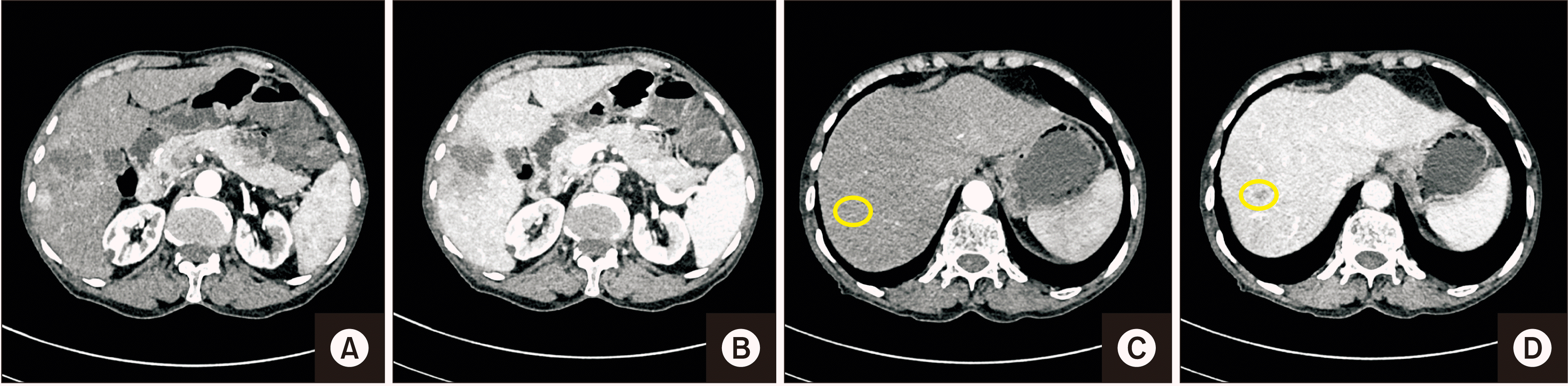
A contrast-enhanced computed tomography (CECT) showed an ill-defined heterogeneous mass lesion of size ~4.8 cm × 3.9 cm × 4.0 cm having a non-rim arterial phase hyperenhancement with washout and enhancing capsule in delayed phase in segment VII of the liver. Another smaller lesion (1 cm) with similar enhancement patterns was noted in segment VIII on a background of the non-cirrhotic liver (Fig. 1). These lesions were further confirmed with a contrast-enhanced magnetic resonance imaging. No other lesions were found. His alpha-fetoprotein level was 3.93 ng/mL. Hepatitis B surface antigen and anti-hepatitis C antibody results were non-reactive. Hence, he was planned to undergo anatomical right hemi-hepatectomy robotically.
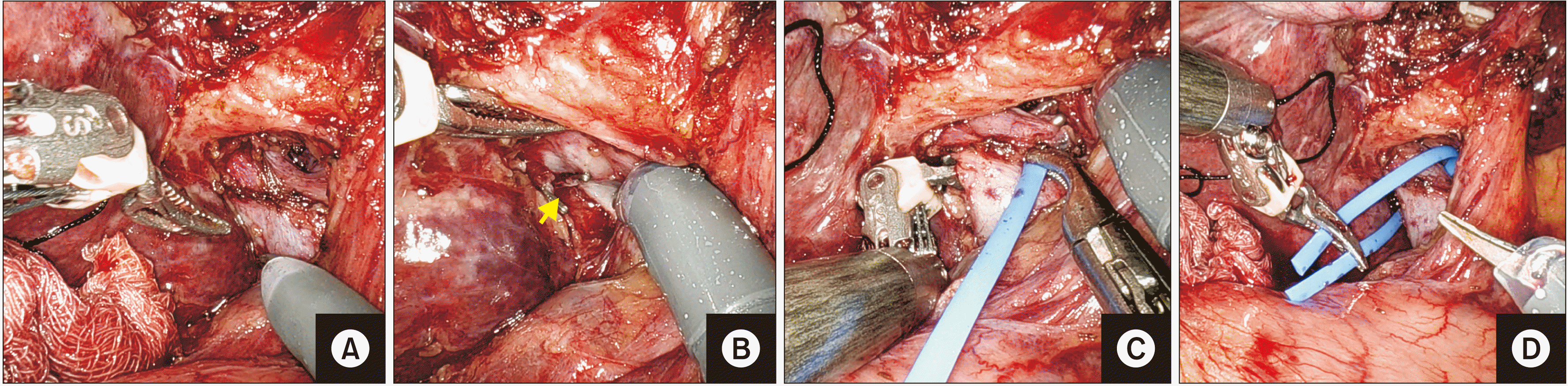
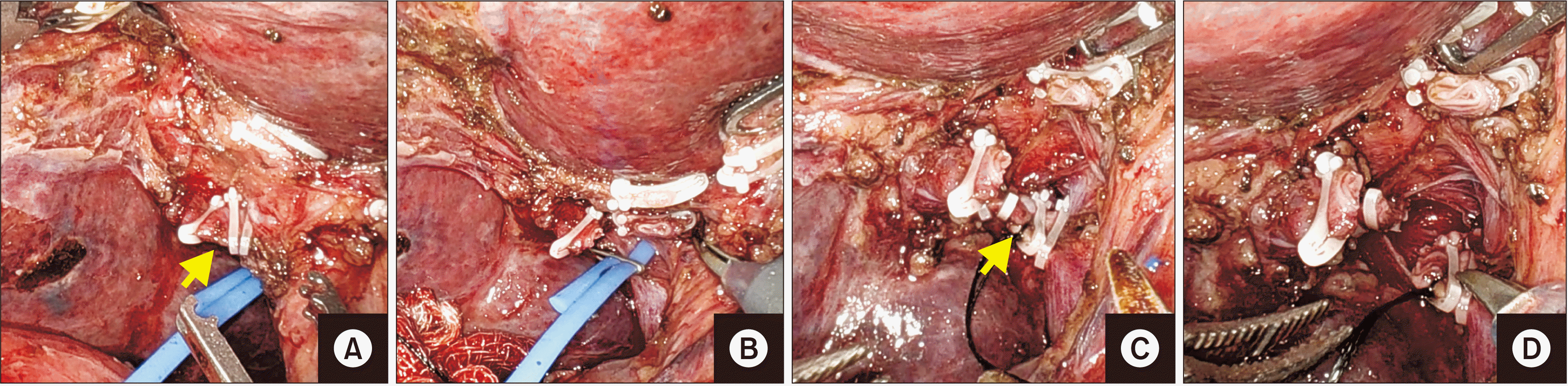
The patient was placed in a supine position with legs split. Pneumoperitoneum was created. Four robotic ports were placed in a straight line at the level of the umbilicus as follows: R1, right anterior axillary line; R2 (camera port) and R3 on either side of the umbilicus at a distance of ~7 cm; and R4 left anterior axillary line 7 cm from R3. Two assistant ports were used: A1, 12 mm infra-umbilical port and an A2, 5 mm port placed between R1 and R2 at the lower level forming a triangle (Fig. 2A–2C). A da Vinci Xi Surgical System (Intuitive Surgical) was docked from the right side. Instruments were placed as follows: MBF in arm R1, VS in arm R3, and Cadière forceps in R4.
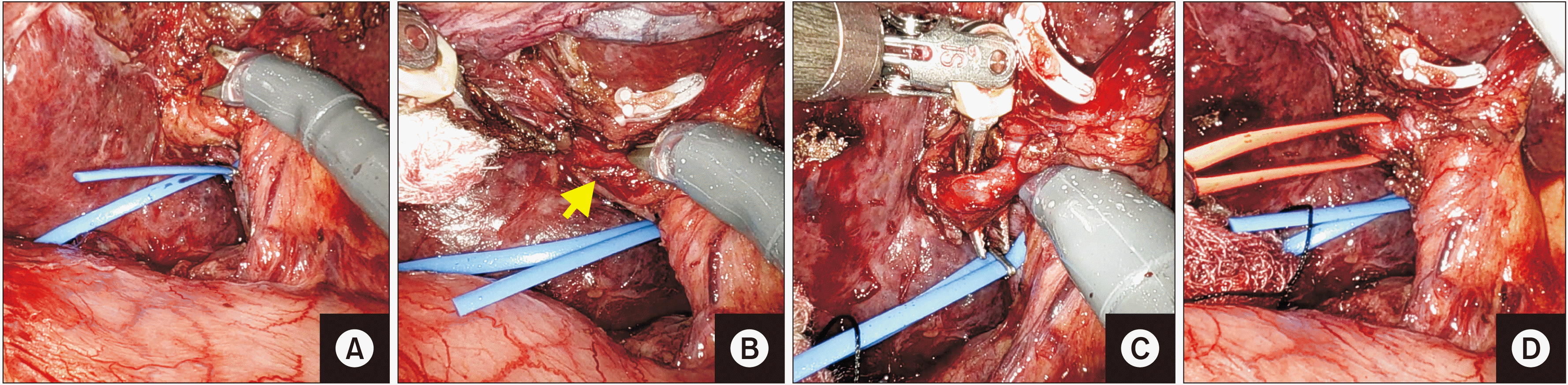
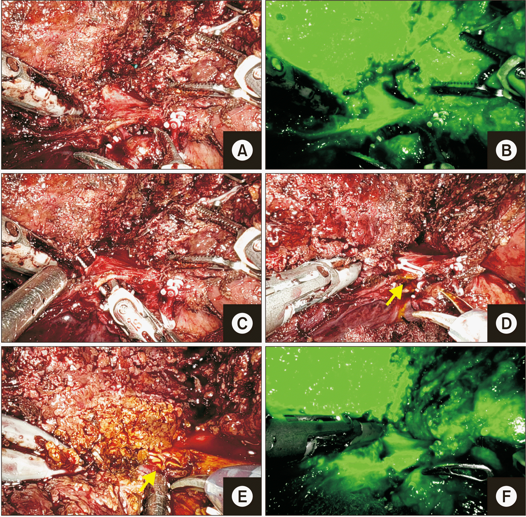
After inspecting the peritoneal cavity for occult metastases, the falciform and right triangular ligament were divided and right hepatic vein (RHV) was identified. Calot’s dissection was done. The gallbladder was dissected out. Inflow control was taken using an intrafascial approach to loop the right portal vein (RPV) and the right hepatic artery (RHA) (Fig. 3, 4). Vascular clamps (bulldog) were applied. The ischemic line was marked and reconfirmed using intravenous indocyanine green dye (Fig. 5). Inferiorly, inferior vena cava (IVC) was identified. Short hepatic veins draining into the IVC were clipped and divided. At the hilar plate, RPV and RHA were clipped and divided (Fig. 6).
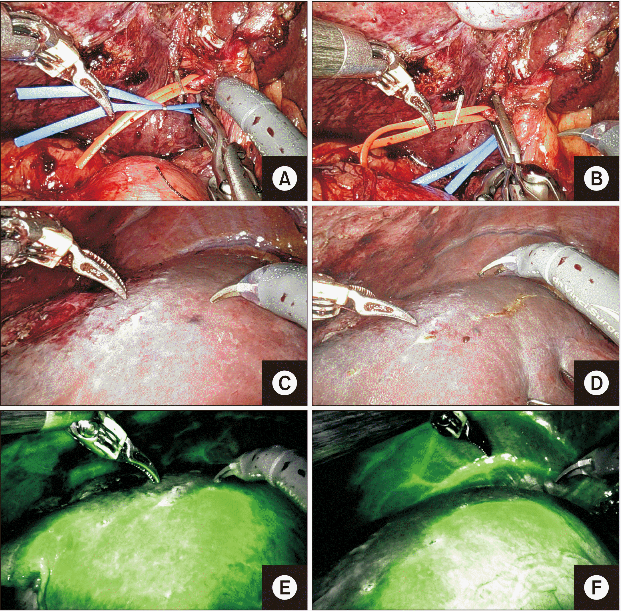
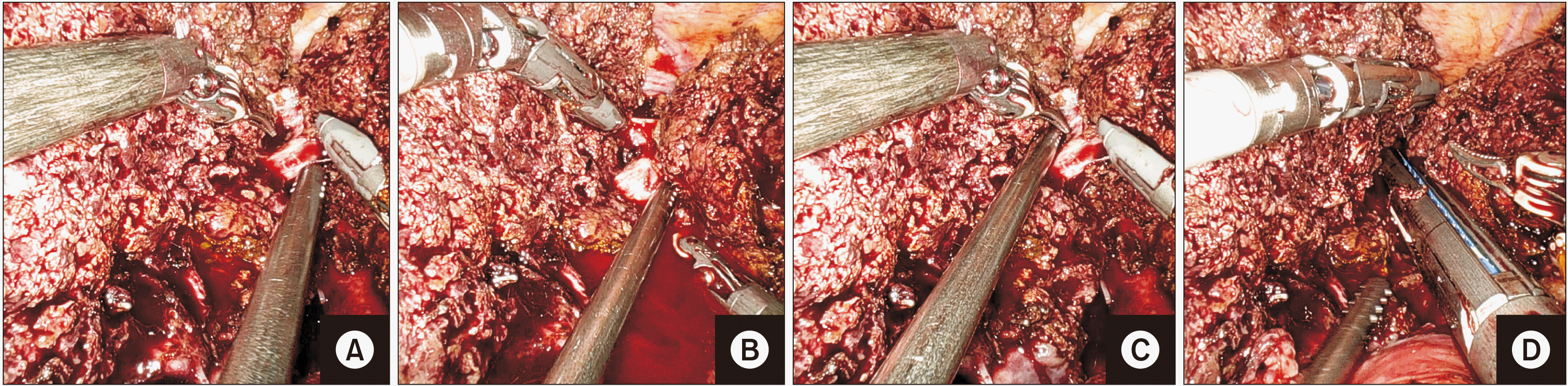
A rubber band technique was used to retract the liver. Parenchyma transection was done using a vessel sealer. MBF and VS were used for crushing the liver parenchyma. The VS was employed to seal all small vessels and glissonian pedicles. Tributaries of the middle hepatic vein from segments V and VIII were clipped and divided between Hem-o-lok clips (Teleflex Inc., Morrisville, NC, USA) (Fig. 7). After that, the right posterior and anterior sectoral ducts were divided using ICG cholangiography (firefly mode) (Fig. 8). Once all structures were divided at the hilum, further parenchymal transection was completed above the IVC in a similar manner. The specimen was left attached to RHV, which was then divided using endovascular staplers (Fig. 9).
The specimen was retrieved using a Pfannenstiel incision (Fig. 2D) and the robot was redocked. Ductal openings were closed with PDS 5-0. The hilar plate was also over-run. There was no apparent bile leak from the cut surface or hilum at the end of the procedure. Total operative time was 520 minutes with a console time of 400 minutes, including 130 minutes for parenchymal transection. Blood loss was ~400 mL without peri-operative transfusion requirement. The patient was discharged on postoperative day five without postoperative complications. Histopathology revealed two well-differentiated hepatocellular carcinomas, concurring with findings of CECT.
DISCUSSION
The utilization of robotic armamentarium is constantly expanding for HPB surgery. Advocates of robotic hepatobiliary surgery claim that depth perception, tremor filtration, improved dexterity, and wrist joint movement make it an inexpensive surgical instrument that can assist in the manipulation of complex bile ducts and blood vessels. The surgeon can sit comfortably on the console, allowing the tissue to be smoothly dissected and sutured accurately without feeling fatigued or stressed. An excellent visualization is the key to control intraoperative bleeding during mobilization and transection of the liver. Hence, a conversion to open surgery is less than a laparoscopic surgery [3].
Since the description of segmental liver resection in 2003 by Giulianotti et al. [1], the number and complexity of robotic liver resections have rapidly increased, especially in the last five years [4]. While earlier indications were limited to segmental, non-anatomical, and wedge resection, surgeons are now performing extended hemi-hepatectomies, hepatectomy for living donor liver transplantation, and segmental resection of posterosuperior segments with increasing experiences [5-8]. The International consensus statement has concluded that robotic hepatectomy is safe and feasible as a conventional open hepatectomy. It has less intra-operative blood loss, shorter hospital stay, and lower intra- and postoperative complications, although it has a longer operation time. Robotic hepatectomy also has effectiveness for liver malignancy lesions comparable to an open approach. There is no statistical difference in radical resection rate, overall survival, or recurrence rate regarding the oncological outcome between the two techniques [4]. The cost has always been the biggest challenge and limitation in robotic surgery. However, recent cost analysis studies of liver resection have suggested that higher intra-operative cost is balanced by shorter postoperative stay and lesser complications, thus reducing the overall cost [9-14].
Various techniques have been described for parenchymal transection during minimally invasive liver resections. The most common and preferred one is the CUSA, which provides precise transection of liver parenchyma, leaving only vascular and biliary structures to be clipped and divided. Apart from being costly, the drawback with CUSA in robotic liver surgery is that an assistant can only use it as it is not compatible with a da Vinci robotic surgical system. Hence, it requires an expert laparoscopic hepatobiliary surgeon to be present on the patient side. Other techniques include the use of a Harmonic scalpel and a robotic bipolar cautery. The problem with both Harmonic and CUSA is that they lack articulation as available in robotic instruments, while bipolar is not suitable for larger transection planes if not being simultaneously used with other energy devices.
An EndoWrist® One™ VS is a good alternative for parenchymal transection, although studies on its use are limited. It is a fully wristed robotic energy device optimal for the division of vessels up to 7 mm in diameter. Kingham et al. [15] have reported a few cases of robotic liver resection using VS. However, outcomes were not discussed for different transection techniques. Nota et al. [2] have used VS exclusively in their series of 70 liver resections. Among them, only 10 (14%) were major hepatectomies with a conversion rate of 7%. They reported that 14% suffered from a major complication [2]. They did not encounter any technical difficulty or instrument failure when using the VS. Thus, they have concluded that it is safe and feasible for parenchymal transection. In our experience, we have performed ten liver resections, including non-anatomical and minor resections using VS without bile leak or any other major complications. Besides liver surgery, VS has also been used in other abdominal surgeries. A retrospective analysis of 72 robotic procedures by Ortenzi et al. [16] has concluded that it is safe with decreased overall operative time. In a study by Kong et al. [17] of 17 patients of robotic gastrectomy for gastric cancer, it was concluded that the use of the VS in robotic gastrectomy was feasible, providing exemplary configuration in the direction of dissection, rapid learning curve, and comparable results between VS and conventional ultrasonic shear groups. At the same time, it can reduce inflammation with less albumin loss as possible benefits of the VS.
A VS can be proposed for dissection of tissue, bipolar coagulation, and transection of vessels and tissue bundles with a minimal lateral spread of thermal energy. Hence, there is minimal use of titanium clips during parenchymal transection. There is less dependency on an assistant surgeon for the same with a rapid learning curve. Besides, it can help dissect tissues in a few places without the need to replace the instrument, thus minimizing the time used in exchanging instruments in a robotic surgery.
The drawback of the proposed VS is that its jaws are broad and bulky. However, retraction of liver parenchyma with the help of a rubber band technique can help accommodate its jaws easily during transection and seal all pedicles. Furthermore, the modern version ‘vessel sealer extend’ or ‘Synchroseal’ has a slimmer jaw profile that will enable more delicate dissection like other robotic instruments.
In our case, we did not encounter any technical problems in using the VS. The patient was discharged with a smooth postoperative course without any complications such as bile leak. Thus, an EndoWrist® One™ VS is a safe and feasible method for parenchymal transection. However, more studies are needed to validate its role in major liver resections and living donor liver hepatectomies in the future.


 XML Download