

 PDF
PDF Citation
 Print
Print


INTRODUCTION
Due to the high metastasis and recurrence rates, lung adenocarcinoma (LUAD) is the leading cause of lung cancer (LC)-associated deaths despite comprising only 40% of the LC cases in 2020 [1]. Despite the immense efforts in LUAD detection and therapy, the overall survival of patients with LUAD is still less than five years [2]. Thus, understanding the key factors associated with LUAD malignancy might offer novel insights for exploring promising fingerprints for LUAD diagnosis and intervention.
Circular RNAs (circRNAs), a subfamily of non-coding RNAs, are distinguished by their covalently closed loop structures and resistance to RNA exonucleases [3]. The dysregulation of circRNAs plays a significant role during the onset and progression of various cancers, including LUAD [4]. For instance, circRNA-002178 upregulates the expression of programmed death-ligand 1, leading to tumor immune escape and permits LUAD development [5]. CircRNA-ENO1 enhances the expression of enolase 1 (ENO1), increasing the risk of LUAD progression [6]. Furthermore, circRNA_102231 is highly expressed in LUAD tissues and closely associated with advanced clinical outcomes [7]. However, the role of other circRNAs in LUAD remains unclear.
Reportedly, the competitive endogenous RNA (ceRNA) mechanism is favored by circRNAs in tumors [4]. The circRNAs recognize miRNAs via miRNA response elements and abrogate miRNA silencing on mRNA expression. For example, the circRNA CircZMYM4 works as a ceRNA for miR-587 to delay LUAD metastasis [8]. CircFBXW7 targets miR-942-5p and arrests LUAD cell proliferation, migration, and invasion [9]. Therefore, we need to understand the mechanism of circRNA as ceRNA activity to uncover the underlying mechanism of LUAD progression.
miRNAs are about 22 nucleotides long small non-coding transcripts. They can silence gene expression by undergoing complementary base pairing at the 3′UTR of a target mRNA [10]. miR-655 plays different roles in tumors. For example, miRNA-665 serves as a tumor suppressor in gastric [11] and ovarian cancer [12], whereas it has an oncogenic role in breast [13] and bladder cancer [14]. However, a discrepant role of miR-665 has been described in ovarian cancer [15,16]. Therefore, the function of miR-665 in tumors is cell-context-dependent. Moreover, miR-665 is a regulator in LUAD [17,18]. Therefore, it is important to investigate the regulatory mechanisms between circ_0129047 and miR-665.
The protein tyrosine phosphatase receptor type B (PTPRB) gene is present at 12q15 and comprises 37 exons. It encodes a member of the protein tyrosine phosphatase (PTP) family—a signaling molecule that regulates a variety of cellular processes, including cell growth, differentiation, mitotic cycle, and oncogenic transformation [19]. Qi et al. [20] showed that PTPRB was downregulated in patients with non-small cell lung cancer (NSCLC) and was related to patient prognosis. Its overexpression reduced proliferation and invasion. However, the effect of miR-665 on PTPRB expression in LUAD remains unclear.
In the present study, we identified a novel circRNA, circ_0129047 that was downregulated in the LUAD cells and tissues. Furthermore, we found an evident cytoplasmic abundance of circ_0129047 in LUAD cells. Therefore, apart from the biofunction of circ_0129047 in LUAD, circ_0129047-miR-665-PTPRB axis is also of interest. This study may provide a theoretical basis for circRNA application in LUAD diagnosis and therapy.
METHODS
Tissue sample collection
From March 2020 to January 2021, 45 paired tumor tissues and corresponding uncancerous tissues were obtained from LUAD patients at the Wuhan Red Cross Hospital. Written informed consent was obtained from all the patients. None of the patients had undergone preoperative radiochemotherapy. Tissues were subjected to pathological verification after surgery. This study was approved by the Institutional Ethics Committee of the Wuhan Red Cross Hospital (approval number: 2020027).
Cell cultivation
Human bronchial epithelium (BEAS-2B, cat#: CRL-9609), LUAD cells (A549, cat#: CRM-CCL-185 derived from the lung tissue of a white, 58-year-old male with LUAD; H1975, CRL-5908 derived from the lung tissue of a nonsmoking female with LUAD; and Calu-3, cat#: HTB-55, derived from the lung tissue of a white, 25-year-old male with LUAD) were obtained from the American Type Culture Collection (ATCC), and PC9 (cat#: 90071810, derived from the lung tissue of an undifferentiated patient with LUAD) was obtained from Sigma-Aldrich. Calu-3 (Eagle’s Minimum Essential medium, Thermo Fisher Scientific), PC9 and H1975 (RPMI-1640 Medium, Thermo Fisher Scientific), A549 (F-12K Medium, Thermo Fisher Scientific), and BEAS-2B (BEBM medium, Thermo Fisher Scientific) were cultivated at 37°C with 5% CO2 in medium containing 10% fetal bovine serum (Thermo Fisher Scientific) and 1% penicillin/streptomycin (Thermo Fisher Scientific).
Cell transfection
Lentivirus‐overexpressing circ_0129047 particles (OE-circ) and their negative control (OE-NC), miR-665 mimic or mimic-NC, empty plasmid (OE-NC), and pcDNA-PTPRB plasmid (OE-PTPRB) were obtained from HanBio Technology Co. Ltd. For transfection of miR-665 mimic or mimic-NC and pcDNA-PTPRB plasmid and their empty plasmid, they were introduced into 80% confluent PC9 and Calu-3 cells using the DharmaFECT1 Transfection Reagent (Horizon) according to the manufacturer’s protocol. For lentivirus transfection, virus particles produced at 10 multiplicity of infection were delivered into PC9 and Calu-3 cells. After 48 h, the cells were transferred in puromycin-containing media. The puromycin-resistant colonies were propagated after a two-week selection. Transfection efficiency was determined using reverse transcription quantitative polymerase chain reaction (RT-qPCR).
RT-qPCR assay
A MolPure Cell RNA Kit (Yeasen) was used to obtain total RNA from the LUAD tissues and cells. RNAs was reverse-transcribed into cDNA using the miRNA 1st Strand cDNA Synthesis Kit (Vazyme) or PrimeScript RT reagent Kit (Takara). The resultant cDNA was amplified and quantified using real-time PCR using a real-time PCR fluorescence quantitative kit (Solarbio) on a 7900HT real-time PCR system. The outcome was normalized to the internal interference (U6 and GDPAH) using the 2-ΔΔCt method [21]. The primers used are listed in Table 1.
Table 1
Real-time PCR primer synthesis list
![]()
Cell nuclear/cytoplasmic fraction isolation
We used a cytoplasmic and nuclear RNA purification kit obtained from Norgen Biotek Corp. USA to prepare the nuclear and cytoplasmic parts of PC9 and Calu-3 cells according to the manufacturer’s instructions. Circ_0129047 in the nuclear and cytoplasmic parts of the cells was quantified using RT-qPCR.
RNase treatment
Total RNA (2 µg) from PC9 and Calu-3 cells was digested with or without RNase for 30 min at 37°C. RT-qPCR was performed to determine the expression of circ_0129047 in PC9 and Calu-3 cells exposed to RNase.
Western blot
Cells were homogenized in radioimmunoprecipitation assay (RIPA) lysis buffer. After centrifugation, the supernatant was quantified with the bicinchoninic acid colorimetric (BCA) method. Next, 30 µg of protein was separated on a 10% SDS-PAGE gel and transferred to PVDF membranes in a pre-assembled transfer sandwich process. After blocking with 5% non-fat blocking grade milk at room temperature for 1 h, the membranes were blotted overnight at 4°C with antibodies against Bax (cat#: PA5-39778, 1:1,000, Thermo Fisher Scientific), Bcl-2 (cat#: 13-8800, 1:1,000, Thermo Fisher Scientific), PTPRB (cat#: PA5-119373, 1:1,000, Thermo Fisher Scientific). Anti-rabbit secondary antibody (cat#: sc-3836, 1:5,000, Santa Cruz Biotechnology) and anti-mouse secondary antibodies (cat#: W4021, 1:10,000; Promega) were used to identify the target proteins on the membranes 24 h later. ECL Plus reagents were used to detect the immunoreactive bands (Amersham).
Cell Counting Kit-8 (CCK-8) assay
PC9 and Calu-3 cells (4 × 10⁴ cells/well) were seeded in 96-well plates. After 24, 48, and 72 h of cultivation, the cells were exposed to 10 µl of CCK-8 reagent (Thermo Fisher Scientific). After 3 h, the optical density (OD) at 450 nm was measured for each well using a microplate reader (BioTek).
Colony formation assay
PC9 and Calu-3 cells were plated in 6-well plates at a density of 500 cells/well. After two weeks of cultivation, the colonies were counted and photographed using a microscope (Olympus) after undergoing fixation with 100% methanol (20 min) and staining with 25% crystal violet (30 min). Colonies containing > 50 cells were counted.
In vivo assay
The Wuhan University Center for Animal Experiment/Animal Biosafety Level III laboratory (ABSL-III lab) provided ten female BALB/C nude mice (4–5 weeks old, 20–25 g). The mice were raised under standard culture conditions for a week for acclimatization. For Xenograft tumorigenicity assays, PC9 cells (4 × 10⁴ cells) infected with lentiviral particles harboring circ_0129047-overexpressing vectors or empty vectors were administered subcutaneously into the lateral tail veins of female BALB/C nude mice. Tumor size was monitored every four days. The mice were euthanized by CO2 inhalation for 28 days, and the final tumor weight was recorded. Tumor volume (mm3) = length (mm) × width2 (mm2) / 2. Our hospital’s Institutional Animal Care and Ethics Committee approved this experiment (approval number: 20210814).
Luciferase reporter assay
To create circ_0129047-wild type (WT) and PTPRB 3′UTR-WT plasmids, the sequences of circ_0129047 and PTPRB 3′UTR binding to miR-665 were ligated into the pmirGLO dual-luciferase miRNA Target Expression Vector to produce WT luciferase reporter vectors. The corresponding mutant (MUT) vectors of circ_0129047 and PTPRB 3′UTR without the binding sites for miR-665 were established using site-directed mutagenesis using the QuikChange Site-Directed Mutagenesis Kit (Invitrogen). The developed WT and MUT luciferase vectors were co-transfected with the miR-665 mimic or NC mimics into PC9 and Calu-3 cells. After 48 h, luciferase density was determined using the Luciferase Reporter System (Promega).
RNA immunoprecipitation (RIP) assay
The RNA-protein pulldown kit (Thermo Fisher Scientific) was used to detect the association between circ_0129047 and miR-665. Briefly, cell lysates were prepared using Thermo Scientific Pierce IP Lysis Buffer and then incubated with magnetic beads conjugated with Argonaute 2 antibody or IgG antibody. Finally, the pulled RNA-Binding Protein Complexes were washed and eluted. RT-qPCR was conducted to determine circ_012904 and miR-665 expression levels.
Statistical analysis
GraphPad Prism 9.0 was utilized (GraphPad Software Inc.) for statistical analyses. Student’s t-test was used to determine statistical significance between two groups, and one-way ANOVA with Tukey’s post-test was used to determine statistical significance between two groups. The relationship between miR-665 expression and circ_0129047 or PTPRB expression was investigated using Pearson analysis. p < 0.05 was considered statistically significant.
RESULTS
Human circular RNA-0129047 (has_circ_0129047) expression was downregulated in LUAD
First, to evaluate the expression of circ_0129047 in LUAD, we measured circ_0129047 levels in LUAD clinical samples and cells. Simultaneous downregulation of circ_0129047 was detected in LUAD tissues (Fig. 1A) compared with that in the normal tissues. Similarly, circ_0129047 expression was also downregulated in LUAD cells (A549, H1975, PC9, and Calu-3) compared with human bronchial epithelium cells BEAS-2B (Fig. 1B). PC9 and Calu-3 cells presented greater downregulation of circ_0129047. Hence, we chose them for subsequent assays. The annular structure of circ_0129047 is shown in Fig. 1C. RT-qPCR analysis was performed to examine the subcellular localization of circ_0129047 and to fully understand its function. The results showed the predominance of cytoplasmic circ_0129047 in PC9 and Calu-3 cells (Fig. 1D). LIF receptor subunit alpha (LIFR) is a linear transcript of circ-0129047. circRNA is resistant to digestion by the 3′–5′ exoribonuclease, RNase R, while linear mRNA is sensitive to RNase R. Therefore, we performed an RNase R treatment assay to identify the circular feature of circ_0129047. The results showed that LIFR was downregulated after RNase R treatment, but RNase R did not affect circ_0129047, further validating the circular feature of circ_0129047 in PC9 and Calu-3 cells (Fig. 1E). These data suggested that circ_0129047 is downregulated and may be involved in LUAD malignancy.
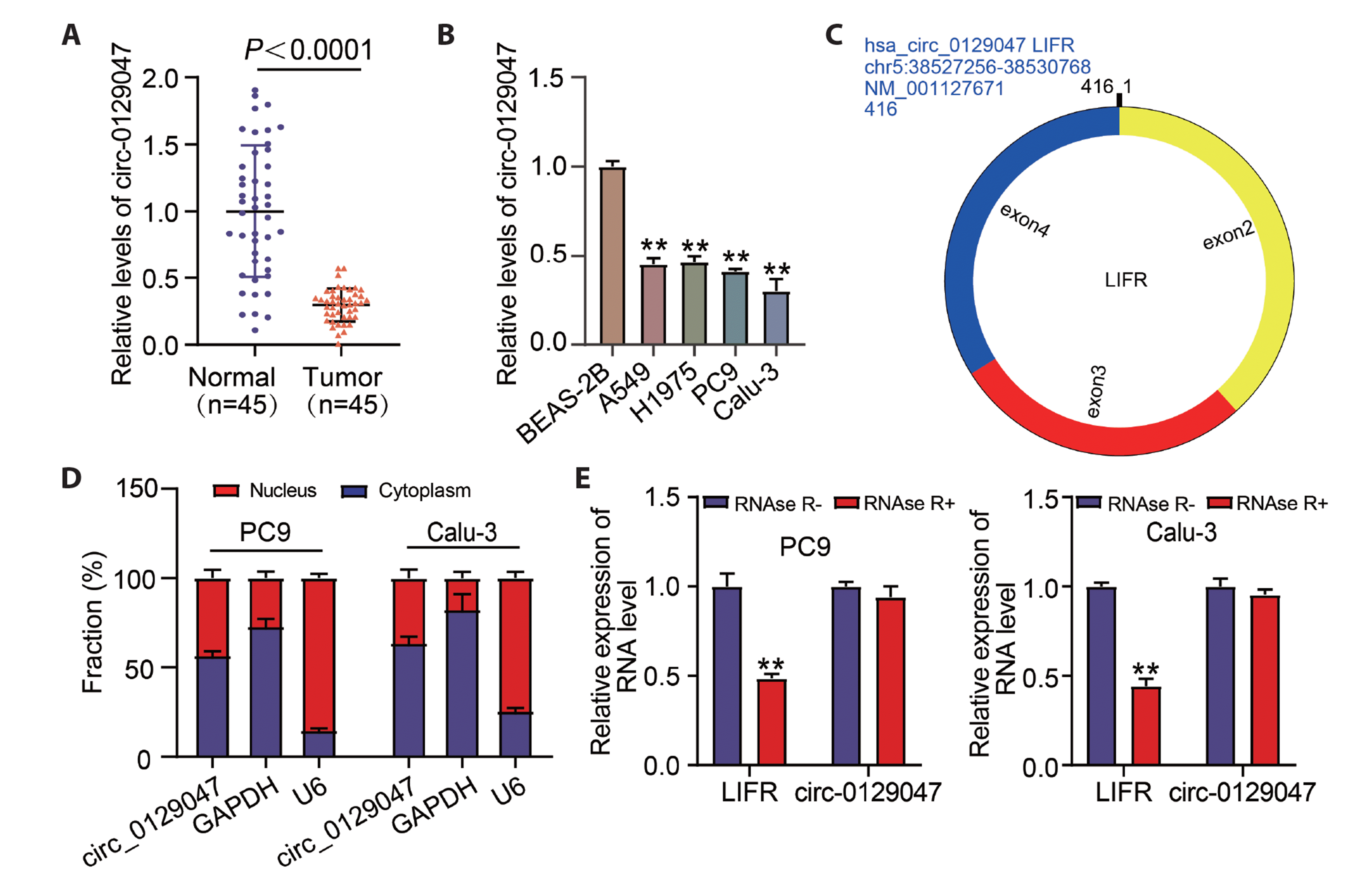
Fig. 1
Hsa_circ_0129047 expression was downregulated in lung adenocarcinoma (LUAD).
(A) RT-qPCR analysis results of circ_0129047 in both LUAD and normal tissues. (B) RT-qPCR analysis of circ_0129047 in human bronchial epithelium (BEAS-2B) and LUAD cells (A549, H1975, PC9 and Calu-3). **p < 0.01 vs. BEAS-2B. (C) Loop structures of Circ_012904. (D) Subcellular localization of circ_0129047 in PC9 and Calu-3 cells. (E) Circ_012904 was resistant to RNase R digestion. **p < 0.01 vs. RNAse R. Values are presented as mean ± SD.
![]()
Augmentation of circ_0129047 expression acts as a tumor suppressor in LUAD in vitro and in vivo
To ascertain whether circ_0129047 plays a role in LUAD malignancy, we manipulated circ_0129047 expression in PC9 and Calu-3 cells by introducing circ_012904-overexpressing vectors. As shown in Fig. 2A, circ_012904 expression significantly increased. Next, the effects of circ_0129047 overexpression on the proliferation, apoptosis, and tumor growth of LUAD cells were detected using in vitro and in vivo functional analyses. In the functional analysis, the OD value was reduced after LUAD cells were transfected with circ_0129047 overexpression vectors, suggesting that LUAD cell proliferation was significantly diminished upon circ_0129047 overexpression (Fig. 2B). Furthermore, we found that circ_0129047 overexpression led to the upregulation of Bax and downregulation of Bcl-2, indicative of apoptosis activation (Fig. 2C). Circ_0129047 overexpression reduced the number of cell colonies formed (Fig. 2D). To clarify whether circ_0129047 has a similar importance in LUAD tumorigenesis in vivo, we developed a PC9 xenograft mouse model. Within four weeks, tumor weight and size were significantly decreased in the circ_0129047-overexpressing group (Fig. 2E). Simultaneously, circ_0129047 expression was significantly upregulated in the circ_0129047-overexpressing group (Supplementary Fig. 1A). These data suggest that downregulation of circ_0129047 promotes LUAD tumorigenesis, both in vitro and in vivo.

Fig. 2
Augmentation of circ_0129047 expression suppresses LUAD tumors in vitro and in vivo.
circ 0129047-overexpressing vectors (OE-circ) and empty vectors were transfected into PC9 and Calu-3 cells (OE-NC). (A) RT-qPCR analysis of circ_0129047 expression 48 h later. (B) Cell proliferation assay using the CCK-8 assay. (C) Expression of apoptosis-related proteins (Bcl-2 and Bax) determined using Western blotting. (D) Colony formation was assessed using colony formation assay (×200). (E) Xenograft tumor growth assay. Values are presented as mean ± SD. LUAD, lung adenocarcinoma; OD, optical density. **p < 0.01 vs. OE-NC.
![]()
Circ_0129047 targets miR-665
Given the cytoplasmic enrichment of circ_0129047, we further explored which miRNAs were sponged by circ_0129047 during LUAD progression. Through an online tool circlnteractome analysis, we found that miR-665 shared a complementary seed sequence with circ_0129047 (Fig. 3A). To confirm the interaction between circ_0129047 and miR-665, RIP and luciferase reporter assays were performed. In the RIP assay, circ_0129047 and miR-665 were predominantly enriched in the AGO-2 groups (Fig. 3B). In the luciferase reporter assay, the miR-665 mimic induced a decrease in luciferase activity in the circ_0129047 WT group, but luciferase activity in the circ_0129047 MUT group remained unaffected (Fig. 3C). Therefore, circ_0129047 may sponge miR-665 to exert its effects. Interestingly, we also found that miR-665 was amplified in LUAD tissues (Fig. 3D) and LUAD cells (Fig. 3E) compared with the normal tissues and BEAS-2B cells. Moreover, the correlation between circ_0129047 and miR-665 was measured, and their negative correlation with Pearson analysis further supported their targeting action (Fig. 3F). In addition, circ_0129047 inhibited the expression of miR-665 in the tumor tissues of nude mice (Supplementary Fig. 1B). Transfection of miR-665 mimic and circ_0129047-overexpressing vectors into PC9 and Calu-3 cells showed that miR-665 expression was enhanced by miR-665 mimic and impaired by circ_0129047-overexpressing vectors. However, the effect of the miR-665 mimic on miR-665 expression was recovered by co-transfection with circ_0129047-overexpressing vectors (Fig. 3G). In summary, circ_0129047 sponges miR-665 and downregulates miR-665 expression.
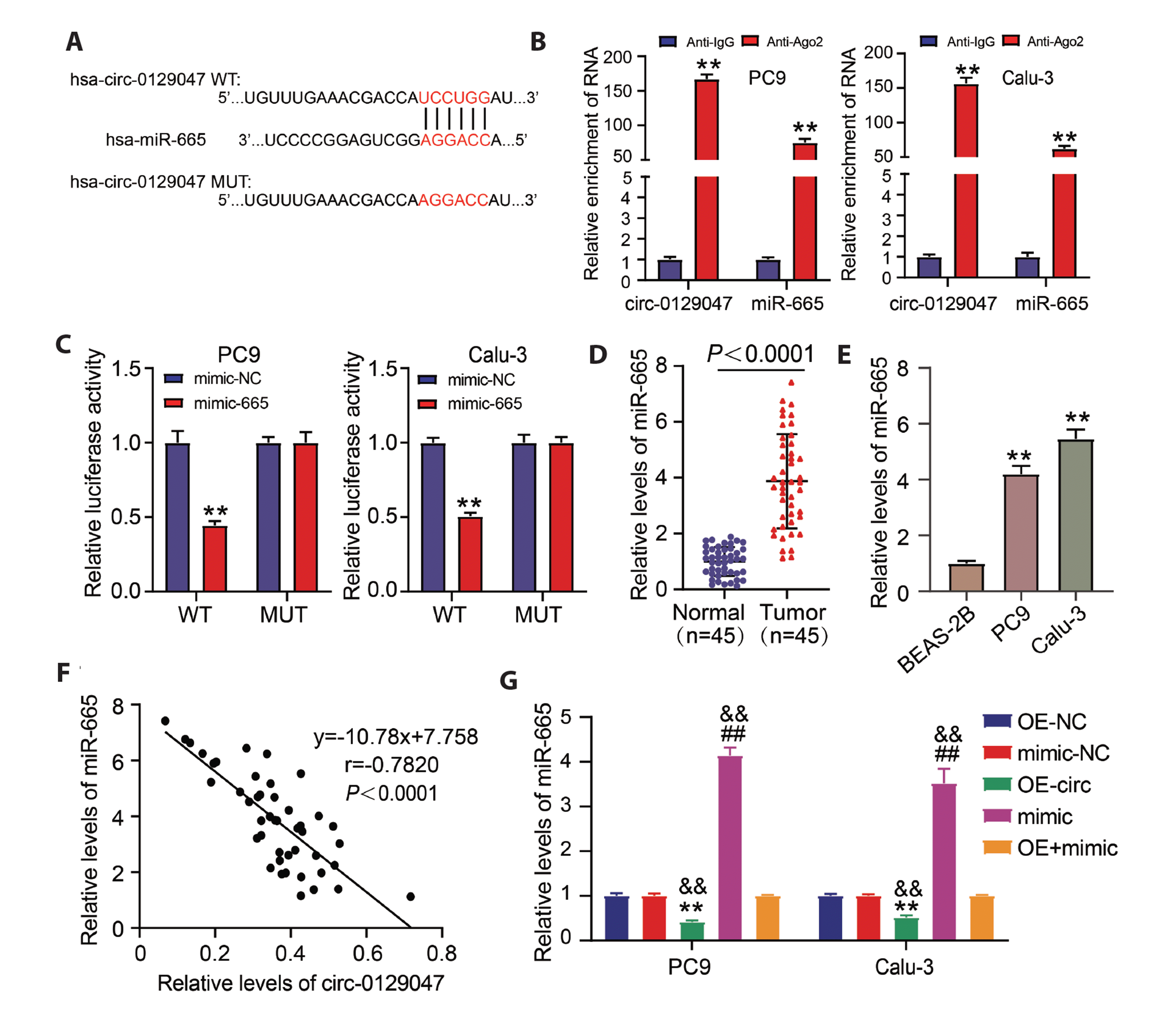
Fig. 3
Circ_0129047 targets miR-665.
(A) MiR-665 is predicted as a target of circ_0129047 through CircInteractome. (B) RIP assay was performed using Ago2 antibody in LUAD cells, and the enrichment of circ_0129047 and miR-665 was detected. **p < 0.01 vs. Anti-IgG. (C) Luciferase activity of Circ_0129047 in LUAD cells transfected with miR-665 mimics, which bind to the circ 0129047 sequence. **p < 0.01 vs. mimic-NC. (D) RT-qPCR analysis of miR-665 in LUAD tissues and normal tissues. (E) RT-qPCR analysis of miR-665 in LUAD cells (PC9 and Calu-3) and normal cells (BEAS-2B). **p < 0.01 vs. BEAS-2B. (F) Pearson analysis of miR-665 expression and circ_0129047 expression in LUAD tissues. (G) circ 0129047-overexpressing vectors (OE-circ), OE-NC, miR-665 mimic (mimic), mimic-NC, and OE+mimic were transfected into PC9 and Calu-3 cells. The expression of miR-665 was measured using RT-qPCR 48 h after transfection. **p < 0.01 vs. OE-NC; ##p < 0.01 vs. mimic-NC; &&p < 0.01 vs. OE+mimic. Values are presented as mean ± SD. LUAD, lung adenocarcinoma; Ago2, Argonaute 2; WT, wild type; MUT, mutant.
![]()
MiR-665 is a critical target via which circ_0129047 mediates LUAD tumorigenesis
Based on the target analysis, we clarified the in vitro association between miR-665 and circ_0129047 using a series of cell functional experiments. After performing the CCK-8 assay, we found that the miR-665 mimic functioned as a proliferation promoter in PC9 and Calu-3 cells. However, its increased proliferation was mitigated by circ_0129047-augmented expression (Fig. 4A). Apoptosis was inactivated in PC9 and Calu-3 cells treated with the miR-665 mimic, as demonstrated by the reduced expression of Bax and increased expression of Bcl-2. However, this inactivation of apoptosis in LUAD cells was re-elicited by the co-transfection of circ_0129047-overexpressing vectors (Fig. 4B). Overexpression of miR-665 also resulted in higher colony formation ability compared with that of the control group, and this was reversed along with circ_0129047 overexpression (Fig. 4C). Taken together, circ_0129047 suppresses the pro-tumorigenic function of miR-665 in LUDA cells by sequestering miR-665.

Fig. 4
MiR-665 is a critical target via which circ_0129047 mediated LUAD tumorigenesis.
PC9 and Calu-3 cells were transfected with circ_0129047 overexpression vectors (OE-circ), OE-NC, miR-665 mimic (mimic) or mimic-NC, and OE+mimic. (A) Cell proliferation was analyzed using the CCK-8 assay. (B) Western blot analysis of anti-Bax and anti-Bcl-2 expression. (C) Colony formation determined using colony formation assay (×200). Values are presented as mean ± SD. LUAD, lung adenocarcinoma; OD, optical density; PTPRB, protein tyrosine phosphatase receptor type B. *p < 0.05, **p < 0.01 vs. OE-NC; #p < 0.05, ##p < 0.01 vs. mimic-NC; &p < 0.05, &&p < 0.01 vs. OE+mimic.
![]()
MiR-665 targets PTPRB
It is known that circRNAs inhibit mRNA silencing by miRNA through ceRNA activity. Since miRNAs modify mRNA expression by mRNA 3′UTR, we conducted TargetScan analysis to mine the downstream mRNA of miR-665. As shown in Fig. 5A, the PTPRB 3′UTR contained a consequential pairing of miR-665. Next, we cloned the consequential pairing region and MUT sequence into luciferase reporter vectors and detected luciferase activity of the PTPRB 3′UTR in LUAD cells. Undoubtedly, PTPRB 3′UTR-mediated luciferase activity was reduced in miR-665 overexpressing LUAD cells, and once PTPRB 3′UTR was mutated, the luciferase activity was restored (Fig. 5B). We found that PTPRB was poorly expressed in the clinical samples (Fig. 5C). Furthermore, the poor expression of PTPRB was negatively correlated with miR-665 (Fig. 5D), consistent with our putative targeting relationship between the PTPRB 3′UTR and miR-665. In addition, PTPRB levels in LUAD tissues were positively correlated with circ_012947 (Fig. 5E), and the nude mice assay further proved that the overexpression of circ_0129047 enhanced the expression of PTPRB in the tumor tissues of nude mice (Supplementary Fig. 1C). Low PTPRB expression was also observed in LUAD cells compared with that in BEAS-2B cells (Fig. 5F). After transfecting the miR-665 mimic and PTPRB overexpression vectors into PC9 and Calu-3 cells, western blotting showed that PTPRB expression was elevated by the PTPRB overexpression vector and reduced by the miR-665 mimic. However, the PTPRB overexpression vector recovered the loss of PTPRB expression in LUAD cells induced by the miR-665 mimic (Fig. 5G). Hence, miR-665 directly targeted PTPRB and silenced its expression in LUAD cells.
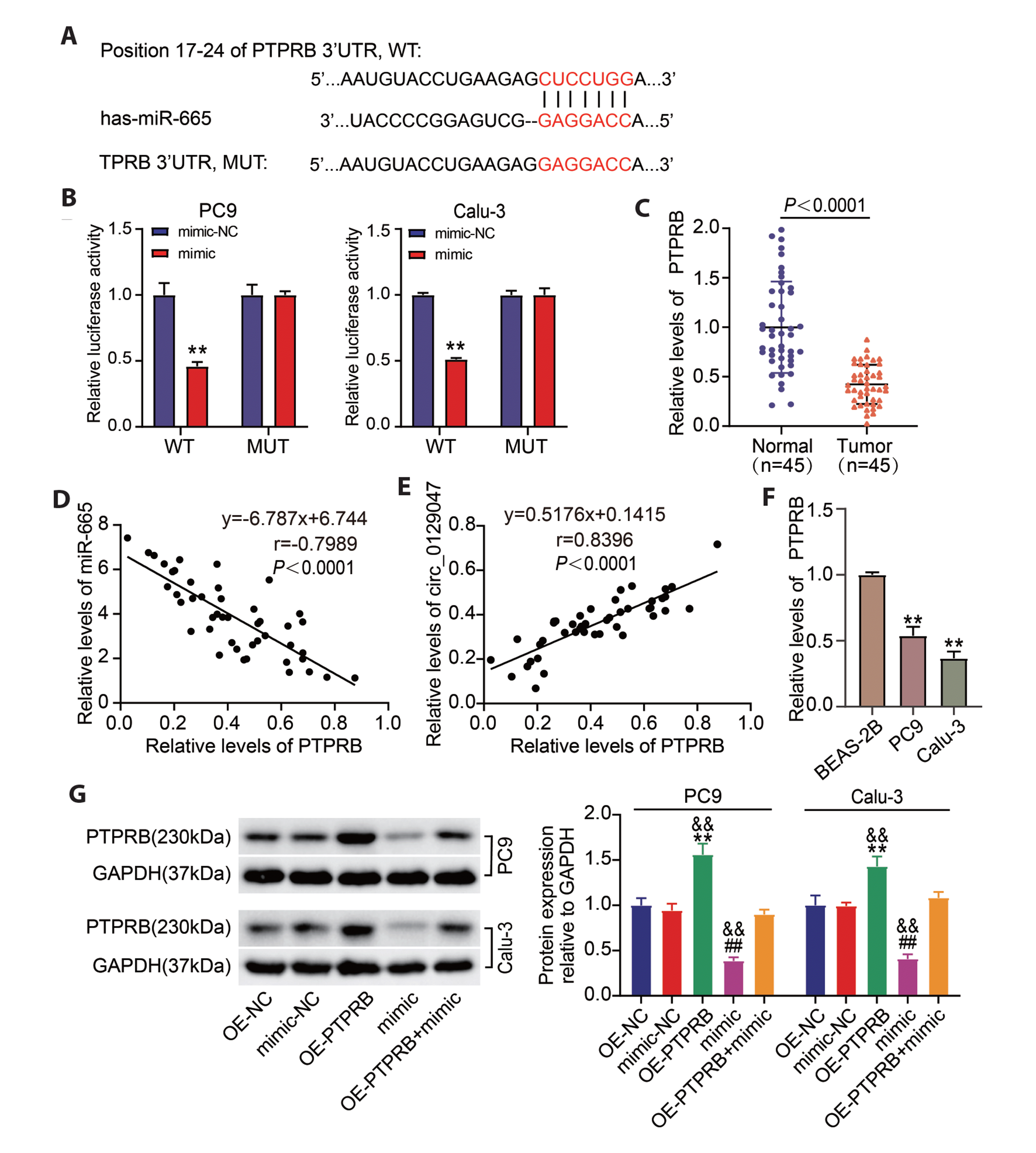
Fig. 5
MiR-665 targets PTPRB.
(A) Predicted miR-665 interactions with PTPRB 3′UTR using miRNA target prediction software based on TargetScan. (B) Luciferase reporter gene assay of PTPRB 3′UTR after miR-665 treatment. **p < 0.01 vs. mimic-NC. (C) PTPRB mRNA expression in LUAD tissues analyzed using RT-qPCR. (D) Pearson correlation analysis was used to analyze the relationship between miR-665 and PTPRB in LUAD tissues. (E) Pearson correlation analysis was used to analyze the relationship between circ_0129047 and PTPRB in LUAD tissues. (F) PTPRB mRNA expression in LUAD cells (PC9 and Calu-3) and normal cells (BEAS-2B) using RT-qPCR. **p < 0.01 vs. BEAS-2B. (G) Western blot analysis determining PTPRB expression in LUAD cells transfected with PTPRB-overexpressing vectors (OE-PTPRB), OE-NC, miR-665 mimic (mimic), mimic-NC, OE-PTPRB+mimic. **p < 0.01 vs. OE-NC; ##p < 0.01 vs. mimic-NC; &&p < 0.01 vs. OE-PTPRB+mimic. Values are presented as mean ± SD. LUAD, lung adenocarcinoma; PTPRB, protein tyrosine phosphatase receptor type B; WT, wild type; MUT, mutant.
![]()
The tumorigenic role of miR-665 may be achieved by targeting PTPRB 3′UTR
Our findings indicate that miR-665 interferes with PTPRB expression by targeting the 3′UTR of PTPRB. Hence, we subsequently investigated the effects of PTPRB on the pro-tumorigenic potential of miR-665 using cell function experiments. In the CCK-8 assay, PTPRB overexpression inhibited LUAD cell proliferation, but this effect was abrogated by co-transfection with the miR-665 mimic (Fig. 6A). Western blotting showed that PTPRB overexpression enhanced Bax expression and reduced Bcl-2 expression, suggesting that PTPRB overexpression elicited apoptosis. However, the promotive effect of PTPRB overexpression on apoptosis was reversed by the miR-665 mimic (Fig. 6B). The colony formation assay revealed that ectopic expression of PTPRB reduced the number of colonies formed, but miR-665 mimic-mediated enhancement of cell colony formation relieved the inhibitory effect of PTPRB ectopic expression on the colony formation (Fig. 6C). Therefore, miR-665 may favor LUAD malignant behaviors by targeting the PTPRB 3′UTR.

Fig. 6
The tumorigenic role of miR-665 may be achieved by targeting PTPRB 3ʹUTR.
LUAD cells were transfected with PTPRB-overexpressing vectors (OE-PTPRB), OE-NC, miR-665 mimic (mimic), mimic-NC, and OE-PTPRB+mimic. (A) Cell proliferation was analyzed using the CCK-8 assay. (B) Western blot analysis of anti-Bax and anti-Bcl-2 expression in the transfected cells. (C) Colony formation was determined using colony formation assay (×200). *p < 0.05, **p < 0.01 vs. OE-NC; #p < 0.05, ##p < 0.01 vs. mimic-NC; &p < 0.05, &&p < 0.01 vs. OE-PTPRB+mimic. Values are presented as mean ± SD. LUAD, lung adenocarcinoma; OD, optical density; PTPRB, protein tyrosine phosphatase receptor type B.
![]()
DISCUSSION
Dysregulation of circRNAs is a common cascade involving many cellular and physiological processes, including ontogenesis [22]. Their biological importance has also been depicted in LUAD [23]. However, circRNAs in LUAD remain largely unknown. Herein, we describe an unrecognized circRNA, circ_0129047, which is downregulated in LUAD. Functional investigation revealed that circ_0129047, a tumor suppressor, inhibited LUAD growth in vitro and in vivo. Further mechanistic exploration revealed a circ_0129047/miR-665/PTPRB ceRNA regulatory network during LUAD in malignant lesions and underscored the potential of circ_0129047 in clinical applications.
Growing evidence has shown that cytoplasmic circRNAs have sponging properties against miRNAs [24]. Intriguingly, we also found subcellular localization of circ_0129047 in the cytoplasm. Therefore, it is reasonable to explore the effects of ceRNA in LUAD. We found that circ_0129047 targets miR-665 through circRNA interactome prediction and validation from luciferase reporter and RIP assays. Interestingly, a paradoxical role for miR-665 has been reported in LUAD. For example, Ying et al. [17] demonstrated that miR-665 targeted by circ-TSPAN4 promotes LUAD metastasis. However, miR-665 targeted by circ-000881 suppresses the malignant behaviors [18]. Therefore, the role of miR-665 in LUAD remains unclear. Our data showed that augmented miR-665 expression strengthened the LUAD cell proliferation and inhibited apoptosis, suggesting its pro-tumorigenic role in LUAD. Further mechanical exploration showed that circ_0129047 attenuated the increase in LUAD cell proliferative potential induced by miR-665. Therefore, circ_0129047 may target miR-665 and downregulate the pro-proliferative potential of miR-665, resulting in LUAD suppression.
PTPRB plays different roles in various cancers. PTPRB overexpression enhances the oncogenic phenotypes of cervical cancer in vitro [25], whereas it is poorly expressed in hepatocellular carcinoma and could weaken oncogenic Hippo signaling, mitigating tumor growth [26]. In addition, PTPRB was found to be a carcinogenic factor in NSCLC [20]. In the present study, we found that PTPRB was downregulated in LUAD tissues, and its overexpression caused a proliferative defect in LUAD cells accompanied by apoptosis activation. Our findings provide evidence for tumor suppression in NSCLC, as evidenced by the fact that PTPRB overexpression abrogates the malignant action of cancer cells [27,28]. Mechanistically, we found that miR-665 silences PTPRB expression. Our findings provide further evidence for the tumor-suppressive role of PTPRB in LUAD.
Our investigation identified a novel circRNA, circ_0129047, in LUAD. However, our study has some limitations. First, the downstream pathway components of PTPRB that weaken the tumorigenic potential of LUAD remain unknown. PTPRB silencing boosts the Src phosphorylation and favors LC progression [20]. Thus, in the near future, we will uncover the detailed mechanisms underlying LUAD progression. Furthermore, owing to the imperfect base pairing, circRNAs are embedded in complex regulatory ceRNA networks, including different miRNAs or mRNAs. Therefore, it is necessary to elucidate the mechanism underlying circ_0129047-mediated LUAD progression.
Our findings revealed that circ_0129047 mitigated LUAD proliferation in vitro and restricted its progression in vivo. Furthermore, circ_0129047 acted as a ceRNA for miR-665 to abrogate the inhibition of miR-665 by PTPRB, thereby interfering with LUAD cell proliferation. Our findings might be helpful in uncovering the mechanism of LUAD progression and suggest that targeting circ_012904 might have promising prospects for LUAD intervention.
SUPPLEMENTARY MATERIALS
Supplementary data including one figure can be found with this article online at https://doi.org/10.4196/kjpp.2023.27.2.131
 XML Download