Journal List > Anat Cell Biol > v.56(1) > 1516081671

Thintharua and Chentanez: Morphological analysis and morphometry of the occipital condyle and its relationship to the foramen magnum, jugular foramen, and hypoglossal canal: implications for craniovertebral junction surgery

Abstract

Anatomical knowledge of the occipital condyle (OC) and its relationships to surrounding structures is important for avoiding injury during craniovertebral junction (CVJ) surgeries. This study was conducted to evaluate the morphology and morphometry of OC and its relationship to foramen magnum, jugular foramen (JF), and hypoglossal canal (HC). Morphometric parameters including length, width, height, and distances from the OC to surrounding structures were measured. The oval-like condyle was the most common OC shape, representing for 33.0% of all samples. The mean length, width and height of OC were 21.3±2.4, 10.5±1.4, and 7.4±1.1 mm, respectively. Moreover, OC was classified into three types based on its length. The most common OC length in both sexes was moderate length or type II (62.5%). The mean distance between anterior tips and posterior tips of OC to basion, and opisthion were 11.5±1.4, 39.1±3.3, 25.2±2.2, and 27.4±2.7 mm, respectively. The location of intracranial orifice of HC was commonly found related to middle 1/3 of OC in 45.0%. JF was related to the anterior 2/3 of OC in 81.0%, the anterior 1/3 of OC in 12.5%, and the entire OC length in 6.5%. These morphological analysis and morphometric data should be taken into consideration before performing surgical operation to avoid CVJ instability and neurovascular structure injury.

Introduction

The craniovertebral junction (CVJ) is an area that connects the cranium to the upper cervical spine, consisting of the first two cervical vertebrae (atlas and axis), foramen magnum (FM) and the lower clivus (LC) that supports the brain stem. It is bounded laterally by the jugular foramen (JF), hypoglossal canal (HC) and occipital condyle (OC) [1-3]. CVJ is a common site for various lesions including neoplasms, vertebral artery aneurysm, rheumatic diseases, malformations, and degenerative pathologies [1, 2, 4-6]. Lesions at CVJ are difficult to manage because of their location and complex anatomic relations [7]. Far lateral approach (FLA) is used to access the ventrolateral part of CVJ and LC by drilling the lateral edge of the FM rim for tumor removal and treatment of vascular lesions [8, 9]. Craniotomy of FLA includes lateral part of the occipital squama to the inferior rim of FM [10]. This process increases the angle of exposure, greater visualization, and enhances access to the ventrolateral and ventral aspects of CVJ [5, 11, 12]. The morphological or morphometric analysis of OC has been described in a majority of CVJ anatomical and biomechanical studies [1, 13-15]. The stability of CVJ depends mainly on the length of OC. Drilling OC in FLA can increase patient risk to develop CVJ instability [1]. HC has been used as a landmark for OC resection. It was claimed to locate in the middle part of OC. Therefore, OC resection should not extend beyond HC to prevent CVJ instability, hypoglossal nerve (CN XII) and meningeal branch of ascending pharyngeal artery injury [16-20]. HC has been reported to relate to the anterior one-third of OC in 85% of cases and middle one-third of OC in 15% of cases [19]. OC resection extended to HC in patients with an anteriorly located HC may increase the risk of CVJ instability [16]. JF is a canal that contains the jugular bulb and cranial nerves including the glossopharyngeal (CN IX), vagus (CN X), and accessory nerve (CN XI) [21, 22]. The JF should be avoided during an OC resection because it may cause damage to neurovascular structures that pass through it [19].
Anatomical knowledge of the relationship of bony landmarks on dry skulls including, FM, HC, JF, and OC are essential for surgical operation in the ventrolateral and ventral aspects of CVJ. Morphometric data can benefit surgeons to be aware of CVJ instability and neurovascular injury after craniotomy of FLA procedure. Therefore, the aim of the present study was to determine the shape and dimension of OC and its distance to the surrounding bony structures in dry skull to improve successful surgical outcome.

Materials and Methods

One hundred adult dry skulls (50 males and 50 females) were examined. They were collected from the Department of Anatomy, Faculty of Medicine, King Chulalongkorn Memorial Hospital, Chulalongkorn University. Skulls with OC fractures and cranial base breaks were excluded. Skulls of undetermined age and sex were obtained, general size and architecture, supraorbital ridges, mastoid process, and external occipital protuberance were used for sex determination of skulls [23, 24]. The sex determination was done by two investigators separately. If there was any conflict, a consensus was made. This study was approved by the Faculty of Medicine, Chulalongkorn University IRB committee (IRB No. 0988/64).

Identification and marking the related bony structures on the base of the skulls
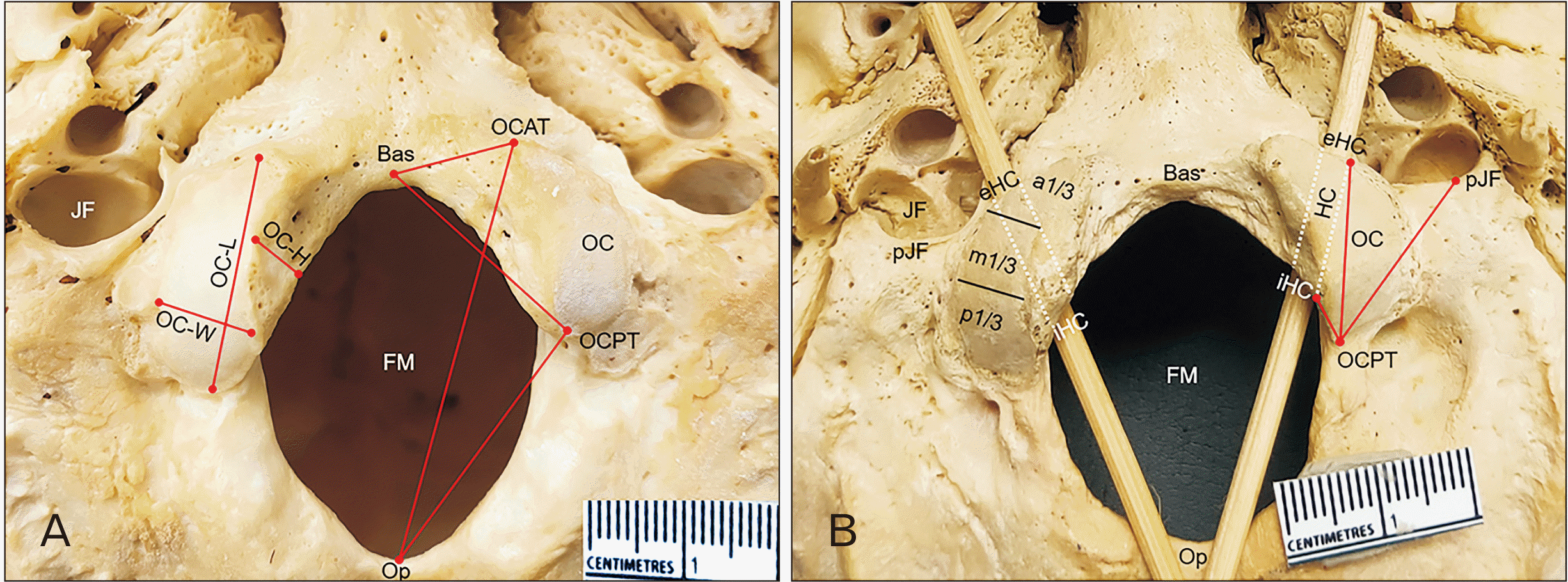
Ten structures on the skull base were identified and marked including the anterior tip of OC (OCAT), basion (Bas), extracranial orifice of HC (eHC), FM, intracranial orifice of HC (iHC), JF, OC, opisthion (Op), the posterior most end of JF (pJF), and the posterior tip of OC (OCPT) (Fig. 1) [4, 25].

Morphological analysis of the shape of OC, location of iHC and eHc, JF-OC relationship

According to Naderi et al. [20], OC shape was classified into eight types: type I; oval-like condyle, type II; kidney-like condyle, type III; S-like condyle, type IV; eight-like condyle, type V; triangle condyle, type VI; ring-like condyle, type VII; two-portioned condyle, and type VIII; deformed condyle (Fig. 2). The location of iHC (L-iHC) and eHC (L-eHC) related to OC was examined in the following areas: location 1 (anterior 1/3 of OC), location 2 (junction between anterior and middle 1/3 of OC), location 3 (middle 1/3 of OC), location 4 (junction between middle and posterior 1/3 of OC), and location 5 (posterior 1/3 of OC) (Fig. 1B). The JF-OC relationship (JF-OC) was determined in terms of its relation to the anterior 1/3, anterior 2/3, and the entire OC length.

Morphometric evaluations of the OC dimensions, distance from iHC and eHC to OCPT, distance from OCAT to Op and Bas, distance from OCPT to Op and Bas, and distance of OCPT to pJF

Ten parameters including: 1) the maximum anteroposterior distance between OCAT and OCPT or length of OC (OC-L), 2) the maximum transverse distance between medial and lateral border of OC or width of OC (OC-W), 3) the maximum vertical distance between upper and lower boundary of medial surface of OC or height of OC (OC-H) (Fig. 1A), 4) the distances from posterior tip of OC (OCPT) to iHC (OCPT-iHC), 5) the distances from OCPT to eHC (OCPT-eHC) (Fig. 1B), 6) the distance between OCAT and Op (OCAT-Op), 7) the distance between OCAT and Bas (OCAT-Bas), 8) the distance between OCPT and Op (OCPT-Op), 9) the distance between OCPT and Bas (OCPT-Bas) (Fig. 1A), 10) the distance from OCPT to the posterior-most end of JF (OCPT-pJF) (Fig. 1B), were measured with a digital vernier caliper (Mitutoyo®; Mitutoyo Co., Kawasaki, Japan, 0–150 mm; range 150 mm, resolution 0.01 mm). All measurements were executed twice by a single investigator to minimize intra and inter observer error.

Statistical analysis

Statistical analysis of the data was performed by IBM SPSS Statistics for Windows, version 22.0 (IBM Co., Armonk, NY, USA). Mean and standard deviation were analyzed in each parameter, paired t-test analysis was used to assess the mean difference in sides, independent t-test analysis was used to assess the mean difference in sex. The Chi-square test was used to investigate a possible correlation between the descriptive variables. The result was considered statistically significant when P-value was less than 0.05. Intraobserver reliability was calculated for each parameter and the least intraobserver intraclass correlation coefficient among all parameter was 0.995.

Results

Shapes and dimension of the OC

Eight types of OC shape were observed (Fig. 2). The prevalence of each OC morphological type is presented in Table 1. The most prevalent shape of OC was S-like condyle (26.0%) in males and oval-like condyle (41.0%) in females. A statistically significant difference of OC shape was found between sex (P=0.037), but not between sides. Symmetrical shape of OC was observed in 46.0% (20.0% from male skulls and 26.0% from female skulls).
The mean and range of length, width, and height of OC were 22.1±2.5 (18.2–28.8), 10.4±1.5 (7.4–15.1), and 7.5±1.1 (5.2–10.8) mm, respectively in males, and 20.5±2.1 (16.0–26.0), 10.6±1.3 (7.4–13.7), and 7.3±1.1 (4.5–9.7) mm, respectively in females (Fig. 1A, Table 2). Statistically significant difference between sex was found in OC-L (P<0.001), but not found in OC-W (P=0.294) and OC-H (P=0.484). Furthermore, a statistically significant difference in OC-H between sides was found in males and females, with P-values of 0.024 and 0.003, respectively.
The OC was classified according to its length [18, 20] into 3 types: type 1 (short: <20.0 mm), type 2 (moderate: 20.0–26.0 mm) and type 3 (long: >26.0 mm) (Table 3). The most prevalent was moderate length, which was reported by 62.5% of both sexes (66.0% of males and 59.0% of females). In comparison, females (41.0%) had a higher prevalence of short OC types than males (25.0%). There was a significant sex difference in the proportions of OC typing by length (P=0.001)

Distance between the tips of OC and FM border

The mean distances of OCAT-Bas, OCAT-Op, OCPT-Bas and OCPT-Op (Fig. 1A) are shown in Table 2. A statistically significant difference between sex was found in OCAT-Op (P<0.001) and OCPT-Bas (P=0.047). There was also a statistically significant difference in OCPT-Bas between female sides (P=0.029).

Locations of HC and its distance to OC

The L-iHC was commonly related to location 3 in 46.0% of male skulls and 44.0% of female skulls. In contrast, L-eHC was related to location 1 in 76.0% and 72.0% of male and female skulls, respectively (Fig. 1B, Table 4). There was no statistically significant difference between sides of L-iHC and L-eHc in female skulls (P=0.292 and 0.585, respectively) and L-eHC of male skulls (P=0.058). However, a statistically significant difference between sides of L-iHC was found in male skulls (P<0.001). Symmetrical L-iHC and L-eHC were found in 64.0% and 36.0% of skulls, respectively. The most common symmetrical L-iHC and L-eHC were location 3 (61.1%) and location 1 (89.1%), respectively. The mean distance of OCPT-iHC was 9.2±1.6 mm in males and 8.8±1.6 mm in females. There was no statistically significant difference between sex (P=0.138), but a statistically significant difference was found between sides (P=0.030). Males had a mean distance of OCPT-eHC of 14.1±2.0 (8.7–18.4) mm while it was 13.3±2.4 (7.0–18.7) mm in females (Table 2). A statistically significant difference in OCPT-eHC was found between sex (P=0.011), but not found between sides (P=0.519).

The relation and distance between OC and JF

JF was related to the anterior 2/3 of OC in 81.0%, the anterior 1/3 of OC in 12.5%, and the entire OC length in 6.5%. Additionally, JF was related to the anterior 1/3 of OC length in 17.0% of male, and 8.0% of female, anterior 2/3 of OC length in 80.0% of male and 82.0% of female, and entire OC length in 3.0% of male and 10.0% of female (Fig. 1B, Table 5). There was a statistically significant difference in JF-OC relation between sides of males (P=0.001), but not in females (P=0.165). The mean distances of OCPT-pJF were 16.4±2.3 and 15.9±2.4 mm in male and female skulls, respectively (Fig. 1B, Table 2). There was no significant difference of the OCPT-pJF between sex (P=0.164) and sides (P=0.102).

Discussion

A detailed knowledge of the morphology and morphometry of OC, as well as its relationship to the surrounding bony structures including the FM, the orifices of HC and the JF in dry skull was evaluated to provide essential data to improve surgical outcomes. The most prevalent shape of OC in this study was oval-like condyle or type I, which agreed with previous findings in Indians [14, 19, 26], and Turks [20, 27]. In contrast, Aragão et al. [13] and Bayat et al. [28], reported that the most common shape in Brazilians was S-like condyle or type III and kidney-like condyle or type II in Iranians. The prevalence of OC shape may be related to ethnicity. The different types of OC, such as the triangle, the deformed, and kidney-like type may require a more extensive condylectomy to reach the ventral lesions. Symmetrical shape of OC was found in 46.0% which was lesser than previously reported by Naderi et al. [20] (51.0%) and Kalthur et al. [26] (62.0%). According to Verma et al. [19] and Naderi et al. [20], the shape of the OC may affect the amount of condylectomy. Different types of OC, such as triangle, deformed, and kidney-like type may require a more extensive condylectomy to reach the ventral lesions. Furthermore, OC screw placement (or nail insertion) for occipitocervical fixation was easier and more convenient to fix in an oval-like OC due to its large surface area, while it would be difficult in a triangle, ring-like, and two-portioned type of OC [19].
The mean length of OC in this study was 21.3±2.4 mm, which was shorter than those determined by Di et al. [1] (23.6±2.0 mm) and Saluja et al. [29] (22.8±2.9 mm). Females had a significantly shorter OC length than males, similar to Rai et al. [30] and Kumar and Nagar [31]. In our analysis, the most prevalent type of OC classified according to its length was the moderate type which was in agreement with previous reports [18, 20]. Previous studies suggested that resection of less than 50.0% of OC-L would not show evidence of CVJ instability [1, 5, 17, 32-34]. However, Bejjani et al. [35] claimed that resection of less than 70.0% of OC-L from the posterior would not reveal evidence of CVJ instability. According to our results, we proposed an appropriated resection of not more than 50.0% of OC-L from the posterior tip of OC which would be not more than 11.1 mm in males and 10.3 mm in females to avoid the CVJ instability. The average OC-W in this study was 10.5±1.4 mm, which was lesser than those reported by Di et al. [1] and Saluja et al. [29]. This data is important in surgery because it can be used to determine how much medially the OC can be resected [26]. Moreover, the average OC-H was 7.4±1.1 mm, which was lesser than those reported previously [1, 30]. This data can be used to determine how deep the OC must be drilled [14] and the successful insertion of screws during occipitocervical fixation [30].
For surgical approach, knowledge of the distance between OCPT and Op is important. The total mean distances of OCAT-Bas, OCAT-Op, OCPT-Bas, and OCPT-Op in our study were 11.5±1.4, 39.1±3.3, 25.2±2.2, and 27.4±2.7 mm, respectively. These distances were shorter than the results reported by lhan et al. [4]. However, the mean distance of OCPT-Op in this study was longer than Naderi et al. [20] and Saluja et al. [29], but was lesser than Kalthur et al. [26]. This distance represents the width of surgical exposure in suboccipital craniotomy and gives a free corridor for posterolateral approach, and a longer corridor will provide more space for FLA [4, 26].
In this study, the majority of L-iHC was related to the middle 1/3 of OC (45.0%), while the majority of L-eHC was related to the anterior 1/3 of OC (74.0%). These findings were similar to Naderi et al. [20] who showed that the majority of L-iHC (56.4%) was related to the junction of the second and third 1/4 of OC, whereas the majority of L-eHC was related to the anterior 1/4 of OC (64.0%). There was no HC orifice related to the posterior 1/3 of OC (location 5). However, Kalthur et al. [26] found that L-iHC was related to the middle 1/3 of OC (location 3) in 100.0% of the cases for both sexes, but the majority of L-eHC was related to the anterior 1/3 of OC (location 1) in 98.0% and 93.7% for males and females, respectively. According to our findings, the location of iHC was more variable than that of eHC. Therefore, iHC was more likely to be injured during OC resection. OC resection can cause a complication with CN XII injury or O-C1 joint instability [18]. Therefore, anatomical localization of the HC is critical to minimize accidental damage to the CN XII [16, 18]. OCPT-iHC and OCPT-eHC represent the HC depth. HC depth is extremely important during transcondylar approach because it indicates the maximal amount of OC resection without reaching the HC and injuring structures in HC [17, 19]. The distance between OCPT and iHC is highly important because iHC is located closer to the OCPT than eHC. The mean OCPT-iHC in our study was 9.0±1.6 mm, which was shorter than those reported by Di et al. [1] (10.2±1.3), Kizilkanat et al. [36] (12.3±2.4), and Parvindokht et al. [37] (11.4±2.5), but it was longer than the other two previous studies [17, 35].
In agreement with previous study [19], we found that the majority of JF was related to the anterior 2/3 of OC in 81.0% of cases and the anterior 1/3 of OC in 12.5% of cases. If the JF was related to the anterior 1/3 of OC, it would result in a lesser neurovascular complication in OC resection. Verma et al. [19] revealed that the OCPT-pJF was 15.7±3.0 mm on the right side and 16.8±2.9 mm on the left side. Similarly, we found that the mean distance of OCPT-pJF was 16.2±2.4 mm. These results could be implicated in performing condylectomy to avoid approaching the JF and causing injury to its neurovascular contents. Limitations of this study are the unknown age and ethnicity of skulls which may affect the evaluated results.
In conclusion, our study population generally have the same OC shape but lesser OC length, OC width and OC height or thickness compared to other populations. Several anatomical parameters of OC, and its relationships to surrounding structures, should be considered during preoperative planning in CVJ surgery to avoid CVJ instability and neurovascular structure injury.

Acknowledgements

The authors would like to express their gratitude to those who have donated their body for medical study and research. Special thanks are extended to the technical staffs of the Department of Anatomy, Faculty of Medicine, Chulalongkorn University for their support.

Notes

Author Contributions

Conceptualization: PT, VC. Data acquisition: PT. Data analysis or interpretation: PT, VC. Drafting of the manuscript: PT. Critical revision of the manuscript: VC. Approval of the final version of the manuscript: all authors.

Conflicts of Interest

No potential conflict of interest relevant to this article was reported.

References

1. Di G, Fang X, Hu Q, Zhou W, Jiang X. 2019; A microanatomical study of the far lateral approach. World Neurosurg. 127:e932–42. DOI: 10.1016/j.wneu.2019.04.004. PMID: 30995558.
crossref
2. Ayoub B. 2011; The far lateral approach for intra-dural anteriorly situated tumours at the craniovertebral junction. Turk Neurosurg. 21:494–8. DOI: 10.5137/1019-5149.JTN.4277-11.2. PMID: 22194106.
crossref
3. Rosa S, Baird JW, Harshfield D, Chehrenama M. 2018. Craniocervical junction syndrome: anatomy of the craniocervical and atlantoaxial junctions and the effect of misalignment on cerebrospinal fluid flow [Internet]. IntechOpen;London: Available from: https://www.intechopen.com/chapters/58783. cited 2022 Oct 18. DOI: 10.5772/intechopen.72890.
4. lhan P, Kayhan B, Erturk M, Sengul G. 2017; Morphological analysis of occipital condyles and foramen magnum as a guide for lateral surgical approaches. MOJ Anat Physiol. 3:188–94. DOI: 10.15406/mojap.2017.03.00117.
5. Mehdi W, Niaz A, Irfan M, Tasdique S, Majeed S. 2020; Far lateral transcondylar approach for anterior foramen magnum lesions. Pak J Neurol Surg. 24:149–55. DOI: 10.36552/pjns.v24i2.454.
crossref
6. Wu A, Zabramski JM, Jittapiromsak P, Wallace RC, Spetzler RF, Preul MC. 2010; Quantitative analysis of variants of the far-lateral approach: condylar fossa and transcondylar exposures. Neurosurgery. 66(6 Suppl Operative):191–8. discussion 198DOI: 10.1227/01.NEU.0000369704.49958.5B. PMID: 20489505.
7. George JR, Francis T, Francis J, Samuel JE. 2019; Morphometric study of dry human occipital bone and its clinical relevance. Int J Anat Res. 7:6230–3. DOI: 10.16965/ijar.2018.447.
crossref
8. Scoville JP, Mazur MD, Couldwell WT. 2020; Unique far-lateral closure technique: technical note. Oper Neurosurg (Hagerstown). 18:384–90. DOI: 10.1093/ons/opz168. PMID: 31236599.
crossref
9. Moscovici S, Umansky F, Spektor S. 2015; "Lazy" far-lateral approach to the anterior foramen magnum and lower clivus. Neurosurg Focus. 38:E14. DOI: 10.3171/2015.2.FOCUS14784. PMID: 25828490.
crossref
10. Lanzino G, Paolini S, Spetzler RF. 2005; Far-lateral approach to the craniocervical junction. Neurosurgery. 57(4 Suppl):367–71. discussion 367–71. DOI: 10.1227/01.NEU.0000176848.05925.80. PMID: 16234687.
crossref
11. Chaddad-Neto F, Doria-Netto HL, Campos Filho JM, Reghin-Neto M, Rothon AL Jr. Oliveira Ed. 2014; The far-lateral craniotomy: tips and tricks. Arq Neuropsiquiatr. 72:699–705. DOI: 10.1590/0004-282X20140130. PMID: 25252234.
crossref
12. Cardoso AC, Fontes RB, Tan LA, Rhoton AL Jr, Roh SW, Fessler RG. 2015; Biomechanical effects of the transcondylar approach on the craniovertebral junction. Clin Anat. 28:683–9. DOI: 10.1002/ca.22551. PMID: 25914225.
crossref
13. Aragão JA, De Santana GM, Aragão ICS, Aragão FMS, Reis PF. Da Cruz de Moraes RZ. 2017; Morphological analysis on the occipital condyles and review of the literature. Int J Morphol. 35:1129–32. DOI: 10.4067/S0717-95022017000300050.
crossref
14. Anjum A, Pandurangam G, Garapati S, Bandarupalli N, Rabbani H, Divya P. 2021; Morphology and morphometric study of occipital condyles. Int J Anat Res. 9:7905–11. DOI: 10.16965/ijar.2021.107.
crossref
15. Karam YR, Traynelis VC. 2010; Occipital condyle fractures. Neurosurgery. 66(3 Suppl):56–9. DOI: 10.1227/01.NEU.0000365751.84075.66. PMID: 20173528. PMCID: PMC8571366.
crossref
16. Mazur MD, Couldwell WT, Cutler A, Shah LM, Brodke DS, Bachus K, Dailey AT. 2017; Occipitocervical instability after far-lateral transcondylar surgery: a biomechanical analysis. Neurosurgery. 80:140–5. DOI: 10.1093/neuros/nyw002. PMID: 28362894.
crossref
17. Lyrtzis C, Piagkou M, Gkioka A, Anastasopoulos N, Apostolidis S, Natsis K. 2017; Foramen magnum, occipital condyles and hypoglossal canals morphometry: anatomical study with clinical implications. Folia Morphol (Warsz). 76:446–57. DOI: 10.5603/FM.a2017.0002. PMID: 28150268.
crossref
18. Cheruiyot I, Mwachaka P, Saidi H. 2018; Morphometry of occipital condyles: implications for transcondylar approach to craniovertebral junction lesions. Anat J Afr. 7:1224–31. DOI: 10.4314/aja.v7i2.174142.
crossref
19. Verma R, Kumar S, Rai AM, Mansoor I, Mehra RD. 2016; The anatomical perspective of human occipital condyle in relation to the hypoglossal canal, condylar canal, and jugular foramen and its surgical significance. J Craniovertebr Junction Spine. 7:243–9. DOI: 10.4103/0974-8237.193258. PMID: 27891034. PMCID: PMC5111326.
crossref
20. Naderi S, Korman E, Citak G, Güvençer M, Arman C, Senoğlu M, Tetik S, Arda MN. 2005; Morphometric analysis of human occipital condyle. Clin Neurol Neurosurg. 107:191–9. DOI: 10.1016/j.clineuro.2004.07.014. PMID: 15823674.
crossref
21. Kaur J, ivastava D Sr, Singh D, Raheja S. 2012; The study of hyperostosic variants: significance of hyperostotic variants of human skulls in anthropology. Anat Cell Biol. 45:268–73. DOI: 10.5115/acb.2012.45.4.268. PMID: 23301194. PMCID: PMC3531590.
crossref
22. Ekanem UI, Olewnik Ł, Porzionato A, Macchi V, Iwanaga J, Loukas M, Dumont AS, Caro R, Tubbs RS. 2022; Morphology of the groove of the inferior petrosal sinus: application to better understanding variations and surgery of the skull base. Anat Cell Biol. 55:135–41. DOI: 10.5115/acb.22.023. PMID: 35773216. PMCID: PMC9256480.
crossref
23. Nagare SP, Chaudhari RS, Birangane RS, Parkarwar PC. 2018; Sex determination in forensic identification, a review. J Forensic Dent Sci. 10:61–6. DOI: 10.4103/jfo.jfds_55_17. PMID: 30745778. PMCID: PMC6344795.
crossref
24. Sangvichien S, Boonkaew K, Chuncharunee A, Komoltri C, Udom C, Chandee T. 2008; Accuracy of cranial and mandible morphological traits for sex determination in Thais. Siriraj Med J. 60:240–3. PMID: 09f90e8725f64b8eae26fd5fe5942626.
25. Gilroy AM, MacPherson BR, Ross LM. 2008. Atlas of anatomy. 3rd ed. Thieme;Stuttgart: p. 742.
26. Kalthur SG, Padmashali S, Gupta C, Dsouza AS. 2014; Anatomic study of the occipital condyle and its surgical implications in transcondylar approach. J Craniovertebr Junction Spine. 5:71–7. DOI: 10.4103/0974-8237.139201. PMID: 25210336. PMCID: PMC4158634.
crossref
27. Ozer MA, Celik S, Govsa F, Ulusoy MO. 2011; Anatomical determination of a safe entry point for occipital condyle screw using three-dimensional landmarks. Eur Spine J. 20:1510–7. DOI: 10.1007/s00586-011-1765-y. PMID: 21416278. PMCID: PMC3175895.
crossref
28. Bayat P, Bagheri M, Ghanbari A, Raoofi A. 2014; Characterization of occipital condyle and comparison of its dimensions with head and foramen magnum circumferences in dry skulls of Iran. Int J Morphol. 32:444–8. DOI: 10.4067/S0717-95022014000200011.
crossref
29. Saluja S, Das SS, Vasudeva N. 2016; Morphometric analysis of the occipital condyle and its surgical importance. J Clin Diagn Res. 10:AC01–4. DOI: 10.7860/JCDR/2016/23278.8800. PMID: 28050351. PMCID: PMC5198304. PMID: 02cb0daeaf6b4696acf783d82f99feef.
30. Rai H, Keluskar V, Patil S, Bagewadi A. 2017; Accuracy of measurements of foramen magnum and occipital condyle as an indicator for sex determination using computed tomography. Indian J Health Sci Biomed Res. 10:80–3. DOI: 10.4103/2349-5006.198595.
crossref
31. Kumar A, Nagar M. 2014; Human adult occipital condyles: a morphometric analysis. Res Rev J Med Health Sci. 3:112–6.
32. Vishteh AG, Crawford NR, Melton MS, Spetzler RF, Sonntag VK, Dickman CA. 1999; Stability of the craniovertebral junction after unilateral occipital condyle resection: a biomechanical study. J Neurosurg. 90(1 Suppl):91–8. DOI: 10.3171/spi.1999.90.1.0091. PMID: 10413132.
crossref
33. Shin H, Barrenechea IJ, Lesser J, Sen C, Perin NI. 2006; Occipitocervical fusion after resection of craniovertebral junction tumors. J Neurosurg Spine. 4:137–44. DOI: 10.3171/spi.2006.4.2.137. PMID: 16506481.
crossref
34. Karasu A, Cansever T, Batay F, Sabanci PA, Al-Mefty O. 2009; The microsurgical anatomy of the hypoglossal canal. Surg Radiol Anat. 31:363–7. DOI: 10.1007/s00276-008-0455-x. PMID: 19148566.
crossref
35. Bejjani GK, Sekhar LN, Riedel CJ. 2000; Occipitocervical fusion following the extreme lateral transcondylar approach. Surg Neurol. 54:109–15. discussion 115–6. DOI: 10.1016/S0090-3019(00)00255-X. PMID: 11077092.
crossref
36. Kizilkanat ED, Boyan N, Soames R, Oguz O. 2006; Morphometry of the hypoglossal canal, occipital condyle, and foramen magnum. Neurosurg Q. 16:121–5. DOI: 10.1097/01.wnq.0000214018.49915.49.
crossref
37. Parvindokht B, Reza DM, Saeid B. 2015; Morphometric analysis of hypoglossal canal of the occipital bone in Iranian dry skulls. J Craniovertebr Junction Spine. 6:111–4. DOI: 10.4103/0974-8237.161591. PMID: 26288545. PMCID: PMC4530509.
crossref

Fig. 1
Inferior view of the skull base. (A) Right occipital condyle showing occipital condyle width (OC-W), occipital condyle length (OC-L), and occipital condyle height (OC-H). Left occipital condyle showing the distances from anterior and posterior tips of occipital condyle (OCAT and OCPT) to basion (Bas) and opisthion (Op). (B) Right occipital condyle showing the relationship of jugular foramen (JF) and occipital condyle, the location of extracranial orifice and intracranial orifice of hypoglossal canal (L-eHC, L-iHC) related to occipital condyle. Left occipital condyle showing the distances from posterior tip of occipital condyle (OCPT) to iHC, eHC and posterior most end of jugular foramen (pJF). FM, foramen magnum; HC, hypoglossal canal; a1/3, anterior one third; m1/3, middle one third; p1/3, posterior one third.
acb-56-1-61-f1.tif
Fig. 2
Photographs showing eight shapes of occipital condyle. (A) Type I, oval-like condyle. (B) Type II, kidney-like condyle. (C) Type III, S-like condyle. (D) Type IV, eight-like condyle. (E) Type V, triangle condyle. (F) Type VI, ring-like condyle. (G) Type VII, two-portioned condyle, and (H) Type VIII, deformed condyle.
acb-56-1-61-f2.tif
Table 1
Prevalence of morphological types of OC
Types Number of occipital condyles
Males (n=100) Females (n=100) Both sexes (n=200)
Left Right Total P-value Left Right Total P-value Total P-value
I 14 (14.0) 11 (11.0) 25 (25.0) 0.886 23 (23.0) 18 (18.0) 41 (41.0) 0.757 66 (33.0) 0.037a
II 4 (4.0) 7 (7.0) 11 (11.0) 9 (9.0) 7 (7.0) 16 (16.0) 27 (13.5)
III 13 (13.0) 13 (13.0) 26 (26.0) 6 (6.0) 8 (8.0) 14 (14.0) 40 (20.0)
IV 6 (6.0) 4 (4.0) 10 (10.0) 1 (1.0) 3 (3.0) 4 (4.0) 14 (7.0)
V 7 (7.0) 8 (8.0) 15 (15.0) 9 (9.0) 10 (10.0) 19 (19.0) 34 (17.0)
VI - 1 (1.0) 1 (1.0) - 1 (1.0) 1 (1.0) 2 (1.0)
VII 3 (3.0) 2 (2.0) 5 (5.0) - 1 (1.0) 1 (1.0) 6 (3.0)
VIII 3 (3.0) 4 (4.0) 7 (7.0) 2 (2.0) 2 (2.0) 4 (4.0) 11 (5.5)
Total 50 (50.0) 50 (50.0) 100 (100.0) 50 (50.0) 50 (50.0) 100 (100.0) 200 (100.0)

Values are presented as number (%). OC, occipital condyle; Type I, oval-like condyle; type II, kidney-like condyle; type III, S-like condyle; type IV, eight-like condyle; type V, triangle condyle; type VI, ring-like condyle; type VII, two-portioned condyle; type VIII, deformed condyle. aStatistically significant difference between group.

Table 2
Morphometric measurement data of OC dimensions, and the distances from OC to basion, opisthion, iHC, eHC, and JF
Parameters Both sexes P-value Males P-value Females P-value
Left (n=50 sides) Right (n=50 sides) Total (n=100 sides) Left (n=50 sides) Right (n=50 sides) Total (n=100 sides)
(n=200 sides)
OC-L (mm) 21.3±2.4
(16.0–28.8)
<0.001a 22.1±2.5
(18.2–28.8)
22.2±2.6
(18.2–28.3)
22.1±2.5
(18.2–28.8)
0.745 20.3±2.0
(16.0–24.8)
20.7±2.2
(16.4–26.0)
20.5±2.1
(16.0–26.0)
0.220
OC-W (mm) 10.5±1.4
(7.4–15.1)
0.294 10.3±1.4
(7.4–15.1)
10.5±1.6
(7.7–14.6)
10.4±1.5
(7.4–15.1)
0.394 10.6±1.2
(7.4–13.6)
10.7±1.5
(8.2–13.7)
10.6±1.3
(7.4–13.7)
0.766
OC-H (mm) 7.4±1.1
(4.5–10.8)
0.484 7.3±1.1
(5.2–10.3)
7.6±1.2
(5.5–10.8)
7.5±1.1
(5.2–10.8)
0.024a 7.1±1.2
(4.5–9.5)
7.5±1.1
(5.3–9.7)
7.3±1.1
(4.5–9.7)
0.003a
OCAT-Bas (mm) 11.5±1.4
(8.3–15.9)
0.961 11.7±1.6
(8.3–15.9)
11.3±1.4
(8.6–14.8)
11.5±1.5
(8.3–15.9)
0.137 11.6±1.3
(8.8–14.3)
11.4±1.4
(8.6–14.3)
11.5±1.4
(8.6–14.3)
0.271
OCAT-Op (mm) 39.1±3.3
(24.5–47.5)
<0.001a 40.3±2.9
(34.7–47.5)
39.9±2.8
(33.8–46.3)
40.1±2.8
(33.8–47.5)
0.091 38.2±3.5
(24.5–44.1)
38.1±3.3
(28.1–44.3)
38.1±3.4
(24.5–44.3)
0.658
OCPT-Bas (mm) 25.2±2.2
(19.9–32.3)
0.047a 25.3±2.3
(20.4–31.2)
25.7±2.2
(21.7–31.6)
25.5±2.3
(20.4–31.6)
0.090 24.6±2.1
(19.9–29.5)
25.2±2.1
(20.2–32.3)
24.9±2.1
(19.9–32.3)
0.029a
OCPT-Op (mm) 27.4±2.7
(21.2–35.5)
0.680 27.5±3.0
(21.5–35.5)
27.5±2.8
(21.5–34.4)
27.5±2.9
(21.5–35.5)
0.976 27.1±2.4
(23.1–32.1)
27.5±2.6
(21.2–32.0)
27.3±2.5
(21.2–32.6)
0.310
OCPT-iHC (mm) 9.0±1.6
(4.4–12.4)
0.138 9.1±1.4
(6.2–12.2)
9.3±1.7
(5.5–12.0)
9.2±1.6
(5.5–12.2)
0.332 8.6±1.7
(4.4–12.1)
9.0±1.6
(6.2–12.4)
8.8±1.6
(4.4–12.4)
0.023a
OCPT-eHC (mm) 13.7±2.2
(7.0–18.7)
0.011a 14.2±2.0
(9.4–18.4)
14.0±2.0
(8.7–17.2)
14.1±2.0
(8.7–18.4)
0.604 13.1±2.2
(7.0–18.7)
13.5±2.6
(8.4–18.0)
13.3±2.4
(7.0–18.7)
0.189
OCPT-pJF (mm) 16.2±2.4
(9.3–21.6)
0.164 16.7±2.4
(12.0–21.6)
16.1±2.2
(10.3–20.1)
16.4±2.3
(10.3–21.6)
0.091 16.1±1.8
(12.3–20.7)
15.8±2.9
(9.3–21.5)
15.9±2.4
(9.3–21.5)
0.485

Values are presented as mean±SD (range). OC, occipital condyle; iHC, intracranial orifice of hypoglossal canal; eHC, extracranial orifice of hypoglossal canal; JF, jugular foramen; OC-L, occipital condyle length; OC-W, occipital condyle width; OC-H, occipital condyle height; OCAT-Bas, distance from the anterior tip of occipital condyle to basion; OCAT-Op, distance from the anterior tip of occipital condyle to opisthion; OCPT-Bas, distance from the posterior tip of occipital condyle to basion; OCPT-Op, distance from the posterior tip of occipital condyle to opisthion; OCPT-iHC, distances from posterior tip of occipital condyle to intracranial orifice of hypoglossal canal; OCPT-eHC, distances from posterior tip of occipital condyle to extracranial orifice of hypoglossal canal; OCPT-pJF, distance from the posterior tip of occipital condyle to the posterior most end of jugular foramen. aStatistically significant difference between group.

Table 3
Prevalence of type of OC-L
Types Classification of occipital condyle according to its length
Males (n=100 condyles) Females (n=100 condyles) Both sexes (n=200 condyles) P-value
Left Right Total Left Right Total
Short 12 (12.0) 13 (13.0) 25 (25.0) 21 (21.0) 20 (20.0) 41 (41.0) 66 (33.0) 0.001a
Moderate 34 (34.0) 32 (32.0) 66 (66.0) 29 (29.0) 30 (30.0) 59 (59.0) 125 (62.5)
Long 4 (4.0) 5 (5.0) 9 (9.0) - - - 9 (4.5)
Total 50 (50.0) 50 (50.0) 100 (100.0) 50 (50.0) 50 (50.0) 100 (100.0) 200 (100.0)

Values are presented as number (%). OC-L, occipital condyle length; short, OC-L <20 mm; moderate, OC-L=20 to 26 mm; long, OC-L >26 mm; –, none. aStatistically significant difference between group.

Table 4
Location of the iHC and eHC (L-iHC and L-eHC) related to part of OC
Locations iHC eHC
Males (N=100 canals) Females (N=100 canals) Males (N=100 canals) Females (N=100 canals)
Left Right Total P-value Left Right Total P-value Left Right Total P-value Left Right Total P-value
1 3
(3.0)
4
(4.0)
7
(7.0)
<0.001a 3
(3.0)
3
(3.0)
6
(6.0)
0.292 43
(43.0)
33
(33.0)
76
(76.0)
0.058 38
(38.0)
34
(34.0)
72
(72.0)
0.585
2 9
(9.0)
28
(28.0)
37
(37.0)
13
(13.0)
22
(22.0)
35
(35.0)
6
(6.0)
13
(13.0)
19
(19.0)
10
(10.0)
12
(12.0)
22
(22.0)
3 29
(29.0)
17
(17.0)
46
(46.0)
25
(25.0)
19
(19.0)
44
(44.0)
1
(1.0)
4
(4.0)
5
(5.0)
2
(2.0)
4
(4.0)
6
(6.0)
4 9
(9.0)
1
(1.0)
10
(10.0)
9
(9.0)
6
(6.0)
15
(15.0)
- - - - - -
5 - - - - - - - - - - - -
Total 50
(50.0)
50
(50.0)
100
(100.0)
50
(50.0)
50
(50.0)
100
(100.0)
50
(50.0)
50
(50.0)
100
(100.0)
50
(50.0)
50
(50.0)
100
(100.0)

Values are presented as number (%). iHC, intracranial orifice of hypoglossal canal; eHC, extracranial orifice of hypoglossal canal; OC, occipital condyle; location 1, anterior 1/3 of OC; location 2, junction between anterior and middle 1/3 of OC; location 3, middle 1/3 of OC; location 4, junction between middle and posterior 1/3 of OC, location 5, posterior 1/3 of OC; –, none. aStatistically significant difference between groups.

Table 5
Extent of JF in relation to OC
JF in relation to OC Males (n=100 foramens) Females (n=100 foramens) Both sexes (n=200 foramens)
Left Right Total P-value Left Right Total P-value
a1/3 15 (15.0) 2 (2.0) 17 (17.0) 0.001a 6 (6.0) 2 (2.0) 8 (8.0) 0.165 25 (12.5)
a2/3 35 (35.0) 45 (45.0) 80 (80.0) 41 (41.0) 41 (41.0) 82 (82.0) 162 (81.0)
Entire OC length - 3 (3.0) 3 (3.0) 3 (3.0) 7 (7.0) 10 (10.0) 13 (6.5)
Total 50 (50.0) 50 (50.0) 100 (100.0) 50 (50.0) 50 (50.0) 100 (100.0) 200 (100.0)

Values are presented as number (%). JF, jugular foramen; OC, occipital condyle; a1/3, anterior 1/3 of OC length; a2/3, anterior 2/3 of OC length; –, none.

aStatistically significant difference between group.

TOOLS
Similar articles