This article has been
cited by other articles in ScienceCentral.
Abstract
The Galenic venous system plays a vital role in the drainage of blood from deeper parts of the brain. This venous system is contributed by many major veins. These veins are located closer to the pineal gland making the surgical approach in this region difficult. Any accidental injury or occlusion of the vein of Galen could lead to devasting results. Thus, studying the dimensions of the vein of Galen is more important. Hence, we aimed to evaluate the morphometry and trajectory to the vein of Galen. About 100 computed tomographic venography records were evaluated and the length, diameter of vein of Galen, angle between straight sinus and vein of Galen and distance from internal occipital protuberance and roof of fourth ventricle to vein of Galen were studied. The mean length and diameter of vein of Galen were 9.8±2.7 and 4.08±1.04 respectively. The mean angle between straight sinus and vein of Galen was 64.2°. The mean distance between external occipital protuberance and roof of fourth ventricle to vein of Galen were 52±6.9 and 33.3±4.5 respectively. No significant morphometric differences were observed between the age groups as well as between the sexs. The results obtained from this study may be helpful for the neurosurgeons in better understanding of the anatomy of the Galenic venous system and to adopt a safe surgical approach to improve the efficacy of the surgeries of the pineal gland and also in the region of vein of Galen.
Keywords: Cerebral veins, Pineal gland, Vein of Galen malformation
Introduction
The deeper areas of the brain around the ventricles and the basal cisterns are drained by the deep venous system. The deep venous system is mainly contributed by the vein of Galen and its tributaries, namely the internal cerebral veins and the basal vein [
1]. The internal cerebral veins are formed by the amalgamation of the thalamostriate and the choroidal veins. These two internal cerebral veins, curve back along the splenium of corpus callosum where they join and the vein of Galen is formed [
2]. The vein of Galen begins just rostral to the pineal gland in the middle of the quadrigeminal cistern. The quadrigeminal cistern is the area where the supratentorial and infratentorial compartments meet together [
3].
Since many veins drain into the vein of Galen, it is also known as “the posterior venous confluent”. The vein of Galen terminates by joining the inferior sagittal sinus to form the straight sinus at the falcotentorial region about 10 mm away from the pineal gland. Sometimes the vein of Galen drains into the lacunae of the straight sinus. The vein of Galen at the junction with straight sinus usually forms acute or right angle facilitating the blood flow in retrograde direction from the vein into the straight sinus [
4].
The pineal region is highly crowded by the tributaries of the vein of Galen in such a way that the injury or sacrificing of one of the tributaries during the approach to the gland is unavoidable. Though there are many safe approaches proposed by the authors, still the removal of the pineal tumor without damaging the vein of Galen and its tributaries is a nightmare for the neurosurgeons. So, a thorough knowledge about the anatomy of the vein of Galen along with its tributaries and the pineal region is essential for the neurosurgeons to avoid complications [
4].
The vein of Galen malformation, though it is a rare condition accounting only about 1% of intracranial venous system malformations, has proven to be fatal if left untreated [
5]. This malformation may result in high cardiac output due to hyperdynamic circulation leading to death within weeks of life. The treatment options of vein of Galen malformations are continuously evolving with the advancement in the intervention radiological techniques [
6]. The amount of literature available for vein of Galen dimensions are significantly less.
The aim of the present study is to determine the morphometry of the vein of Galen and trajectory to the vein of Galen from nearby landmarks using computed tomographic venography (CTV). Thus the findings of the present study would serve as the much needed data to the neurosurgical approach to the vein of Galen and also provides reference points that can be used during neurosurgical interventions.
Materials and Methods
The current study was a descriptive study with data collected retrospectively. The study was done in the Department of Radiology and Anatomy in Jawaharlal Institute of Postgraduate Medical Education and Research, Puducherry between 2020–2022. The normal CTV studies of patients evaluated for various conditions of the brain were taken from the Department of Radiology through Picture Archiving and Communication System (PACS), from the year 2017–2020. Approval from both the Departmental Postgraduate Research Monitoring Committee and the Institute Human Ethics Committee was obtained.
Inclusion criteria
All cerebral CTV studies done during 2017–2020, between the age group of 18 to 60 years which were reported as normal by a neuro-radiologist were included in the study.
Exclusion criteria
Poor quality of CTV due to motion artifacts or inadequate contrast opacification
Pathologies affecting the normal venous anatomy like venous thrombosis, tumor invading vein of Galen
CTV records of patients satisfying inclusion criteria were used in the study. The CTV studies of the brain were retrieved from PACS, Department of Radiology, after obtaining permission. The CTV studies from 2017–2020 were analysed.
The CTV data sets were loaded in SIEMENS SYNGO VIA SERVER WORKSTATION and the venographic images obtained in axial plane were reconstructed by Multiplanar reformation and the morphometric measurements were taken in the appropriate plane. The measurements like length, width of the vein of Galen and angle formed between vein of Galen and Straight sinus were taken. The trajectory of vein of Galen, the distance between vein of Galen and the external occipital protuberance (EOP) and the distance between vein of Galen and roof of fourth ventricle were noted. All measurements were done by a single investigator thrice and its mean was taken as final. The first consecutive 25 CTV images evaluated by the principal investigator was repeated again after a month and the intraobserver variability was checked. Another 25 CTV images were selected randomly and measured independently by another observer and the interobserver variability was checked. The demographic data like age and sex were noted. The records were divided into five groups based on the age as ≤20, 21–30, 31–40, 41–50, 51–60 and the data were evaluated.
Statistical analysis
Continuous data were analysed for normality distribution using Shapiro wilk test. Independent student t-test was done to compare sex difference between the above-mentioned parameters. One way ANOVA was done to compare the variables between different age groups. Post-hoc Tukey test was done to compare between each age group.
P-value<0.05 was considered as significant. IBM SPSS Statistics for Windows, Version 19.0 (IBM Co., Armonk, NY, USA).
Results
Dimensions of vein of Galen
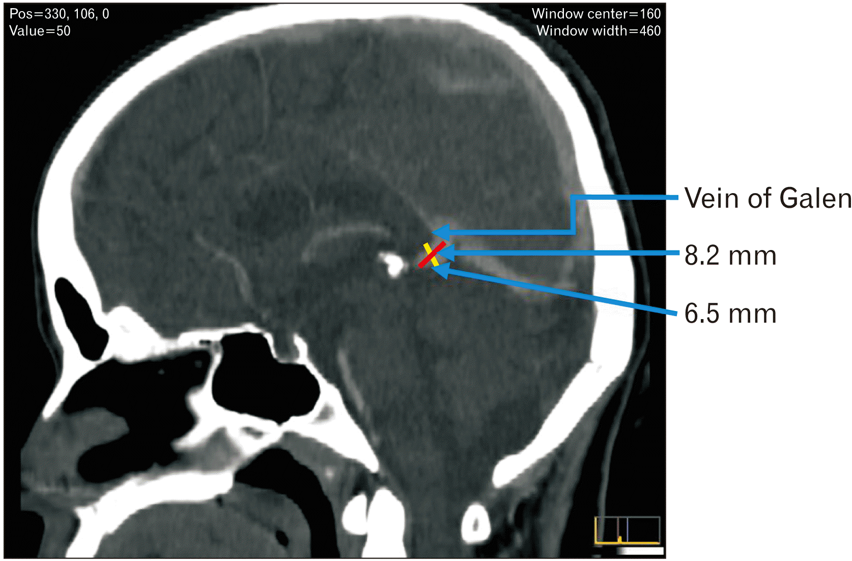
The vein of Galen was visualized in all 100 CTV records. The mean length of the vein of Galen was 9.8±2.7 mm with the maximum length of 15.9 mm and minimum length of 3.5 mm, whereas the mean diameter was 4.08±1.04 mm with the maximum diameter of 6.8 mm and minimum diameter of 2 mm. The CTV image showing the measurement of length and diameter of the vein of Galen is given in
Fig. 1.
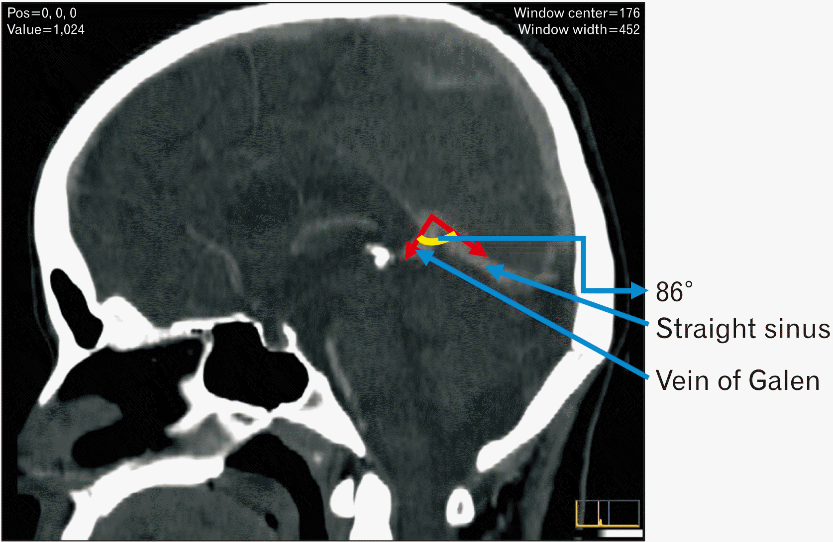
The mean angle between the straight sinus and the vein of Galen was 64.2° with the maximum angle of 99° and minimum angle of 24°. The CTV image showing the measurement of angle between the vein of Galen and the straight sinus is given in
Fig. 2. The dimensions of vein of Galen were compared between the sex and the age groups. However, there was no significant difference in dimensions between the sex and the age groups. The overall dimensions and comparison of dimensions of vein of Galen between sex and age groups are given in
Table 1.
Trajectory to vein of Galen
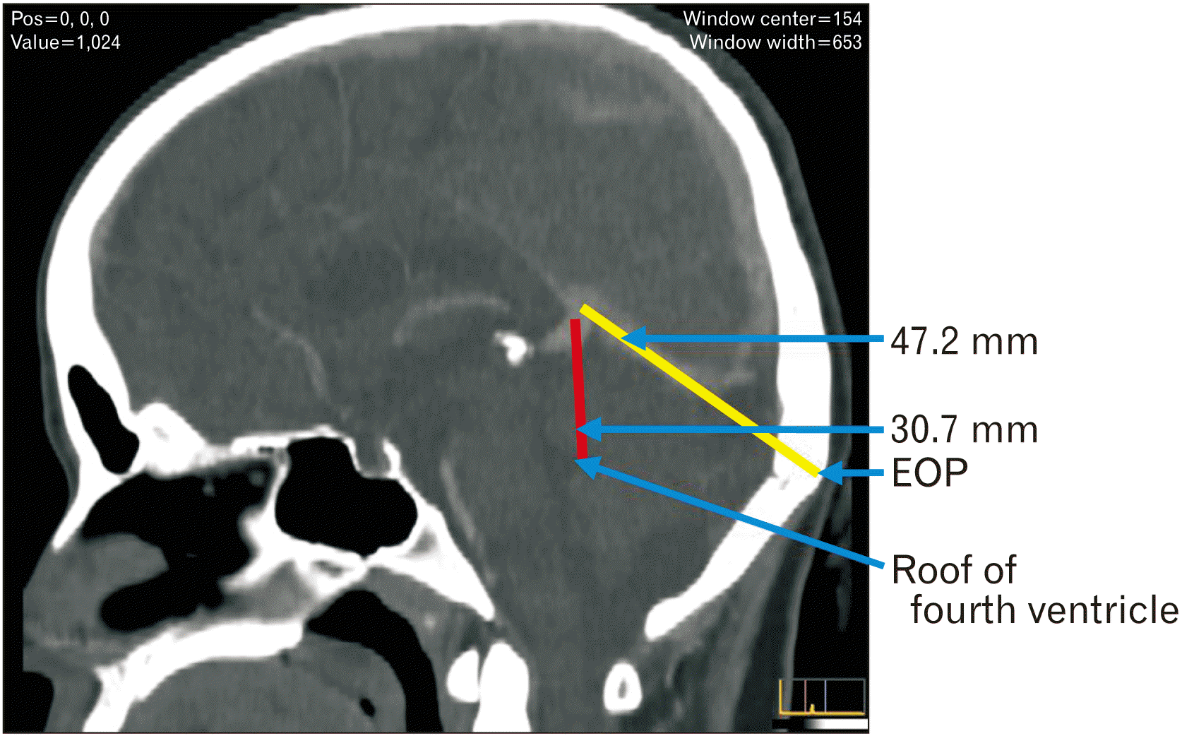
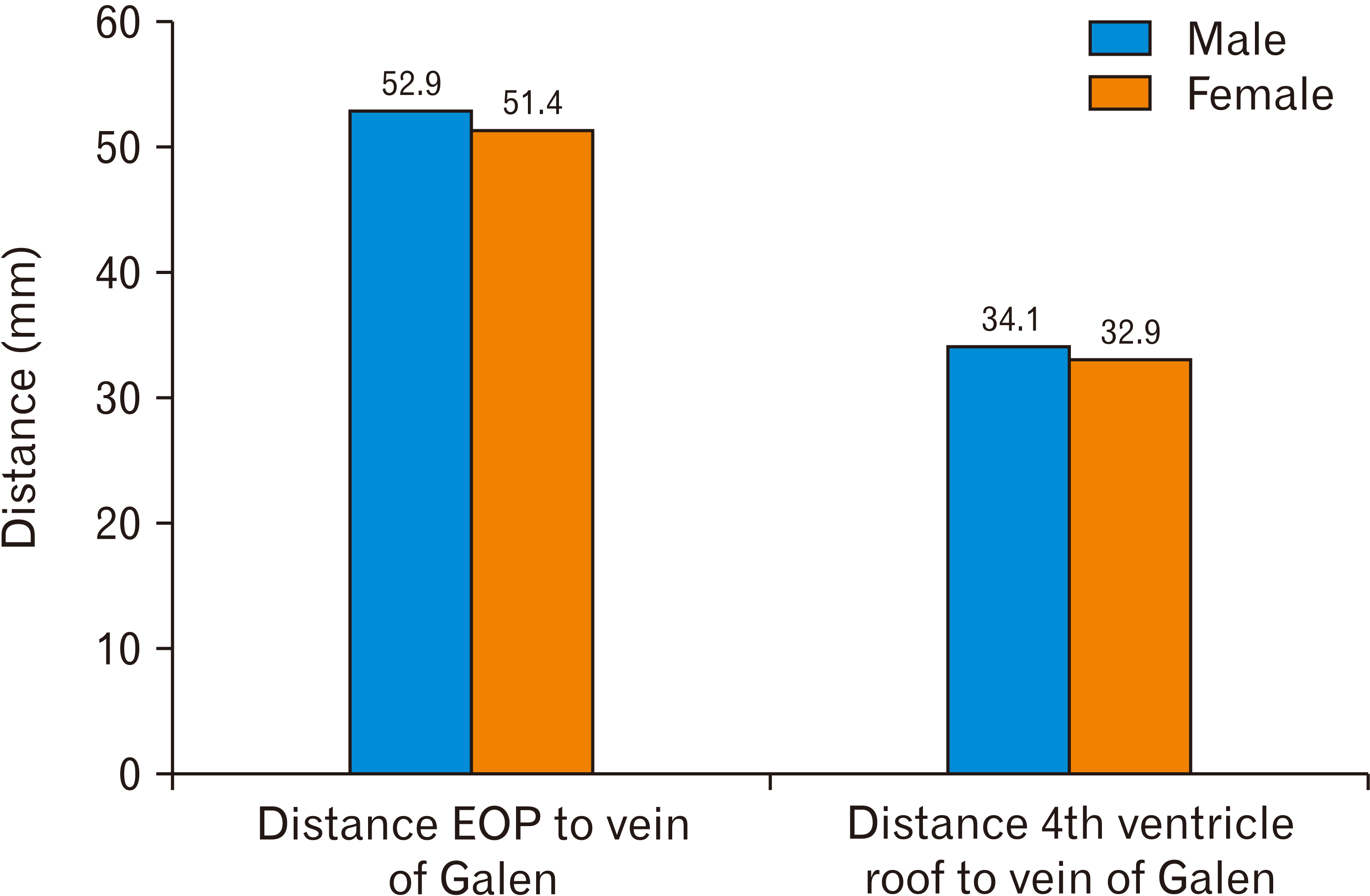
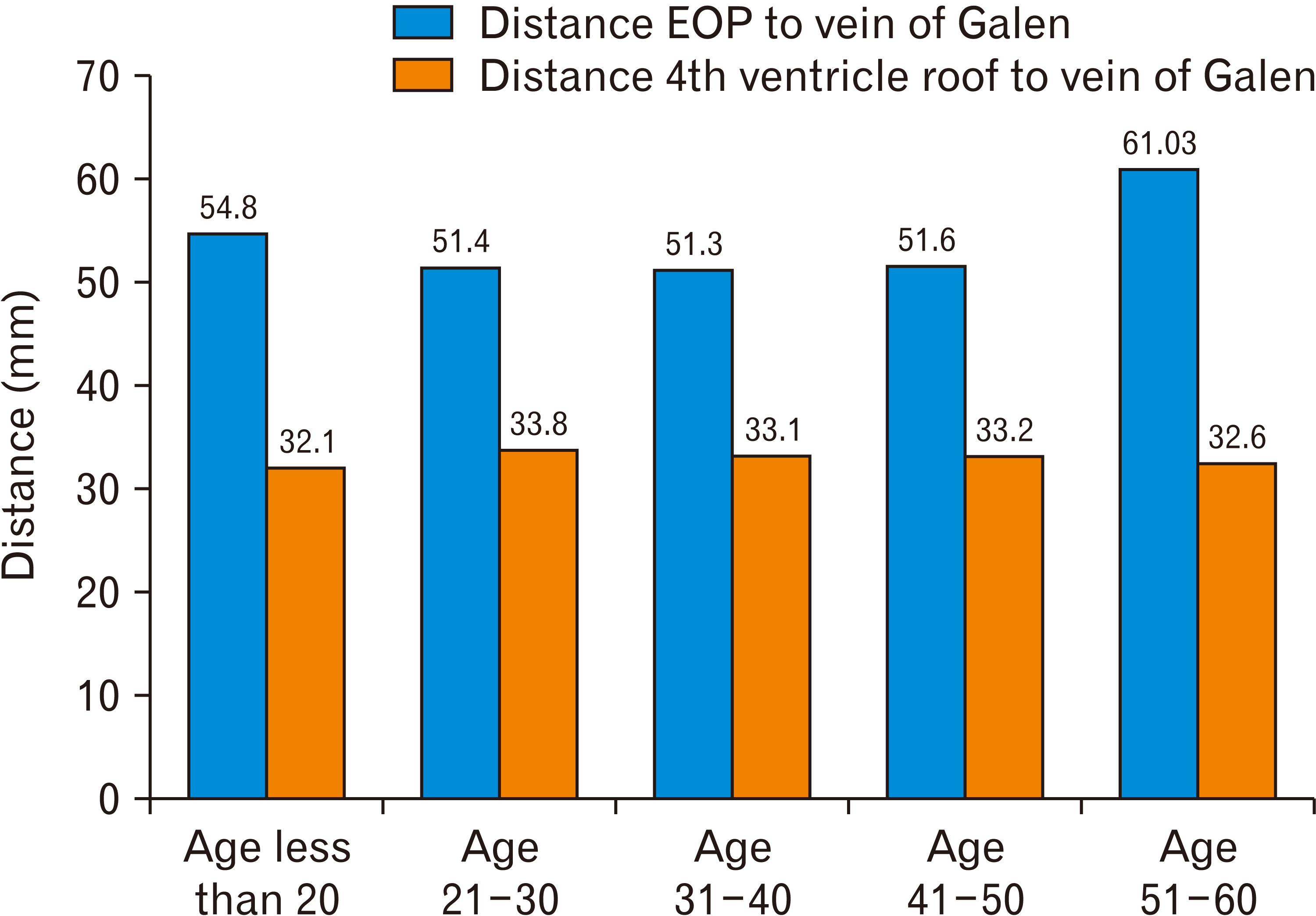
The trajectory to vein of Galen, mean distance from two reference points EOP and the roof of the fourth ventricle were 52±6.9 and 33.3±4.5 mm respectively. The measurements are shown in the
Fig. 3. There was no significant difference between the sex and the age groups. The overall trajectory measurements of vein of Galen are given in
Table 2. The comparison of trajectory to vein of Galen between sex and age groups are given in
Figs. 4 and
5 respectively.
Discussion
In the present study about 100 CTV records were studied and the dimensions of vein of Galen were noted. Though many authors across the globe have tried studying the dimensions of the vein of Galen, there is paucity in the available literature, especially in the radiological studies. The various studies showing the dimensions of the vein of Galen are summarised in the
Table 3.
In a cadaveric study by Chaynes [
4], the mean length of the vein of Galen was 10.33 mm. According to the author, the vein of Galen was found to be shorter if they are formed above the splenium of corpus callosum and it is said to be longer if the vein is formed below the splenium of corpus callosum [
4]. While Ono et al. [
1] measured the mean size of the vein of Galen at the point of its termination which was about 7.3 mm. In other cadaveric studies by Browder et al. [
7] and Isao Yamamomo and Kageyama [
8] the mean length was 15 mm and 12 mm respectively. The mean length of the vein of Galen in the present study using CTV was found to be 9.8±2.7 mm.
In the study using magnetic resonance venography (MRV), the mean diameter of the vein of Galen was 4.39 mm using three-dimensional spoiled gradient recalled (SPGR) Echo MRV, and it was 4.04 mm using two-dimensional time of flight (TOF) MRV [
9]. Great vein received many tributaries as it coursed, increasing its width along the course. The mean diameter of the vein of Galen in the present study using CTV was found to be 4.08±1.04 mm.
In our study we have measured the angle between the vein of Galen and the straight sinus using CTV. The mean angle was 64.2°. In a cadaveric study by Chaynes [
4]. the author measured the angle between the vein of Galen and the straight sinus using a goniometer in 25 cadavers. The angle ranged from 16°–117° with a mean of 75.25°. Further the author classified the angle as acute, obtuse and right angle based on their range. Angle was sharp in the range of 16°–62°. This acute angle was noticed in 9 cases by the author when the flow of vein of Galen was along the curve of the splenium of the corpus callosum. The angle was said to be right angle when the range is from 75°–104°. This type of angulation was seen in 15 cases where the vein of Galen was not in close proximity to the splenium of the corpus callosum. Obtuse angle was noticed when the vein did not follow splenium of corpus callosum rather; it had a horizontal course. The angle was about 117°, seen in a single case. The angle also varied depending on the location of the apex of the tentorium [
4]. The tentorial apex may be located either above or underneath the corpus callosum, making the angle acute or flat, respectively [
1]. In another cadaveric study by Ghali et al. [
10] the minimum angle was 60° and maximum angle was 80°.
Further in this study we have used two different reference points from where the distance to the vein of Galen is measured. The first reference point used was the EOP. The mean distance from the EOP was 52±6.9 mm. The second reference point used was the roof of the fourth ventricle. The mean distance from the roof of the fourth ventricle was 33.3±4.5 mm. These distances will give an idea of approximate location of the vein of Galen. This would help the neurosurgeons in planning the approach to the vein of Galen during various neurosurgical procedures like in the removal of pineal tumour.
The various surgical approaches used for the removal of the pineal tumour are supra- cerebellar-infratentorial, occipital interhemispheric transtentorial, posterior transcallosal posterior trans ventricular and combined supra and infra tentorial approach. The approaches are selected based on the location of the pineal tumour [
1,
3,
4]. The various surgical approaches to the pineal gland tumour is given in
Table 4. Though there were many approaches provided in the literature the supracerebellar infratentorial is the most effective, safe and commonly used, because this method provides the complete removal of the tumour with low morbidity and good histopathological diagnosis [
11]. The occipital interhemispheric transtentorial approach is found suitable for vascular malformations [
11]. This occipital transtentorial approach gives a good visibility of the tentorial notch paving the way for dissection of large tumours. Even though there are lot of approaches suggested the iatrogenic injury to the deep veins and their tributaries are inevitable leading to disturbances in consciousness, visual impairment, hemiplegia and death. Thus it is imperative to know about the skull base vascular anatomy before any surgical approaches [
12].
Embryology of vein of Galen (Fig. 6)
An accessory straight sinus called falcine sinus appears at the falx cerebri around the fifth month of gestational life. This falcine sinus appears transiently above the straight sinus connecting the superior sagittal sinus and Galen’s vein [
13].
The remanent of this falcine sinus appears as bulbous prominence. Widjaja and Griffiths [
13] reported two such bulbous prominences in their study using MRV. These prominences are normal variants in the absence of a fistula, provided the patient is asymptomatic [
13]. This necessitates the importance of differentiating these bulbous prominences in the absence of fistula, as they mimic aneurysm of great vein of Galen.
Clinical importance
Vein of Galen malformation and aneurysm
The vein of Galen is developmentally the remanent of the caudal portion of the median prosencephalic vein of Markowski. This median prosencephalic vein usually regresses, but here, in case of malformation, instead of regression, there is a continuous enlargement of median prosencephalic vein leading to the aneurysmal formation in the vein of Galen malformation. The endovascular embolization using liquid embolic agents like Onyx or N-butyl-cyanoacrylate along with multidisciplinary approaches like shunting for hydrocephalus after embolization, has improved the prognosis [
14]. There was increased incidence of post-procedure haemorrhage in transvenous approach when compared to the transarterial approach. This is because of the connections of the bulbous enlargements in malformations to the superficial subependymal veins through choroidal veins or to the deeper veins through underdeveloped internal cerebral veins. Stereotactic radiotherapy can be used in older people after staged endovascular embolization [
5].
Aneurysm in the vein of Galen more or less has the same presentation as that of the vein of Galen malformation. This necessitates us to differentiate between the vein of Galen malformation and aneurysm. Aneurysmal dilatation of the vein of Galen occurs in a normally developed vein of Galen as a part of dural arteriovenous fistula. There is dilatation in the vein due to the blockage or stenosis in the outflow [
14]. With the advancement in neuroradiological techniques, diagnosis has become fast and easier. Multimodality management involves surgical removal and endovascular embolization [
15]. Thus the findings of the present study would give a rough idea for the neurosurgeons about the location of the vein of Galen and its dimensions. This might be helpful for approaching the malformations or aneurysm during endovascular procedures.
In conclusion, the anatomy in and around the vein of Galen is very complex. So, the knowledge of the morphometry and trajectory to the vein of Galen, is of utmost importance for the neurosurgeons for opting the definite surgical approach to the clinical conditions arising in and around the vein of Galen.


 PDF
PDF Citation
 Print
Print


 XML Download