

 PDF
PDF Citation
 Print
Print


Introduction
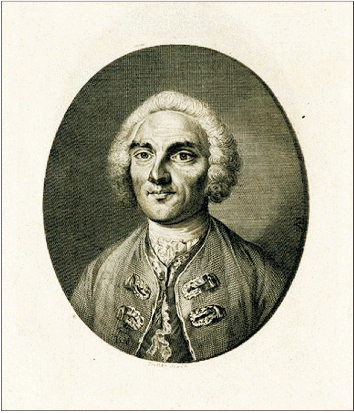
Johannes Nathanael Lieberkühn (1711–1756) was a German anatomist (Fig. 1) whose fundamental work in understanding the details of the digestive system at microscopic level was pivotal in advancement of anatomical knowledge during eighteenth century [1]. His keen interest in mechanics as well as iatromathematics and their application in the realm of medical science constituted the core element of his body of work [2]. Although histological staining techniques were yet to be introduced in anatomical practice during his time, Lieberkühn was able to comprehend minute details pertaining to the glands present in relation to villi of small intestine and the arrangement of blood vessels in the vicinity of a villi [3, 4]. His extraordinary flair in the field of mechanics guided him to assemble meticulous experimental models through which he could empirically describe the function of tubular glands in small intestine (glands of Lieberkühn) which were located in epithelial invaginations at the base of each villi (crypts of Lieberkühn) [1]. It is worthwhile to mention here that his exemplary work in the field of anatomical science was achieved through the application of inventions made by none other than Lieberkühn himself in the field of optics [5, 6]. To be precise, Lieberkühn’s contributions were seminal in the field of anatomy and possibly catalysed the advent of histological staining methods which revolutionized the field of microscopic anatomy as a novel epistemological method [3]. In modern times, when anatomical practice is directed to reveal relevant details at nano level, it would be prudent to revisit the achievements of this luminary scholar whose noteworthy contributions constitute significant landmarks in the eventful evolutionary process of the discipline.
Review
Methods
An extensive literature search was undertaken for this study and indexed databases such as Medline, PubMed, Scopus, EMBASE, CINAHL, Google Scholar as well as popular search platforms such as Wikipedia and standard Google search engine were referred to for relevant published materials. The following terms were used during literature search: “Johannes Nathanael Lieberkühn”, “Lieberkühn”, “Lieberkühn Biography”, “Lieberkühn and anatomy”, “Lieberkühn and microscopy”, “Microscopic anatomy in eighteenth century”, “Anatomical discoveries in 18th century”, “Lieberkühn and digestive system”, “Crypts of Lieberkühn”, “Glands of Lieberkühn” and “Lieberkühn and vascular system”. Published texts of Lieberkühn and their translations in English were consulted from online libraries while conducting the present study and wherever applicable have been appropriately referenced. The images used in the text were procured from the internet and it was ensured that all the figures included in this study are in public domain i.e. free from copyright issues.
Inclination towards iatromathematics, mechanics and medicine early in his career
Johannes Nathanael Lieberkühn (1711–1756) was a noted German anatomist and physician of early eighteenth century. He was born in Berlin and his father Johannes Christianus Lieberkühn was a goldsmith by profession. Lieberkühn fulfilled his father’s wish (he wanted his son to devote himself to religious practice) and enrolled himself in a theology course in the prestigious Friedrich-Schiller University at Jena, Germany [2]. However, Lieberkühn was himself interested to pursue a career in science and he added mathematics, physics and philosophy in his academic repertoire at Jena [7]. Lieberkühn’s stay at Jena and his association with scholars across fields proved decisive in his eventual career in medicine. He was deeply influenced by the works of Georg Erhard Hamberger (1697–1755) who along with being a physician was also a noted iatromathematician of that period [1]. It may be mentioned here, that iatromathematics was a popular discipline that has its roots in 17th century Italy, whereby physicians tried to apply laws of mathematics and mechanics in order to understand the functioning of human body [8]. Lieberkühn’s interest in medicine was triggered by his intriguing experience with Hamberger and later on he applied this knowledge towards application of laws of mechanics in his scientific experiments. Accordingly, he continued with his tryst of exploring multiple disciplines and went on to study chemistry, anatomy and physiology [9]. During this time, his perceptions were influenced by Johann Adolph Wedel (1675–1747), who was a physician and the professor of medicine at University of Jena and Hermann Friedrich Teichmayer (1685–1746), who was the professor of experimental physics, medicine and botany at the same institute [7].
Graduation in medicine from Leiden
In 1733, Lieberkühn completed his studies in Jena and moved to Rostock, a city on the German Baltic coast, to undertake activities as a religious preacher [10]. However, he realized very soon that his actual place was in the laboratory, where he could pursue his quest for knowledge in the realm of medical science [2]. In this context, he was aptly guided by Johann Gustav Reinbeck (1683–1741), German theologian and philosopher, as he recognized Lieberkühn’s aptitude for medical science [10]. Reinbeck introduced Lieberkühn to the Prussian king, Frederick William I. The king personally interviewed Lieberkühn and upon realizing his potential in science and medicine, relieved him of his religious duties [11]. The chain of events paved the way for Lieberkühn to follow his dream of pursuing a career in medicine. Shortly after, Lieberkühn was incorporated as a fellow by the Berlin Academy of Sciences in recognition of his earlier academic exploits at Jena [1]. Being motivated by the turn of events, Lieberkühn returned to Jena in 1735 and embarked on an academic journey that would prove to be seminal in the advancement of anatomical sciences [7]. His odyssey in quest of knowledge continued as he completed his sojourn in Jena and arrived at Erfurt, where he was inducted as a fellow of the imperial Natural Sciences Academy [9]. He left his motherland and enrolled in the University of Leiden, The Netherlands for further studies in medical sciences [2]. It may be mentioned here that Leiden was already established as a centre of excellence for medical studies in Europe [12]. His stay in Leiden proved to be decisive in developing his perspectives for his future academic exploits. As a medical student, he came in contact with prominent figures in medical sciences such as Herman Boerhaave (1668–1738), Bernhard Siegfried Albinus (1697–1770) and Gerard van Swieten (1700–1772) among many others. He completed his medical studies in Leiden and was awarded his medical degree in 1739 [13].
Devoting his life to anatomical practice as a physician in berlin
After completing his medical studies, Lieberkühn travelled to London and Paris (both were established and preeminent centres for advances in studies within the realm of medical sciences) to apprise himself with the recent headways in medicine and effectual areas of research pertaining to anatomical sciences [9, 14]. The following year i.e. in 1740, Lieberkühn came back to his homeland and settled down in Berlin [2]. In the same year, he was honoured through induction as a member of the Collegium Medico-Chirurgieum, a scientific body which was entrusted with improving the practice of medicine in the Holy Roman Empire [11]. From this time onwards till his untimely death in 1756 (at the age of 45), Lieberkühn worked as a physician in Berlin and devoted himself to exploring the hitherto unravelled facets pertaining to anatomical details of the human body [2]. His accomplishments in the field of anatomical sciences involved his unparalleled inputs resulting in invention of highly revered variants of light microscopes. The microscopes so designed and invented by Lieberkühn became the very basis of his ground breaking exploits involving micro-anatomical details pertaining to the field of digestive system and vascular system.
Fundamental inventions pertaining to optics in microscopy
Microscopic anatomy was established as an integral element of anatomical sciences by late 17th century courtesy of extraordinary works of luminary scientists like Marcello Malpighi (1628–1694) and many others [3, 15]. It may be mentioned here, that compound microscope (that was used by anatomists during 17th century) was discovered by Galileo way back in 1609 [16]. The instrument underwent minor modifications and adjustments as per the needs of the anatomical practitioners and the trend continued into first half of 18th century [17]. Lieberkühn, owing to his initial training in iatromathematics as well as mechanics in Jena, developed deep interest in the application of his knowledge in refining optical details of the microscope [18]. In accordance with his ongoing experiments with optical aspects of microscopes, Lieberkühn invented an improved device for illuminating specimens for microscopic examination in 1738 while continuing with his medical graduation in Leiden. He designed a reflector (basically a concave mirror) for studying tissue specimens under microscope by applying the principle of epi-illumination, whereby the light source and detectors are positioned on the same side of tissue at a particular angulation [19]. The reflector (often termed as Lieberkühn reflector) was made of silver and other highly polished metal and considerably increased the amount of light illuminating the tissue specimen. Consequently the reflector devised by Lieberkühn contributed to illuminating an opaque object placed at almost any distance from the observer while using the microscope [20]. The introduction of the reflector significantly contributed towards his success in anatomical discoveries.
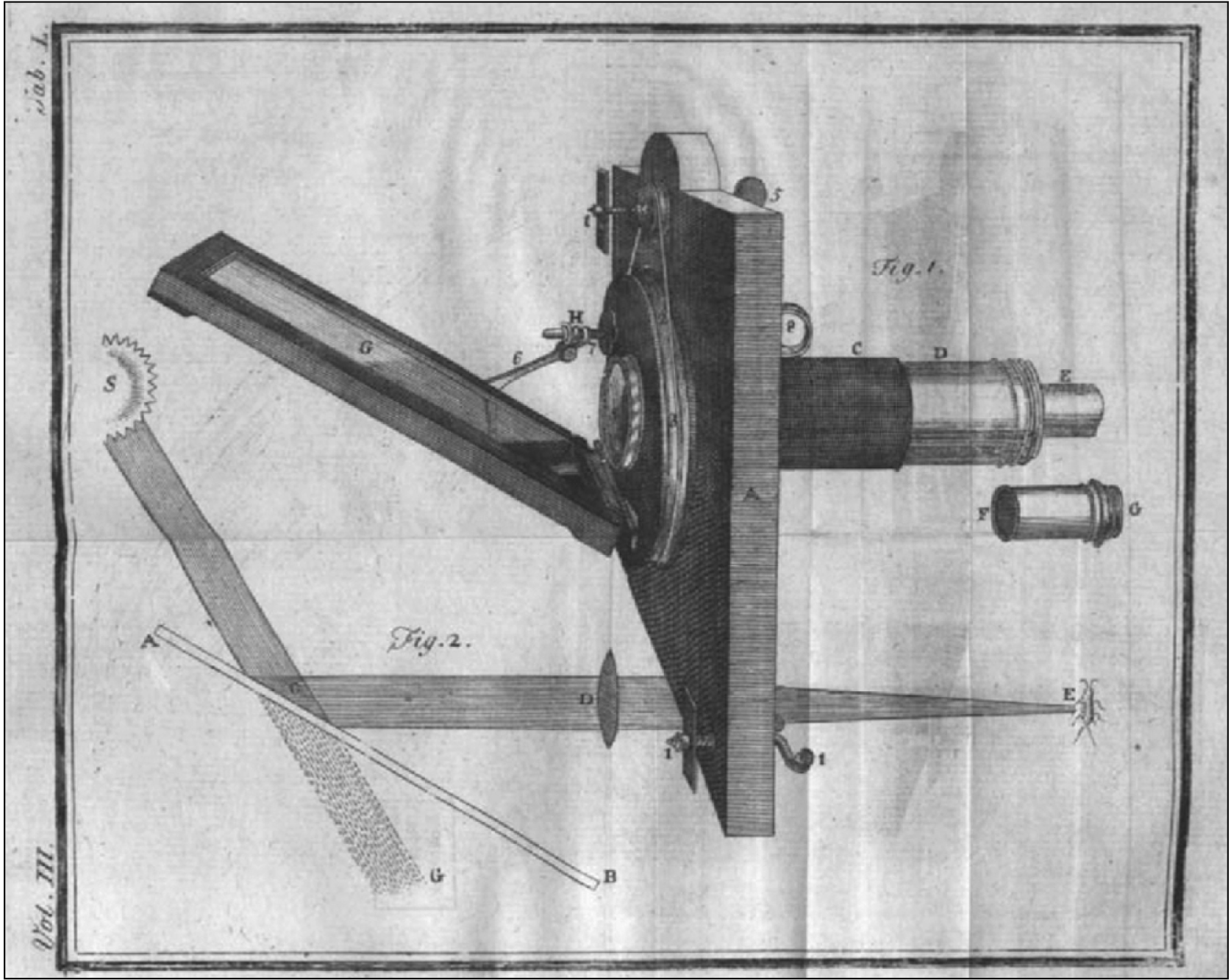
Lieberkühn’s tryst with pioneering contributions towards refining the optical efficiency of microscope continued as he invented the solar microscope in 1740 after he had settled in Berlin [2]. The solar microscope was an improvised device that required very bright light (such as sunlight) and an intense reflection of the same was focussed on the transparent specimen by the highly polished reflector (designed by Lieberkühn himself) to provide excellent resolution of the minute anatomical details to the viewer [21]. To be precise, this device provided the viewer a highly magnified image in sufficiently high resolution so as to appreciate the micro-anatomical details (Fig. 2). The distinct advantage of solar microscope over contemporary light microscope was that it provided a better resolution of the specimen examined. The solar microscope formed an important element in the repertoire of Lieberkühn for his scientific exploits. Lieberkühn’s efforts were pivotal in his pursuit of anatomical knowledge through experiment based methods, a trend prevalent among European anatomists since seventeenth century [3].
Discovery of intestinal crypts and glands in relation to villi
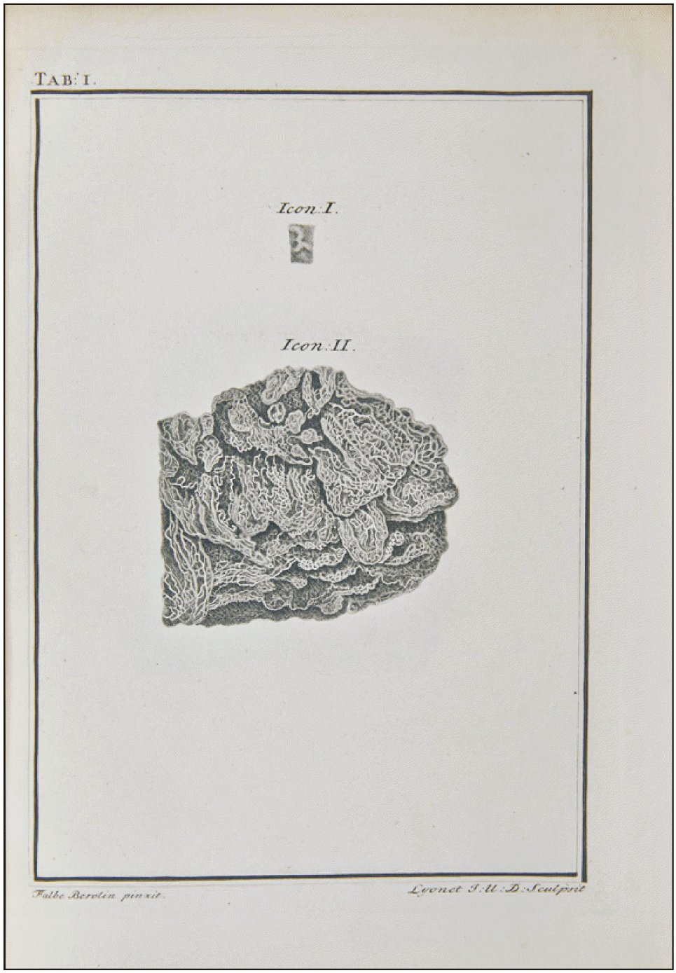
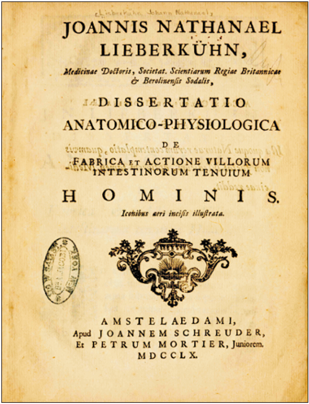
Lieberkühn focussed on exploring the anatomical details of the digestive system while undertaking his medical graduation in Leiden. This is evident from the topic of his dissertation “De valvula coli et usu processus vermicularis” where he ventured to elicit the anatomical details of the ileocecal valve (Fig. 3) [22]. His work was appreciated by his mentors Boerhaave and Swieten, who were noted physicians in Leiden. Incidentally, anatomy of the ileocecal valve was first described by Swiss anatomist Caspar Bauhin (1560–1624) in 1588 and was later detailed by Dutch physician Nicolas Tulp (1593–1674) [23, 24]. By this time, Lieberkühn had started working on the anatomical details of the glands attached to the villi of small intestine. Lieberkühn observed that the base of each villus in the small intestine is surrounded by multiple epithelial invaginations (crypts). Lieberkühn made these crypts comprehensible for anatomical observations through meticulous and proficient injections comprising of a mixture of wax, turpentine and dark resin. The wax mixture also possibly provided support to the delicate anatomical structures and was of considerable assistance during microscopic observation. With the advent of histological staining techniques almost a century later, the use of wax became an integral component of microscopic studies [3]. Lieberkühn’s endeavour was an ardent example of empirical practice in anatomical science as he relied on experimental data for validation of his findings rather than focussing on theoretical ideas. Through microscopic observations he further elucidated that these epithelial invaginations (crypts) contain numerous tubular glands that secrete digestive juices having enzymes such as amylase, protease and lipase (Fig. 4). These digestive juices are released in the form of mucous rich secretions, where the mucin comes from the goblet cells of intestinal mucosa. From the functional viewpoint, it is the digestive juices secreted by glands located in crypts of Lieberkühn that convers the chyme to chyle in the small intestine. Lieberkühn further constructed a model with the help of simple laboratory instruments such as brass tubes, glass tubes and metallic cones to demonstrate the complex pathway for the movement of chyle in the small intestine. He demonstrated the flow of chyle (whitish in appearance) from intestinal lacteals to the villi and flow of lymph (clear fluid) from the villus into the collecting lymphatic vessels in the mesentery [25]. His demonstrations through methodically designed experimental set-up were highly appreciated by the scientific community. As a recognition of his exemplary contributions towards understanding the intricacies of the human digestive system, Lieberkühn was inducted as a fellow of the Royal Society of London in 1740 [26]. Later on, Lieberkühn published his detailed observations with regards to structure and function of gland attached to the villi of small intestine (glands of Lieberkühn) as well as the specifics of structure and function of villi of small intestine in his text, De fabrica et actione villorum intestinorum tenuium hominis, which was published in 1745 (Fig. 5) [25].
Observations on arrangement of vessels juxtaposed with villi
During his extensive studies pertaining to glands related to villi of small intestine, Lieberkühn got interested in the details of circulatory vessels and the mechanics involved in the movement of fluid in these vessels [9]. As he was already working on experimental models involving the digestive system, hence he focussed his attention towards mucosal vascular architecture of small intestine [27]. He used animal models (mostly amphibians) for this purpose [28]. He noted that the arterioles related to a villi ramify to the under surface of the mucosa and eventually terminate in the capillary plexus surrounding the Crypts of Lieberkühn. He also observed that the cryptic capillary plexus gave rise to the entire villus capillary net [25, 29]. His findings were later validated and it was found that the circulation system for a single villus began with a central villous arteriole and terminated by draining into a single central villous vein [30]. It is quite remarkable that Lieberkühn was able to undertake such accurate observations of fine anatomical details without advanced histological techniques at his disposal. The implications of his scientific exploits were way ahead of his time and are a testimony of his exemplary scientific skills.
Grandiose collection of specimens on vascular anatomy
His volume of work in vascular anatomy can be gauged by the fact that in his lifetime, he had accumulated more than 400 specimens of vascular tissues of various animals. Such enormous collection was built around his tireless desire to expose and preserve vascular tissues for subsequent exploration of anatomical details [2, 31]. His collection comprised of 3 types of anatomical preparations: wet specimens preserved in transparent fluid, dry specimens injected and hardened and tissue sections hardened through fixation for viewing under microscope [4, 9]. After his death, his wife Catherine donated the entire Lieberkühn collection to the Russian Medical Military Academy’s Museum of Anatomy, where they remain in public display till present day [32]. Lieberkühn’s collection of anatomical specimens was in accordance with the rapidly evolving epistemological method of building anatomy museums, which has its roots in early eighteenth century Europe [3, 33].
Conclusion
Lieberkühn was a prodigious anatomist and an ardent practitioner of optical mechanics relevant to the field of microscopy. His ground breaking inventions in the field of microscopy aptly complemented his quest for anatomical knowledge at microscopic level. His meticulous descriptions and deep insights in relation to the anatomy of intestinal glands, their functioning and intricate association with network of blood vessels juxtaposed with the intestinal villi is unparalleled in medical science. His tireless efforts helped in comprehending the anatomy of digestive system and paved the way for further enhancement of knowledge with the advent of histological staining methods almost a century later.
 XML Download