

 PDF
PDF Citation
 Print
Print


Introduction
Situs inversus totalis (SIT) is a congenital condition in which there is a complete transposition of the organs and vessels along the longitudinal axis of the body. The reported incidence is 1 in 25,000 live births, with no sex or racial predilections. Genetic and environmental factors have been implicated as the cause of this condition [1].
Although the condition remains asymptomatic, a few cases of concomitant vascular variation in SIT have been reported. As far as the existing literature is concerned, any vessel is prone to be affected and can prove as a surgical dilemma [2]. Generally, vascular anomalies in SIT have been reported from classical cadaveric dissections and targeted radiological imaging in the living. Limited studies exist that have attempted to describe the vascular system and its variations in SIT from a whole-body computed tomographic (CT) angiography perspective. We herein report a case of SIT in which a whole-body postmortem CT (PMCT) was performed as a part of the forensic death investigation process, and discuss the results based on the radiological findings.
Go to : 
Case Report
Case history
A man in his 30s of Japanese ethnicity was found dead on his bed in the house. The police brought the deceased’s body to our university for a forensic autopsy. His medical records showed that he was a known patient of SIT but were otherwise unremarkable. He was not known to consume alcohol or smoked cigarettes in his lifetime.
Postmortem CT angiography (PMCTA)
Before the autopsy, we performed the PMCTA procedure using the multiphase postmortem CT angiography (MPMCTA) technique, following their standardized protocol [3]. Briefly, the right femoral vessels were exposed and cannulated. The contrast mixture was prepared using 210 ml of Angiofil® (Fumidica, Muri, Switzerland) as the main contrast agent and 3,500 ml of paraffin oil (as solvent). The angiographic procedure was then performed by injecting the contrast mixture in three phases (arterial phase, 1,200 ml; venous phase, 1,800 ml; and dynamic phase, 500 ml) using the perfusion device Virtangio® (Fumidica). A 64-slice multidetector (Aquilion 64; Toshiba, Otawara, Japan) CT scanner was used to acquire the CT images by coordinating with the above perfusion device. The CT scan parameters were as follows, tube voltage, 120 kV; tube current, 200–400 mA; and a slice thickness of 1.25 mm (arterial and venous phase), and 2.50 mm (dynamic phase). A three-dimensional (3D) workstation (AquariusNet; TeraRecon, San Mateo, CA, USA) was used to reconstruct the MPMCTA images. The Anatomist cross-checked the radiological findings observed during the conventional autopsy.
General and pathological findings
The apex of the heart was pointing toward the right, with the right ventricle giving rise to the aorta (Fig. 1). Three aortic sinuses were visible, with the left coronary artery (LCA) emerging from the anterior sinus and the right coronary artery (RCA) arising from the right-posterior sinus. The left main coronary artery descended between the left atrium and ventricle (coronary sulcus) and divided into two main branches, one, which ran along the base of the heart moving toward the apex, and the other continuing as a posterior descending artery (PDA). The RCA branched into two branches, corresponding to the circumflex artery (CA) and anterior descending artery. Interruptions of contrast flow were noted along the right coronary vessels, suggesting a blockage in the lumen (Fig. 2).
 | Fig. 13D VR reconstruction of the heart and great vessels. A dynamic phase of MPMCTA. Right-sided heart, the apex pointing towards the right (star) and AA emerging from the right ventricle. AA, ascending aorta; VR, virtual reconstruction; MPMCTA, multiphase postmortem computed tomographic angiography; 3D, three-dimensional.
|
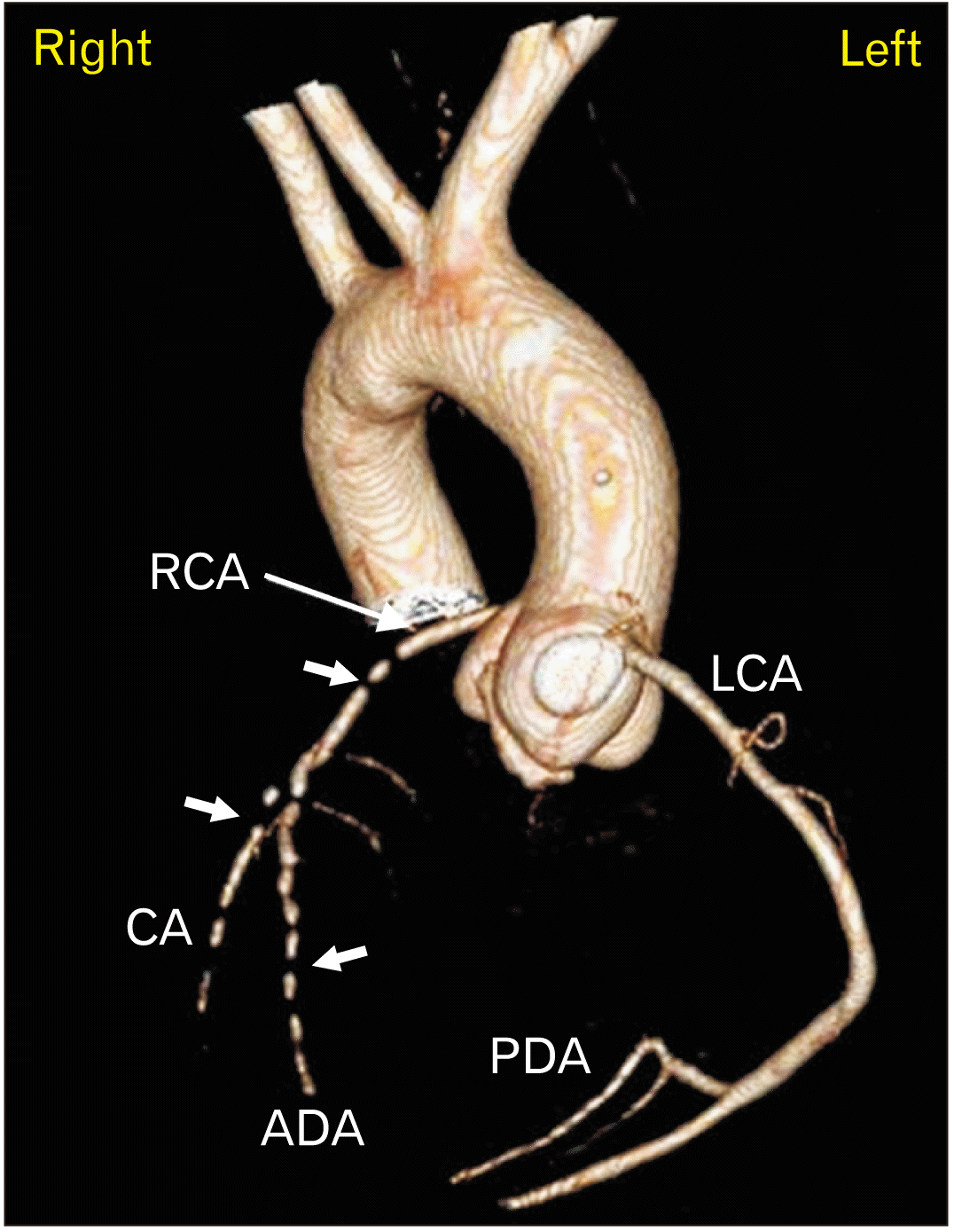 | Fig. 23D VR reconstruction of coronary arteries. Arterial phase of MPMCTA. Note the contrast flow interruptions shown as filling defects (short arrows). VR, virtual reconstruction; MPMCTA, multiphase postmortem computed tomographic angiography; RCA, right coronary artery; LCA, left coronary artery; CA, circumflex artery; ADA, anterior descending artery; PDA, posterior descending artery; 3D, three-dimensional.
|
Almost all major arterial branches including the intracranial vessels and drainage of veins depicted the complete inversion of the Situs solitus, but it was otherwise normal. Similarly, abdominal organs were completely reversed in their position and anatomy, but with normal morphology. No radiological abnormality in the sinuses and airways was noted.
Vascular variations
Right internal mammary artery (IMA)
The right IMA emerged from a common trunk with other branches, namely, the suprascapular artery (SSA), and transverse cervical artery (TCA), anterolaterally to the thyrocervical trunk (ThyCT) (Fig. 3).
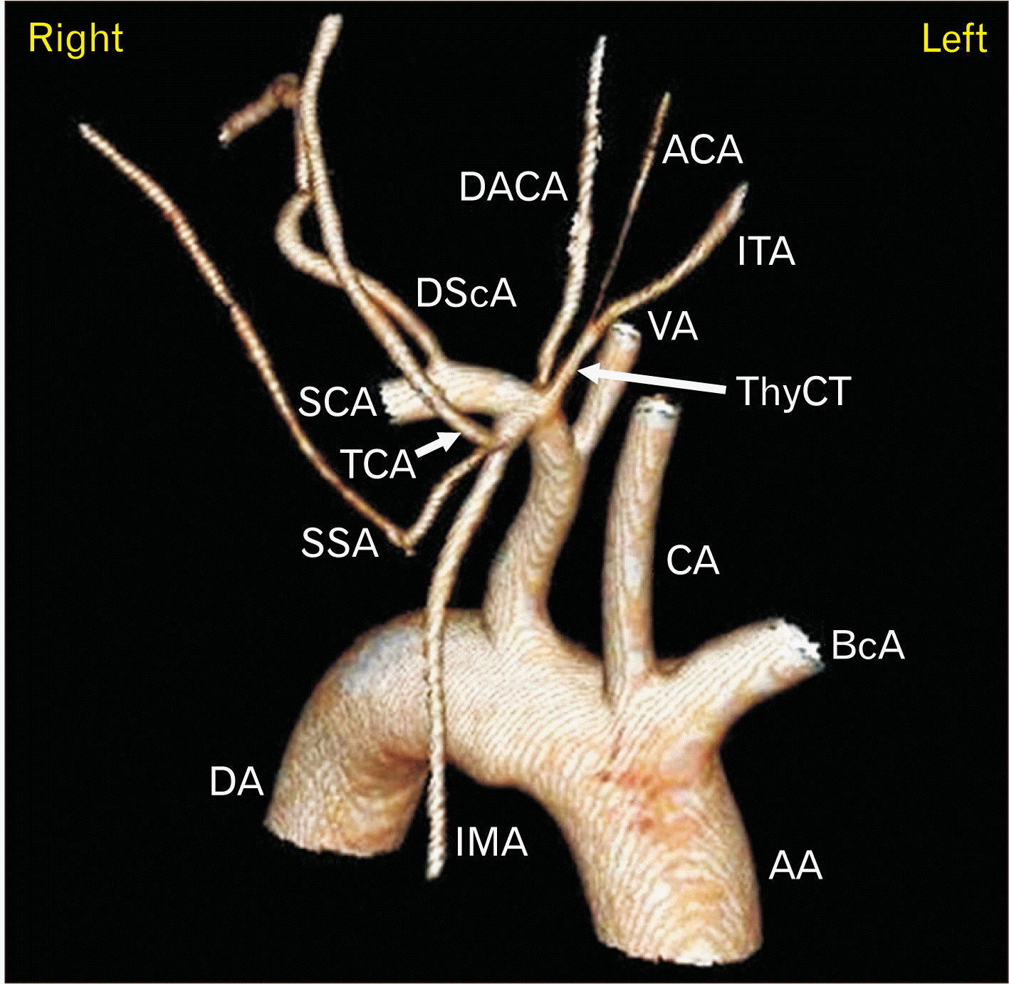 | Fig. 33D VR reconstruction of the right anterior neck. Arterial phase of MPMCTA. Note the common origin of the right IMA with other arterial branches, namely the TCA and SSA. VR, virtual reconstruction; MPMCTA, multiphase postmortem computed tomographic angiography; IMA, internal mammary artery; TCA, transverse cervical artery; SSA, suprascapular artery; AA, ascending aorta; BcA, brachiocephalic artery; CA, common carotid artery; VA, vertebral artery; ThyCT, thyrocervical trunk; ITA, inferior thyroid artery; ACA, ascending cervical artery; DACA, deep ascending cervical artery; DScA, dorsal scapular artery; SCA, subclavian artery; DA, descending aorta; 3D, three-dimensional.
|
Thyrocervical trunks
Unlike in the right, the left ThyCT emerged from a common trunk with another atypical branch (Fig. 4). The left ThyCT was smaller in caliber compared to the atypical branch. The atypical branch immediately gave a smaller branch corresponding to a SSA, continued further to give rise to the dorsal scapular artery (DScA), and finally divided into several superficial cervical arteries. Both the right and left ThyCT gave rise to only two branches, namely, the inferior thyroid artery (ITA) and ascending cervical artery (ACA).
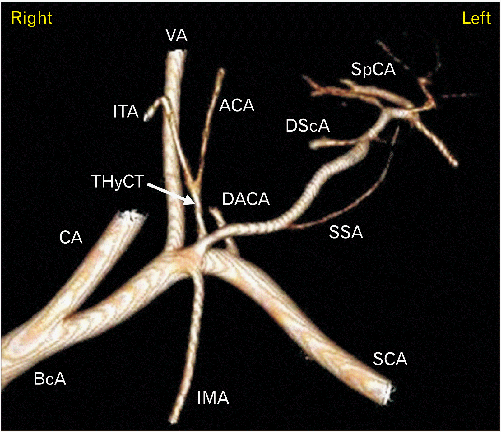 | Fig. 43D VR reconstruction of the left anterior neck. Arterial phase of MPMCTA. Note the common origin of the left ThyCT with another atypical arterial trunk, which is comparatively large. VR, virtual reconstruction; MPMCTA, multiphase postmortem computed tomographic angiography; ThyCT, thyrocervical trunk; BcA, Brachiocephalic artery; CA, carotid artery; VA, vertebral artery; IMA, internal mammary artery; ITA, inferior thyroid artery; ACA, ascending cervical artery; DACA, deep ascending cervical artery; SSA, suprascapular artery; DScA, dorsal scapular artery; SpCA, superficial cervical artery; SCA, subclavian artery; 3D, three-dimensional.
|
Drainage of the testicular veins
The right testicular vein drained into the vesicle venous plexus, whereas the left testicular vein, which appeared grossly dilated, drained into the left renal vein at a right angle to it (Fig. 5).
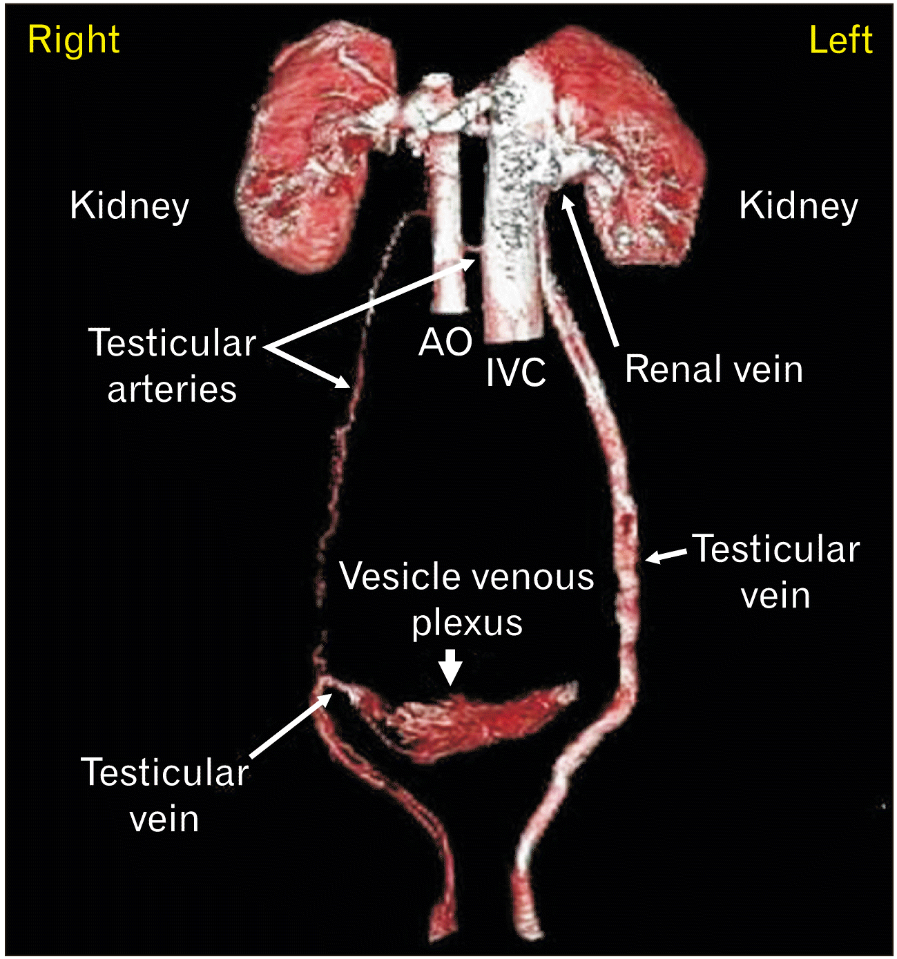 | Fig. 53D VR reconstruction to separate the testicular vessels. A dynamic phase of MPMCTA. Notice dilated & tortuous (varicocele) left testicular vein draining into the left renal vein. Right testicular vein coursing inwards and draining into vesicle venous plexus. Normal testicular arteries. VR, virtual reconstruction; MPMCTA, multiphase postmortem computed tomographic angiography; AO, abdominal aorta; IVC, inferior vena cava; 3D, three-dimensional.
|
Go to :
Discussion
Unlike in other laterality defects (Situs ambiguous, Situs inversus abdominis, etc.), the incidence of vascular and gross organ abnormalities in SIT is less common [4]. Plausibly, in the current case, almost all the major vessels (except a few, mentioned below) were completely inverted, yet with normal morphology. Few authors have reported on the reversal of intracranial structures in SIT [5]. We also observed a similar inverted morphology of intracranial vessels, which is in line with their reports.
The coronary circulation was left dominant. The PDA branched off from the LCA and supplied the posterior interventricular septum. In Situs solitus, coronary circulation is 80%–85% right dominant [6]. However, in the case of SIT, the exact prevalence and nature of cardiac dominance are unknown. There was an interruption of contrast flow in the branches of the RCA (Fig. 2), which suggested the possibility of vessel occlusions, upon which an autopsy revealed more than 90% of its lumen being occluded by atheromatic plagues at several locations. Although no risk factors for ischemic heart disease could be found in the deceased, we diagnosed the cause of death as ischemic heart disease. Perhaps, the incidence of coronary artery disease in these patients may be similar to that in the general population [7]. Whereas much higher incidences of congenital cardiac anomalies (SIT, 41%; other laterality defects, 90.2%) could occur in SIT [4], which was not present in this case, however.
SIT is also reported to be associated with another medical condition called Kartagener syndrome (>50% of cases), in which defective ciliary movements in them lead to chronic sinusitis, bronchiectasis, and infertility [8]. The current case did not have any such features in either the radiological findings or the autopsy.
The IMA’s anatomy and course in the body have been studied extensively owing to their clinical relevance, the arteries being used as a conduit for the bypass graft in coronary bypass surgeries and autologous breast reconstructions [9]. The common origins of IMA with other branches, with the thyrocervical trunk, scapular artery, DScA, and thyroid artery have been reported in Situs solitus [10]. However, anomalies in the origin of IMA with SSA and TCA in SIT have never been reported before. Origin of right IMA with a common trunk in a patient with SIT may need a modified or completely different surgical approach.
In Solitus solitus, the ThyCT arises from the first segment of the subclavian artery, has a relatively shorter length, and it generally divides into the ITA, TCA and SSA. Perhaps, the ThyCT shows more variations in its anatomy and branches than other branches of the subclavian artery [11]. And, as expected, variations of the ThyCT have also been documented in the case of SIT. For example, González-Castillo et al. [12] found two ITA originating from the left ThyCT in a cadaver with SIT. In this study, we did notice the left ThyCT emerging from a common trunk with an atypical branch, virtually in a smaller caliber (Fig. 2). Also, in the current case, both the right and left ThyCT gave rise to only two branches, namely, ITA and ACA.
In a Situs solitus, the left testicular vein drains into the left renal vein, whereas the right testicular vein drains into the inferior vena cava. In SIT, the morphology of gonadal vessels is expected to be inverted. However, in the current case, the right testicular vein drained into the vesicle venous plexus, while the left testicular vein drained into the left renal vein at a right angle, possibly resulting in tortuosity and varicocele of the vessel. Variations of gonadal veins in Situs solitus are common with a reported incidence of 45% [13], yet only one study has described the abnormal testicular veins in SIT [14]. Knowledge about the anomalies in gonadal vessels is essential for it can be a reliable donor graft in reconstructive abdominal surgeries and to better understand the related pathologies that cause the varicocele of the testicular veins, for example. It is hypothesized that these vascular variations occur because of abnormal embryogenesis of vascular channels, and due to erratic ‘pathfinding signals’ during organogenesis in the embryo of SIT [15].
In conclusion, using PMCTA findings we could describe the vascular anatomy, its relevant pathology, and a few vascular variations in a deceased known to have SIT. Our findings might be helpful to clinicians and surgeons alike and add to the body of literature on SIT.
Go to :
 XML Download