

 PDF
PDF Citation
 Print
Print


Introduction
The variations of the muscles of the forearm flexor compartment are not uncommon, which can lead to a variety of clinical disorders that impair forearm and hand function [1]. The anterior compartment contains the flexor muscles of the forearm. These are arranged in superficial and deep groups. Flexor digitorum profundus (FDP) is one of the deep muscles of this compartment. FDP arises from the proximal three-quarters of the anterior and medial surfaces of the ulna, in addition it also takes origin from coronoid process, from the proximal three-quarters of the posterior ulnar border and from the anterior surface of the ulnar half of the interosseous membrane. The muscle ends in four tendons that run deep to the tendons of flexor digitorum superficialis and the flexor retinaculum. The part of the muscle that acts on the index finger is usually distinct throughout, while the tendons for the other fingers are interconnected by areolar tissue and tendinous slips as far as the palm. Ventral to their proximal phalanges, the tendons pass through the tendons of flexor digitorum superficialis to insert on the palmar aspects of the bases of the distal phalanges. The tendons of the FDP undergo fascicular rearrangement as they pass through those of superficialis. Studies suggest that FDP may be joined by accessory slips from the radius (which act on the index finger), flexor digitorum superficialis, flexor pollicis longus (FPL), the medial epicondyle or the coronoid process [2].
Neurovascular compressions are thought to be caused by muscle variations in the forearm. The majority of these variations are discovered during normal anatomical dissections or surgeries. For a clinician to comprehend the atypical symptoms and indications caused by nerve compression, they must have a full understanding of these variants [3]. A comprehensive grasp of this kind of variation is required for radiologists and physicians, as well as hand surgeons for diagnostic purposes and while performing corrective surgical procedures [4]. The purpose of this case report is to address the clinical and embryological significance of the presence of a unilateral auxiliary muscle in the forearm.
Go to : 
Case Report
We report the unilateral presence of an accessory muscle in front of the forearm, accessory to the flexor FDP muscle. During the deep dissection of the front of the forearm of a 69-year-old female cadaver, as a part of practical sessions of first-year medical undergraduates, an anomalous accessory muscle in relation to the FDP muscle was encountered only in the right forearm. The left limb didn’t show any atypical presence of accessory muscles.
An anomalous muscle was approximately 17 cm long and consisted of a spindle-shaped muscle belly with a long tendon (muscle belly: 7 cm and tendon: 10 cm) and was 1.0 cm wide, observed underneath the FPL muscle. The muscle belly arose from the lower third of the anterior surface of the radius, close to the FDP origin (Fig. 1). As a result, we called this muscle accessory muscle (AcM) to FDP. However, no fibrous connection appeared to exist between the AcM and the FDP.When followed distally, the accessory muscle tendon (AcMTn) was found lateral to the FDP tendon for the index finger (Fig. 2), and entered the palm under the flexor retinaculum (as content of the carpal tunnel). AcMTn ran superficial to the adductor pollicis muscle in the palm, and we noticed the first lumbrical muscle as a bipennate muscle taking origin from the adjacent sides of the middle 1.5 cm of the tendons of FDP and AcMTn (Fig. 3). Length of the first bipennate lumbrical muscle was approximately 3.5 cm, formed small tendon, which passing lateral to the tendon of FDP and AcMTn got inserted as usual to lateral side of extensor expansion. Other three lumbricals did not show any variations. After giving origin to first lumbrical muscle AcMTn got merged with the tendon of FDP for index finger. Innervation of the AcM was from a branch of anterior interosseous nerve. The bipennate first lumbrical got its nerve supply from a branch of lateral division of the median nerve in hand. The morphology of adjacent muscles in the flexor compartment was normal. The findings observed by other authors with the present variation are summarized in Table 1.
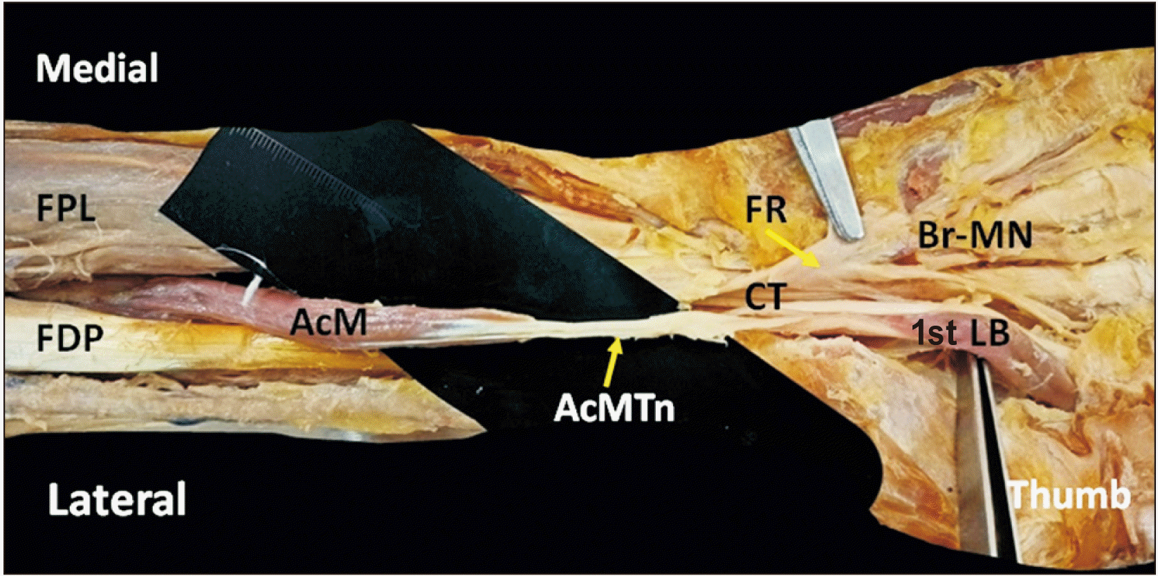 | Fig. 1Showing accessory muscle of right flexor compartment of the forearm with half belly and half tendon. AcM, Accessory muscle; FDP, flexor digitorum profundus; FPL, flexor pollicis longus; FR, flexor retinaculum; CT, carpal tunnel; AcMTn, accessory muscle tendon; 1st LB, first lumbrical; Br-MN, branch of median nerve.
|
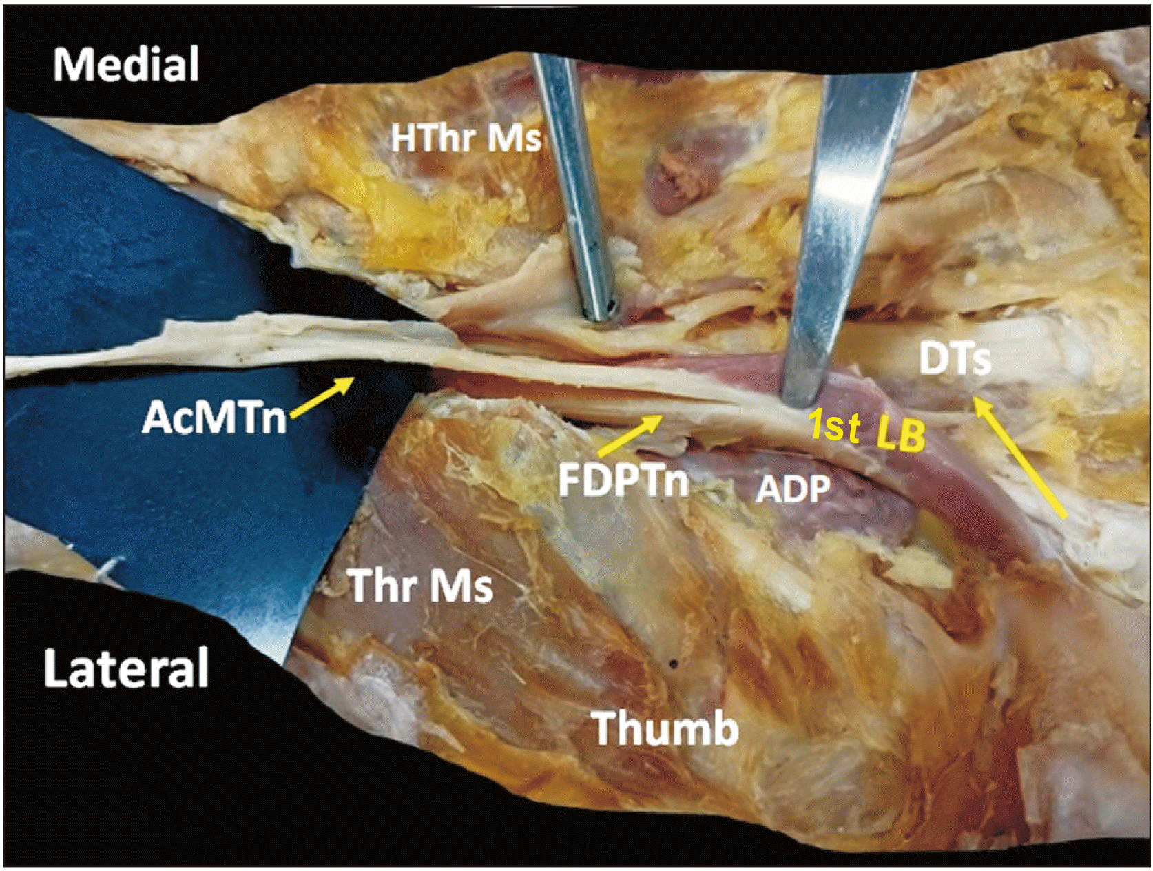 | Fig. 2Showing AcMTn of front of right forearm, lying lateral to the FDPTn. Attachment of bipennate 1st LB can be seen from its deeper aspect lying superficial to ADP. AcMTn, accessory muscle tendon; FDPTn, tendon of the Flexor Digitorum Profundus; 1st LB, first lumbrical; ADP, adductor pollicis; DTs, digital tendons; HThr Ms, hypothenar muscles; Thr Ms, thenar muscles.
|
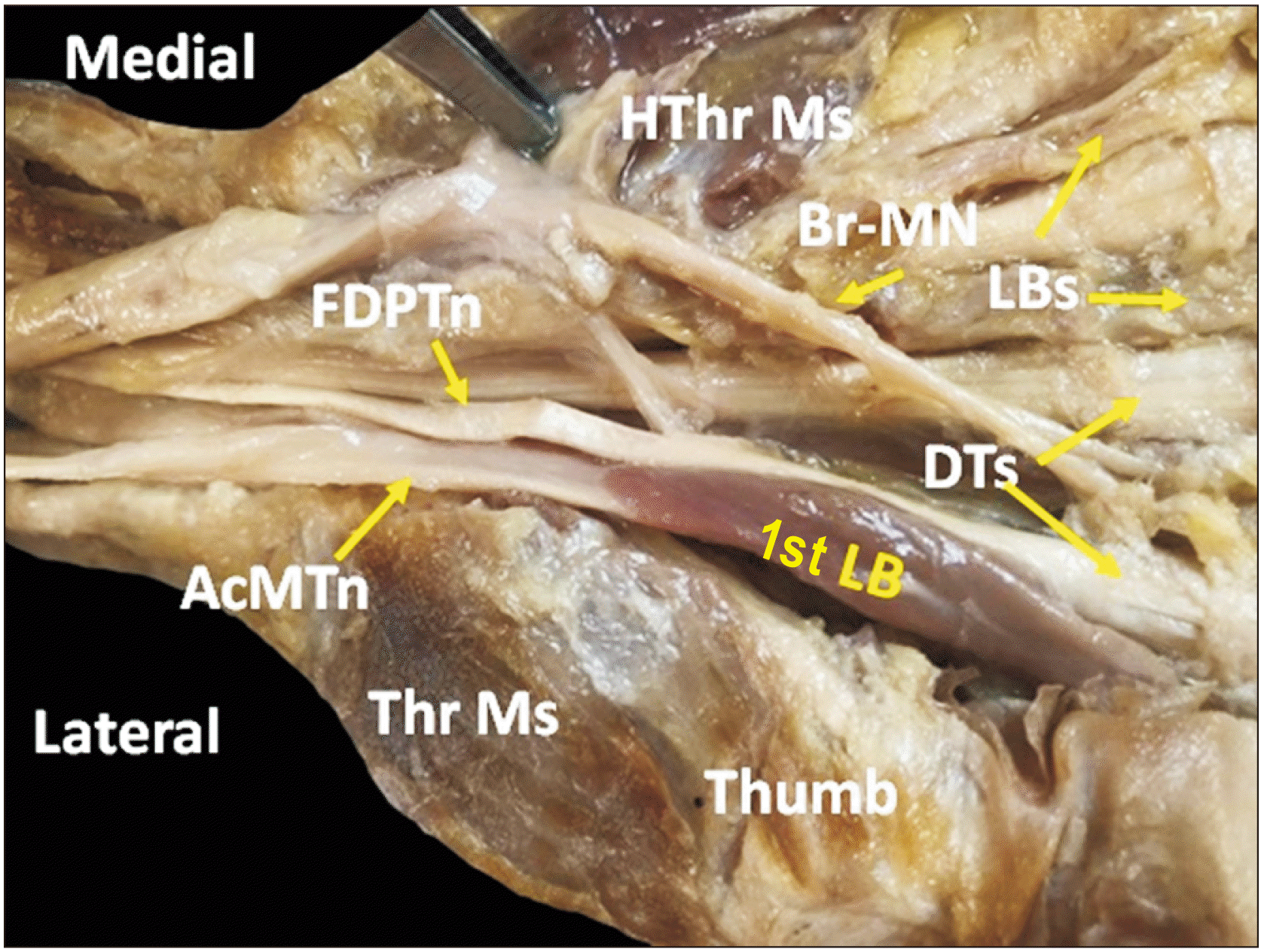 | Fig. 3Showing distal part of the AcMTn (right limb), lying lateral to FDPTn. Bipennate 1st LB taking origin from tendon of FDP and tendon of accessory muscle, passing lateral to these tendons to the dorsum of the hand. AcMTn, accessory muscle tendon; FDPTn, tendon of the Flexor Digitorum Profundus; 1st LB, first lumbrical; DTs, digital tendons; Thr Ms, thenar muscles; ADP, adductor pollicis; HThr Ms, hypothenar muscles; LB, other lumbricals; Br-MN, branch of median nerve.
|
Table 1
Showing comparison of findings observed by other authors with the present variation
| No. | Author, year of publication | Origin | Insertion | Shape of the muscle belly | Innervation |
|---|---|---|---|---|---|
| 1 | Jones et al. [5], 1997 | Deeper surface of FDS | One or several tendons of FDP | 54±5% specimens, triangular in 36±4% specimens and voluminous 9±1% specimens | Anterior interosseous nerve |
| 2 | Wood et al. [7], 1868 | Deeper surface of FDS | Rounded tapering | ||
| 3 | Macalister [9], 1875 | Deeper surface of FDS | 9 different possible ways into FDP tendons | Slim | |
| 4 | Le Double [10], 1897 | Deeper surface of FDS | Musculotendinous fascicle | ||
| 5 | Parson [11], 1898 | Deeper surface of FDS | |||
| 6 | Kida [8], 1988 | Coronoid process | Median nerve | ||
| 7 | Bhavya et al. [12], 2016 | Coronoid process | One of the tendons of FDP | ||
| 8 | El Domiaty et al. [13], 2008 | Pronator teres | Tendon of FDP to index, middle or ring finger | ||
| 9 | Testut [14], 1884 | Slender, long | |||
| 10 | Present case report, 2022 | Lower third of the anterior surface of the radius, close to the FDP origin | After giving origin to first lumbrical muscle AcMTn got merged with the tendon of FDP for index finger. | Spindle shaped | Anterior interosseous nerve |
![]()
Go to :
Discussion
The occurrence of aberrant muscles in the forearm's flexor compartment is not very common. Most of the time, these muscles go unrecognized since they do not cause any symptoms in the person. However, sometimes by compressing the neurovascular structures, these aberrant muscles may induce functional impairments. Surgical intervention is required in such circumstances. In 1813, Gantzer described two accessory muscles in the forearm–the more frequently occurring flexor digitorum pollicis and the less frequently occurring FDP [5]. The prevalence of the auxiliary FDP has been reported to be varying. Mangini [6] reported the prevalence of accessory FDP to be 2.9%, whereas Wood et al. [7], and Kida [8], reported them to be 18.6% and 35.2% respectively.
In our finding, the accessory muscle of FDP originated from the lower third of the anterior surface of the radius, close to the FDP origin, underneath the FPL muscle. Previous studies in the literature have reported the accessory FDP mostly arising from the deeper surface of FDS [5, 7, 9-11]. A few others have reported accessory head of FDP arising from the coronoid process [8, 12]. Similar other studies have documented the origin of accessory FDP from the pronator teres [13].
In our case report, AcMTn got merged with the lateral aspect of tendon of FDP for index finger after giving origin to first lumbrical. El Domiaty et al. [13], in their study, observed that accessory FDP tendon joined the tendons of the FDP muscle to the index or middle and ring fingers. In a case study done by Bhavya et al. [12], they documented that in most of the cases the tendon of the accessory muscle ended in the middle of forearm and got inserted into one of the tendons of FDP. Macalister [9], described the insertion of the accessory muscle into the main muscle in 9 different possible ways: into the index tendon; into the middle tendon; into the ring tendon; into the 5th finger tendon; into the ring and 5th finger tendons; into the index and ring tendons; into the index and 5th finger tendons; into the middle, ring and 5th finger tendons; into the index, middle and ring tendons. Jones et al. [5], in their study found the accessory tendon getting inserted into the one or several tendons of FDP.
The shape of the belly of the accessory muscle in our report is spindle shaped. The shape of this specific muscle has been earlier described as a slim tendon [9], a rounded tapering muscular slip [7], a slender long tendon [14], and a musculotendinous fascicle [10]. Jones et al. [5], in their study observed that the shape of the accessory FDP is slender in 54±5% specimens, triangular in 36±4% specimens and voluminous 9±1% specimens. In the present case report, innervation to the accessory muscle was from a branch given by anterior interosseous nerve. Kida [8], in their study observed that the innervation to the accessory FDP was directly from median nerve whereas Jones et al. [5], in their study observed that the innervation to the accessory muscle was from anterior interosseous nerve.
What makes the present study more interesting is the presence of bipennate first lumbrical which takes it origin from the contiguous side of the accessory tendon and tendon of FDP to the index finger. The lateral belly of the first lumbrical arises from the medial aspect of AcMT and the medial belly of first lumbrical arises from the radial side of the tendon of FDP to the index finger and the muscle is innervated by a twig from the median nerve. Nayak [15], reported a similar finding where two anomalous muscles originated from the undersurface of FDS–the muscle on the radial side merged with that of FPL and the muscle on the ulnar side formed an independent tendon for the middle finger and the tendon of the additional muscle and FDP tendon for the middle finger gave origin to the second lumbrical in the carpal tunnel.
Somatic mesoderm colonises the limb buds during 4th week of development, forming ventral and dorsal condensations. The upper limb flexors and pronators are formed through ventral condensation. The forearm flexor muscles that grow from flexor mass are organised into two layers - superficial and deep. Deep layer gives rise to the FDS, FDP and FPL. The imprecise cleavage of the deep layer of the flexor mass during development, which indicates an atavistic aspect, might explain the presence of auxiliary muscles joining the flexor muscles [5].
Defining the anatomical characteristics for clinical diagnosis and surgical treatments requires recognizing the important anatomical variations. An abnormal muscle might mimic a ganglion or a soft tissue tumour, or it can induce pressure neuritis and symptoms like carpal tunnel syndrome if it is adjacent to a nerve [12, 13]. The additional flexor tendon in the present case increases the volume of the carpal tunnel and may cause carpal tunnel syndrome.
In conclusion, the presence of extra muscles and tendons may cause surgeons to become confused while gaining access to the forearm and hand. The current findings will add to our understanding of muscle differences in the antebrachial and carpal areas, and they should also be taken into account when determining the cause of carpal tunnel syndrome.
Go to :
 XML Download