

 PDF
PDF Citation
 Print
Print


Introduction
Most of the renal vascular variations are attributed to the development of the kidney and the three pairs of large embryonic veins namely posterior cardinal veins, subcardinal veins and supracardinal veins. The right kidney is usually supplied by one renal artery (RA) and one renal vein (RV). Right RA is the branch of abdominal aorta and the right RV drains into the inferior vena cava (IVC). RV variations are less common compared to the renal arteries. However, they are more common on the right side than the left side. The reported variations include accessory renal veins, and unusual tributaries like right gonadal vein. We report unique, combined variations of right renal vessels and discuss about their clinical, functional and developmental aspects in this report.
Case Report
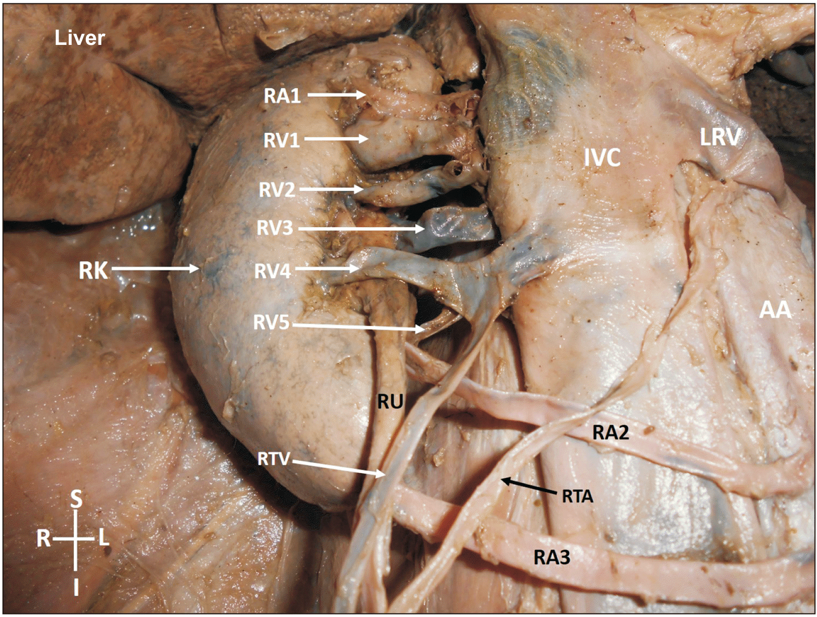
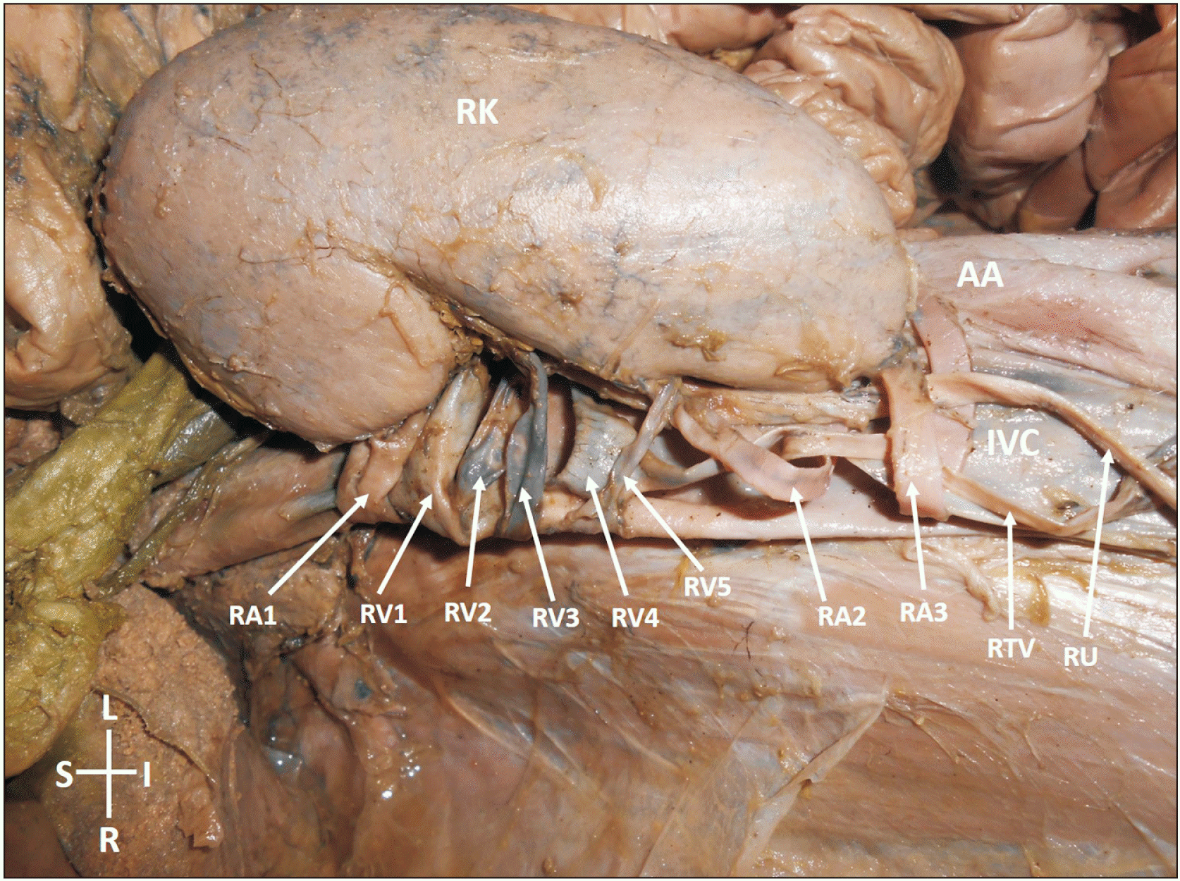
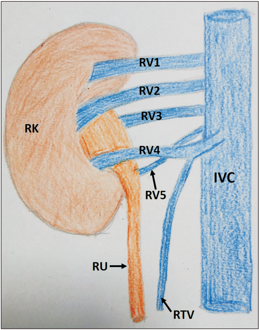
During routine dissection classes, we observed multiple renal vessels supplying the right kidney in an adult male cadaver aged about 75 years. The right kidney was apparently healthy and had normal dimensions and position. However, its hilum was twisted towards the anterior surface. The right kidney was supplied by three renal arteries. The main (normal) right RA arose from the lateral side of the abdominal aorta at the level of second lumbar vertebra, passed behind the IVC and entered the kidney through the uppermost part of the hilum. There were two accessory right renal arteries, which took origin from the anterior wall of the abdominal aorta, at the level of fourth lumbar vertebra, travelled to the right in front of the IVC and entered the right kidney. Among the two accessory arteries, the upper one entered the right kidney through the lowest part of the hilum and the lower one entered the right kidney by piercing its lower pole as the lower polar artery. Ureter was situated anterior to both the accessory right renal arteries. There were five right renal veins, all of which emerged from the hilum of the right kidney. Among them, only the fourth vein from above, emerged in front of the renal pelvis. First and second veins emerged above the renal pelvis, while the third and fifth veins emerged behind the renal pelvis. The upper four veins were of almost equal size, while the lowest vein was half the size of other veins. The fourth vein was peculiar as it passed in front of the renal pelvis, received the right testicular vein and opened into the IVC through its anterior wall. The other veins opened into the IVC through its posterolateral aspect. The hilum of the right kidney was congested with the five veins, two arteries and the renal pelvis. The variant vessels have been shown in Figs. 1, 2. A simplified schematic diagram of the pattern of right renal veins has been shown in Fig. 3.
Discussion
Right renal vessels show frequent variations, most of which go unnoticed as they do not cause any functional disturbance. Many cadaveric, radiologic and surgical studies have been conducted about the renal vessels. The commonest type of RA variation is the presence of accessary renal arteries. The accessory renal arteries can arise from the aorta above or below the level of first and second lumbar vertebrae. In one of the angiographic studies conducted, right RA variations were noted in 16% cases [1]. In another recent angiographic study, accessory renal arteries were found in 10% of cases [2]. Inferior polar arteries usually arise from the aorta or other arteries below the level of the main RA. However, rarely, they arise from the aorta, above the level of the main RA and enter the kidney through its lower pole [3]. In the current case, the inferior polar artery arose from the aorta, just above its termination. In most of the cases, the right kidney is supplied by a single RA. However, this right RA may divide into various number of branches on the way to the hilum. Division of the RA up to eight prehilar branches and formation of an arterial glomerulus near the hium have been reported already [4].
Accessory renal arteries could be the cause of hypertension when they undergo stenosis due to the hilar congestion [5]. They may cause problems during renal transplant surgeries and retroperitoneal lymphadenectomy. Pre-operative renal embolization is one of the safe techniques to minimise bleeding during laparoscopic nephrectomies. However, when the accessory renal arteries are present, there could be unexpected bleeding [6].
Variations of the renal veins are also very common. The left RV is more constant than the right renal vein. Most of the studies show a very high percentage of variations of right renal vein. In a recent meta-analysis about the variations of the renal veins, right RV variations were found in 16.7% and left 2.1% of cases [7]. Zhu et al. [8], conducted a study with multidetector computed tomography and classified the right renal veins into three types. Type 1 had multiple right renal veins (more than one in number, opening separately into the IVC). Type 2 had a normal vein from the renal hilum and another vein arising from the upper or lower pole of the right kidney. The Type 3 had more than one veins arising from the right kidney and one of them draining into the left renal vein. The maximum numbers of renal veins observed by them was 4 (in 4.2% cases). Apart from variations in the number of veins, a few rare types of right RV variations have been reported in the literature. Lavy et al. [9], have reported the presence of a ‘Y’ shaped vein which arose as a single vein from the right kidney and after bifurcation, opened into the IVC as two separate veins. Kim et al. [10] have reported a circum-aortic right RV. To the best of our knowledge, there are no reports on five veins emerging from the hilum of the right kidney and opening separately into the IVC. Our case was also special as one of the renal veins opened into the anterior aspect of the IVC and received the right testicular vein. Opening of a RV into the anterior wall of the vena cava is also a rare observation.
The development of the renal veins is closely related to the development of the IVC [11]. The renal segment of the IVC develops from the right subcardinal vein and the subcardinal-supracardinal anastomosis. Human definitive kidney develops from the metanephros. During the development, one metanephric vein drains the right kidney into the right subcardinal vein. This vein in the future, becomes the right renal vein. If there are more than one metanephric veins in the foetal life, and if all of them persist, that would lead to the formation of multiple renal veins.
Termination of the right testicular vein into the RV instead of the IVC is not an uncommon event. There are reports on opening of the right testicular vein into the RV or accessory renal veins [12, 13]. Its duplication and triplication have also been reported where, one of the veins terminated into the RV [14].
Knowledge about the presence of five veins in the renal hilum, their course and termination are of importance to radiologists, renal transplant surgeons and even general surgeons performing retroperitoneal surgeries. During the transfer of right kidney, the right RV is grafted along with a segment of vena cava [15]. When there is more than one RV as in the current case, a large segment of vena cava has to be grafted. Additional renal veins could also result in iatrogenic bleeding during retroperitoneal surgeries and renal transplant surgeries.
On a functional view point, the current case where, five veins and two arteries were clogging the hilum of the right kidney could result in hemodynamic changes of the kidney at some stage of life. This in turn could result in altered kidney functions. Termination of the right gonadal vein vertically into one of the renal veins could also lead to right sided varicocele. Accessory renal arteries could result in compression of the IVC as they pass anterior to IVC.
In conclusion, concurrent variations of the right renal and gonadal vessels are rare. This case is unique in having five veins emerging from the hilum of the right kidney and opening separately into the IVC, opening of right testicular vein into one of the renal veins and having the presence of two accessory renal arteries. Knowledge of this variation could be of use to radiologists, nephrologists and other medical disciplines.
 XML Download