

 PDF
PDF Citation
 Print
Print


Introduction
Depression, is a widespread mental upset, exhibiting with depressed mood, anhedonia, disturbed sleep or appetite, sluggishness and poor concentration, globally, 5% of adults suffer from depression. It is estimated by the World Health Organization that depression influences around 280 million people worldwide is associated with increasing suicide [1].
Antidepressants (ADs) play a key role in alleviating depression. Selective serotonin reuptake inhibitors (SSRIs) are a novel AD that has befit as a premier-line therapy for depression and abundant psychiatric imperfections because of their efficiency, safety, and bearability [2].
SSRIs, comprising escitalopram, citalopram, sertraline, and fluoxetine are vastly used in the United States and accounting for 62% of ADs consumed [3]. Recently; regard of the safety of SSRIs is growing.
Escitalopram is a SSRI, sold under the brand name cipralex, used to restore serotonergic function in the treatment of depression and anxiety [4].
Pancreas is a pivotal endocrine and exocrine digestive gland displays numerous hormones, as insulin that is concerned with regulation of carbohydrate metabolism and preserves glucose path across the cell membrane, and enzymes, that assist in carbohydrates, lipids, and proteins digestion [5, 6]. Thence, any alteration in its function may inspire the achievement and the physiological function of the body [7].
Pancreatitis at most comprises two types; acute and chronic [8]. Acute pancreatitis (AP) is distinguished by death of acinar cell with local and systemic inflammation [9], and a universal prevalence of thirty-four cases per one hundred thousand persons [10]. Chronic pancreatitis (CP) is a heterogeneous group of diseases manifested by long-lasting pancreatic inflammation resulting in fibrosis, gradual loss of pancreatic function [11] and eminent parenchymal destruction. Also an increased risk of pancreatic cancer exists in CP [12].
One of the agreeable theories is that repeated episodes of AP results in CP [13].
Drug-induced pancreatitis records about 2% of all cases of pancreatitis. Several drugs cause pancreatitis, including SSRIs [14]. Also some studies demonstrated that SSRIs may be linked with an elevated incidence of AP [15, 16], hyperglycemia and type II diabetes mellitus but with inconsistent opinion [17-19].
Plurality of patients have low subjection and refuse to take ADs due to their side effects. Therefore, the identification of unprecedented drug or treatment combination with drugs that augment the efficacy of AD is still needed.
Medicinal plants have a pivotal role in maintaining human health, amending quality of life, curing, and prevailing different diseases due to their integrity, efficacy, tolerability, and cheapness [20, 21].
Silymarin (SIL), a free radical scavenger, belongs to the flavonolignan family and is a main constituent of Silybum marianum [22, 23]. Silybum marianum has been applied as a medicinal plant to remedy liver and gallbladder diseases in ancient Greek, and to preserve the liver from chemicals and toxins in Europe and Asia [24, 25].
Recently SIL reported an antidiabetic, anticancer, anti-oxidant, anti-inflammatory, anti-fibrotic, and osteoprotective activities [26-28].
The mechanism of cipralex toxicity on pancreas is not still completely understood. Therefore, the hypothesis evaluates, for the first time, the effect of AD cipralex on the structure of the endocrine and exocrine portions of the pancreas of adult male albino rats and assess the beneficial safeguarding effects of SIL on these changes by means of its anti-inflammatory, anti-oxidant, and anti-apoptotic impacts, and also by increasing the anti-insulin antibody expression.
Go to : 
Materials and Methods
Animals
Forty-five adult male albino rats weighing 180–200 g, were housed at Alexandria University, Egypt in Medical Research Institute’s animal house. Rats had caged in standardized room conditions and permitted indefinite access to chow and water. The experiment started after one week of caging. The approved the entire process by the values, principles and ethics of scientific research Committee, Faculty of Medicine, Menoufia University, Egypt (Clearance no. 9/2022 ANAT5).
Experimental substances
Cipralex (traditional name of Escitalopram); tablets (H-Lundbeck A/S, Valby, Denmark) were obtained from a local pharmacy, Egypt. Each tablet contains 10 mg escitalopram. The experimental dose is 10 mg/kg/day dissolved in (0.1 ml/100 g) 0.9% saline solution and was approached orally by gavage [29].
SIL powder; was obtained from a local distributer (Sigma chemical), Egypt. The experimental dose is 100 mg/kg dissolved in (0.1 ml/100 g) distilled water and was presented daily orally by gavage [30].
Experimental design
Rats were randomly categorized into three equal groups, each included 15 animals.
Drugs were approached daily orally by gavage for four weeks as follows:
Group I (control group): consisted of 15 rats divided into three subgroups (n=5 per subgroup).
Subgroup Ia: consisted of 5 rats that obtained only standard diet and ordinary drinking water (negative control).
Subgroup Ib: consisted of 5 rats that obtained 0.1 ml/100 g 0.9% saline solution (as a cipralex vehicle).
Subgroup Ic: consisted of 5 rats that obtained SIL (100 mg/kg/day).
Group II (cipralex group): consisted of 15 rats that administered cipralex (10 mg/kg/day).
Group III (cipralex plus SIL group): consisted of 15 rats that administered SIL (100 mg/kg/day) concomitantly with cipralex (10 mg/kg/day).
Evaluation methods
Pancreatic weight/rat weight ratio analysis
All rats were weighed before being sacrificed, and pentobarbital was administered intraperitoneally to anesthetize the rats. The pancreases were removed immediately after drawing the blood sample, trimmed from fat and weighed. The ratio of Pancreatic weight (PW)/rat weight (RW) was calculated for each rat as follow; PW (g) was divided by RW (g) and multiplied by 1000 to obtain the ratio as a natural number [31].
Biochemical analysis
At the termination of the 4th week, intracardiac blood samples were drawn after nocturnal fasting and split into two sections. One was obtained, using the Accu-chek Active (Roch Diagnostics, Mannheim, Germany), to assess the fasting blood glucose level (mg/dl) using the glucose oxidase method [32]. The remaining portion was conveyed to a tube without EDTA addition, allowed to clot, and then centrifuged at 3,000 rpm for 10 minutes to get sera that were kept at −20°C and utilized to measure the levels of insulin and amylase. An enzyme-linked immunosorbent test kit (Mercodia, Uppsala, Sweden) was used to determine the insulin level (U/l). The serum amylase level (U/l) was measured using an automatic biochemical analyzer (Olympus AU5400; Olympus Co., Tokyo, Japan) [33].
Histological and immunohistochemical assessments
The pancreases were dissected out and specimens were fixed in 10% formol saline for 24 hours and they were then processed to create 5-μm-thick paraffin sections for light microscopic analysis.
Histological study [34]
1. Hematoxylin and Eosin to set the histological details.
2. Masson’s trichrome stain to elucidate the collagen fibers.
Immunohistochemical study
A conventional avidin–biotin immune peroxidase technique was performed [35], After rinsing the 5-μm deparaffinised and rehydrated pancreatic sections in phosphate-buffered saline (PBS), they were blocked for an hour by incubation in 10% normal goat serum. After that, the sections were incubated with the primary antibodies which included; inducible nitric oxide synthase (iNOS), 1:500, ThermoScientific; tumour necrosis factor-alpha (TNF-α), 1:500; a rabbit polyclonal antibody (purchased as caspase-3 ab-4, rabbit polyclonal antibody, RB-1197-PO, Lot: 1197P10100; NeoMarkers, Fremont, CA, USA); primary proliferating cell nuclear antigen (PCNA) antibody (PC10; Abcam, Cambridge, United Kingdom); and with anti-sera containing polyclonal primary antibodies for rat insulin (Bio-Genex, cas no.: AR.295-R.) for 1h at ambient temperature. Following a 20-minute incubation with the secondary goat anti-rabbit biotinylated antibody (1:200, Vector Labs, Burlingame, CA, USA, BA-1000), PBS was used to rinse the sections. The conjugate solution of streptavidin and horseradish peroxidase was then performed for 10 minutes. The slides were then counterstained with 100 μl haematoxylin, washed with distilled water and dehydrated in graded ethanol for 5 minutes, clarified in xylene and a coverslip was mounted on it. Brown colored cytoplasmic with some nuclear immunostaining cells considered a positive reaction. Negative control was performed after omitting the primary antibody [36, 37].
Morphometrical studies
1. Number and diameter of islets of Langerhans were counted via light microscopic examination of H&E stain photographs ×400.
2. Mean area percentage of collagen fibers were counted via light microscopic examination of the Masson’s trichrome stain photographs ×400.
3. The numbers of iNOS, TNF-α, caspase-3, and PCNA positive immunoreactive cells in the islets of Langerhans and in the pancreatic acinar cells were counted via light microscopic examination of the immune-stained photographs ×400.
4. The number of insulin positive β-cells of all groups were counted via light microscopic examination of the anti-insulin antibodies immune-stained photographs ×400.
All measurements were quantified in 5 high-power fields and five separate sections for each group employing the image analyzer (Leica Q 500 MC program; Leica, Wetzlar, Germany) at the Menoufia University, Faculty of Medicine, Department of Histology.
Statistical analysis
On an IBM compatible computer, the data was gathered, tabulated, and examined using SPSS Statistics for Windows, Version 20.0 (IBM Co., Armonk, NY, USA). The mean, standard deviation, and range of the quantitative data were described and compared between each two groups using the student t-test and Mann Whitney U-test for normally distributed and abnormally distributed data, respectively. Significance was determined by a P-value of less than 0.05, highly significance by a P-value of less than 0.001, and non-significance by a P-value of more than 0.05. Graphs were used to display all of the resulting data.
Go to :
Results
There was no significant difference in all the examined parameters between the three subgroups of group I (control group). So, group I (control group) applied to them.
Pancreatic weight (PW)/Rat weight (RW) ratio results
As shown in (Table 1, Fig. 1), the PW, RW, and PW/RT ratio in cipralex group demonstrated a highly significant decrease (P<0.001) compared to control group, while in cipralex plus SIL group, these are significantly increased when compared to cipralex group. There was no significant difference in all weights between control group and cipralex plus SIL.
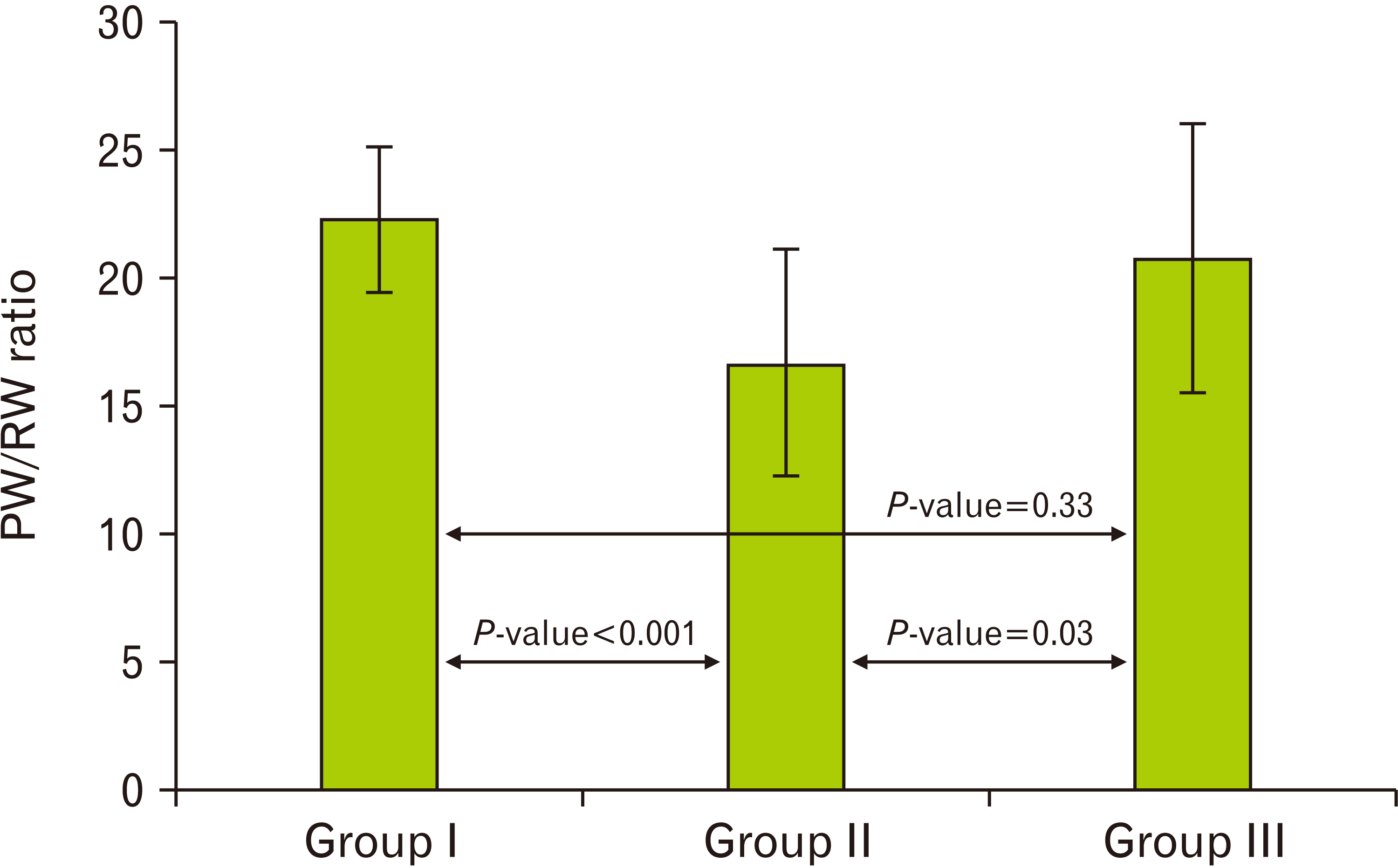 | Fig. 1A histogram demonstrating a highly significant decrease (P<0.001) in the PW/BW ratio in the cipralex group compared to the control group, while the cipralex plus SIL group, illustrating a significant increase compared to the cipralex group, with no significant difference between control and cipralex plus SIL groups. PW, pancreatic weight; RW, rat weight; SIL, silymarin.
|
Table 1
PW, RW, and PW/RW ratio among the studied groups
| Weight parameter |
Group I (control) n=15 |
Group II (cipralex) n=15 |
Group III (cipralex plus SIL) n=15 |
t-test | P-value |
|---|---|---|---|---|---|
| PW | 5.06 | <0.001a | |||
| 4.17±0.64 | 2.72±0.65 | 3.43±0.87 | 1.95 | 0.06b | |
| 3.1–5.0 | 1.6–3.6 | 2.2–4.6 | 2.78 | 0.01c | |
| RW | 4.83 | <0.001a | |||
| 186.93±11.53 | 167.07±10.98 | 178.47±13.24 | 1.87 | 0.07b | |
| 165–210 | 150–185 | 155–194 | 2.57 | 0.02c | |
| PW/RW ratio | 4.13 | <0.001a | |||
| 22.29±2.83 | 16.66±4.46 | 20.76±5.28 | 0.98 | 0.33b | |
| 18.24–26.74 | 8.65–23.78 | 11.58–29.68 | 2.30 | 0.03c | |
![]()
Biochemical results
Regarding fasting blood glucose level, there was a highly significant increase (P<0.001) in cipralex group compared to control group. However, cipralex plus SIL group exhibited a high significant decrease compared to cipralex group (Table 2, Fig. 2).
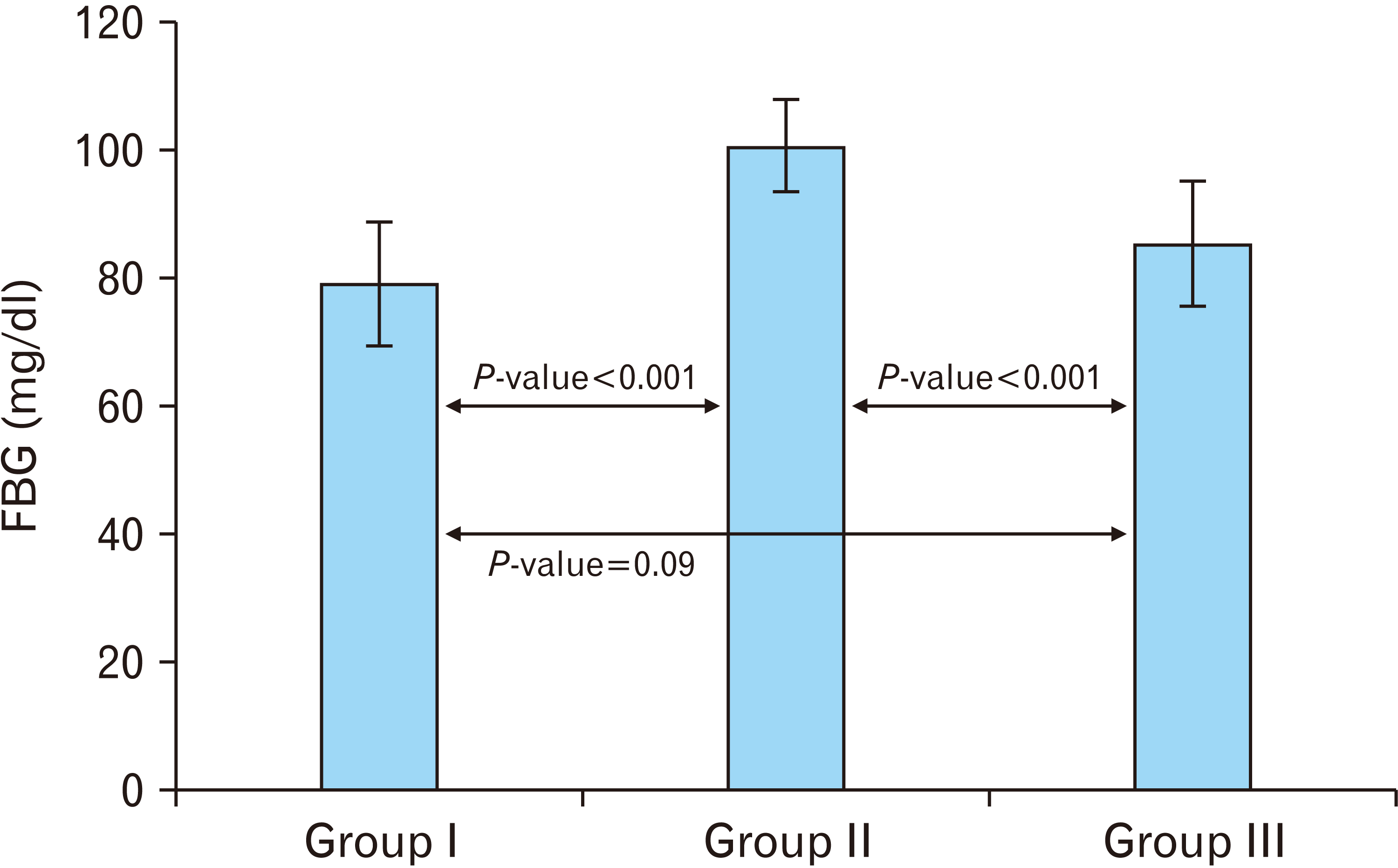 | Fig. 2A histogram illustrating a highly significant increase (P<0.001) in FBG in the cipralex group compared to the control group, while the cipralex plus SIL group, illustrating a high significant decrease compared to the cipralex group, with no significance between control and cipralex plus SIL groups. SIL, silymarin; FBG, fasting blood glucose.
|
Table 2
Biochemical analysis among the studied groups
| Biochemical analysis parameter |
Group I (control) n=15 |
Group II (cipralex) n=15 |
Group III (cipralex plus SIL) n=15 |
t-test | P-value |
|---|---|---|---|---|---|
| Fasting blood glucose (mg/dl) | 6.80 | <0.001a | |||
| 79.13±9.62 | 100.53±7.21 | 85.33±9.66 | 1.74 | 0.09b | |
| 60–95 | 88–111 | 70–100 | 4.88 | <0.001c | |
| Insulin level (U/l) | 4.18 | <0.001a | |||
| 2.39±0.29 | 1.93±0.32 | 2.22±0.34 | 1.51 | 0.14b | |
| 1.9–2.8 | 1.5–2.5 | 1.3–2.6 | 2.42 | 0.02c | |
| Serum amylase level (U/l) | 12.19 | <0.001a | |||
| 763.73±37.41 | 586.07±42.26 | 757.47±27.83 | 0.52 | 0.61b | |
| 666–800 | 499–678 | 708–808 | 13.12 | <0.001c |
![]()
Regarding insulin level, cipralex group illustrated a highly significant decrease (P<0.001) compared to control group. However, cipralex plus SIL group demonstrated a significant increase compared to cipralex group (Table 2, Fig. 3).
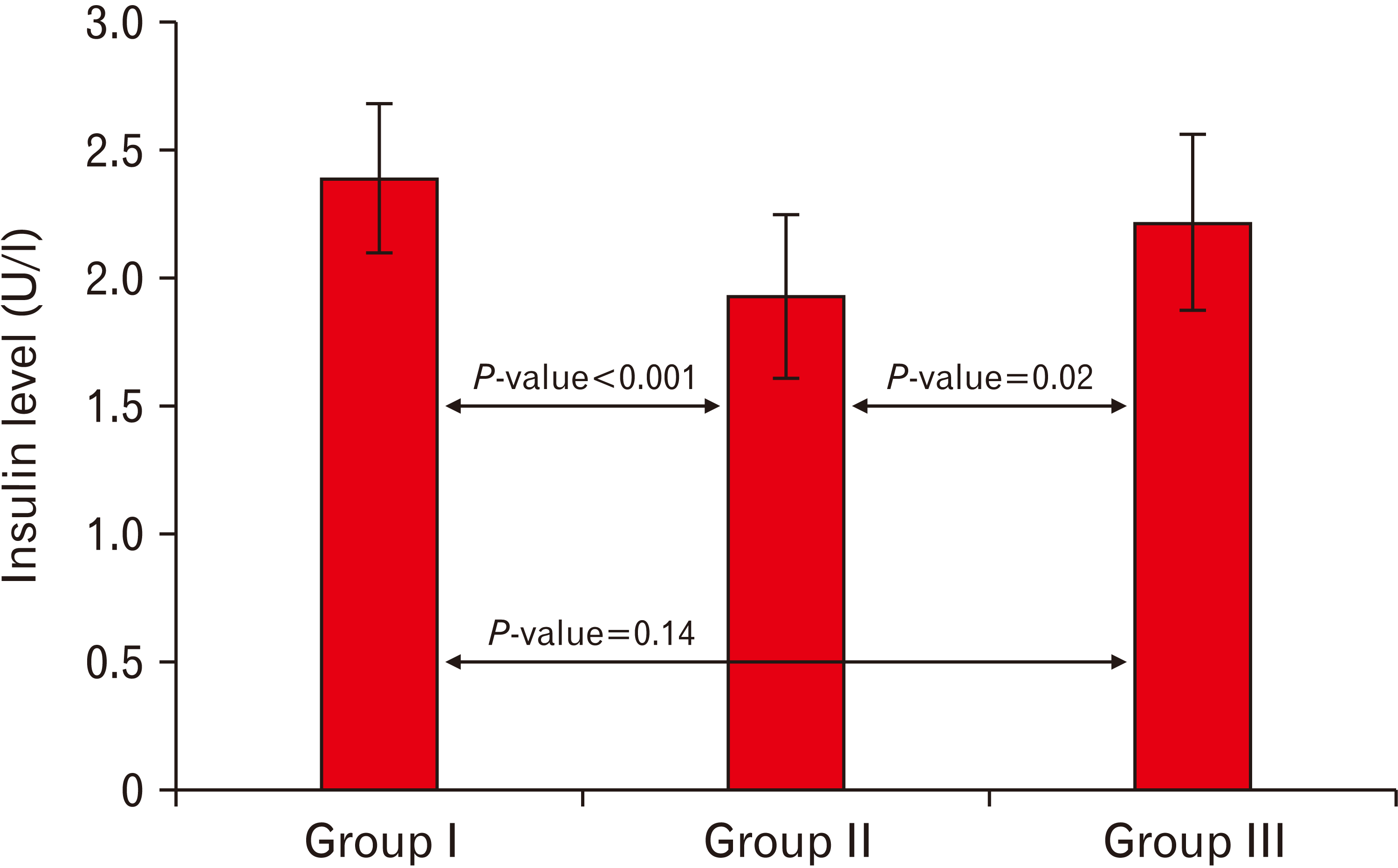 | Fig. 3A histogram showing a highly significant decrease (P<0.001) in the insulin level in the cipralex group compared to the control group, while the cipralex plus SIL group, illustrating a significant increase compared to the cipralex group, with no significant difference between control and cipralex plus SIL groups. SIL, silymarin.
|
Regarding amylase level, there was a highly significant decrease (P<0.001) in cipralex group compared to control group. However, cipralex plus SIL group demonstrated a high significant increase compared to cipralex group (Table 2, Fig. 4).
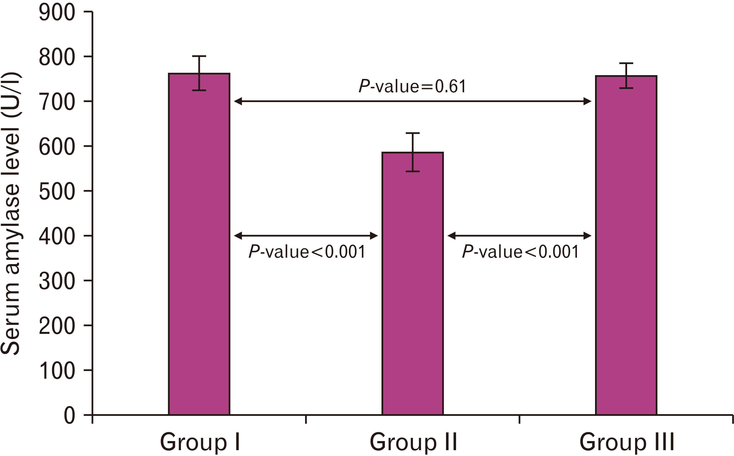 | Fig. 4A histogram demonstrating a highly significant decrease (P<0.001) in the amylase level in the cipralex group compared to the control group, while the cipralex plus SIL group, illustrating a high significant increase compared to the cipralex group, with no significance between control and cipralex plus SIL groups. SIL, silymarin.
|
There wasn’t any significant difference between control and cipralex plus SIL groups in all biochemical parameters.
Light microscopic examination
Hematoxylin and eosin stain results
Sections of group I (control group) stained with H&E demonstrated that the pancreas was formed of lobules of different sizes and shapes separated by thin connective tissue septa. Interlobular ducts and blood vessels were observed. Each lobule was formed of highly packed darkly stained acini (the exocrine portion of the pancreas). The islet of Langerhans (the endocrine portion of the pancreas) appeared as large defined pale stained area scattered between the darkly stained pancreatic acini (Fig. 5A). On the other hand, sections of group II (cipralex group) exhibited marked destruction of the normal pancreatic architecture of the exocrine part, shrunken and vaguely identified pancreatic islet. Congested interlobular blood vessel and distorted fat cells were observed. Distorted acini and inflammatory cell infiltration were also seen (Fig. 5B, C). Compared with the cipralex group, H&E sections of group III (cipralex plus SIL group) showed apparent restoration of the nearly normal architecture of the exocrine part and endocrine part. Interlobular blood vessel and duct appeared normal in between the cells (Fig. 5D).
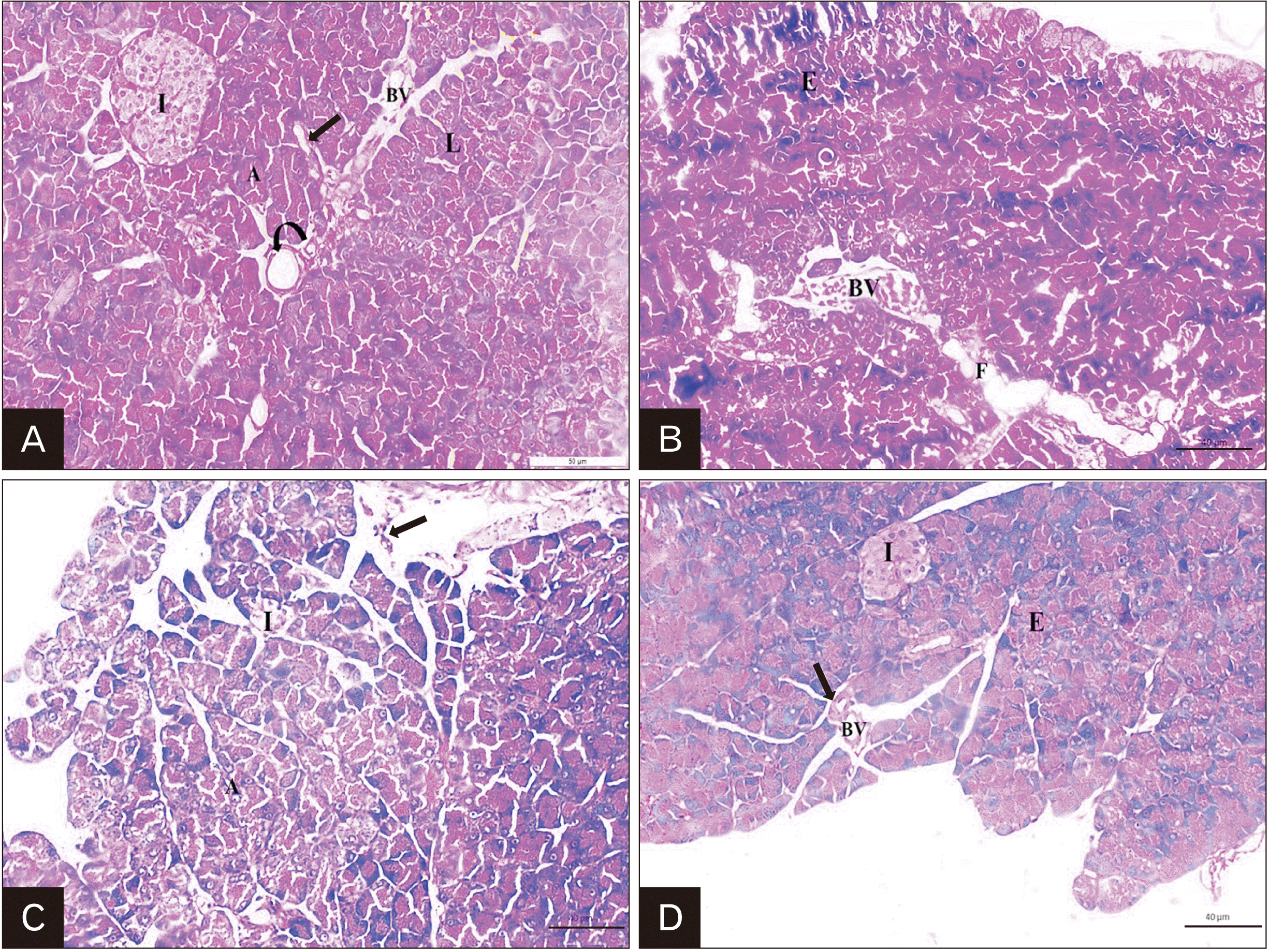 | Fig. 5Representative H&E staining of rat pancreas; (A) Control group: demonstrating that the pancreas is formed of lobules (L) of different sizes and shapes separated by thin connective tissue septa (arrow). Interlobular duct (curved arrow) and blood vessel (BV) are observed. Each lobule is formed of highly packed darkly stained acini (A) (the exocrine portion of the pancreas). The islet of Langerhans (I) (the endocrine portion of the pancreas) is appeared as large defined pale stained area scattered between the darkly stained pancreatic acini. (B) Cipralex group: showing destruction of the normal pancreatic architecture of the exocrine part (E), congested interlobular BV and distorted fat cells (F) are observed. (C) Cipralex group also illustrating shrunken and vaguely identified pancreatic islet (I), distorted acini (A). Inflammatory cell infiltration (arrow) are also observed. (D) Cipralex plus SIL group: showing restoration of the normal architecture of the E and endocrine part (I). Interlobular BV and duct (arrow) appear normal in between the cells (×200, scale bar=40 μm).
|
Also, control group revealed that the pancreatic acini has basal basophilic cytoplasm containing rounded nuclei and apical acidophilic zymogen granules. The islet of Langerhans composed of cords of secretory cells separated by blood capillaries. Two types of cells were recognized; central beta (β) cells with large rounded nuclei and peripheral alpha (α) cells with smaller nuclei (Fig. 6A). Cipralex group showed that most of acinar cells had pyknotic nuclei and cytoplasmic vacuolation. Distorted shape of islet cells with loss of many cells of islets of Langerhans was also a prominent feature and congested blood capillaries were noticed. Empty spaces were found between pancreatic acini. Marked cytoplasmic vacuolations of beta cells were observed. Thickening in the wall of pancreatic duct and thickening of the interlobular septa were observed (Fig. 6B, C). Cipralex plus SIL group showed few acinar cells with pyknotic nuclei and some cells had mild vacuolated cytoplasm. Few beta cells appeared vacuolated (Fig. 6D).
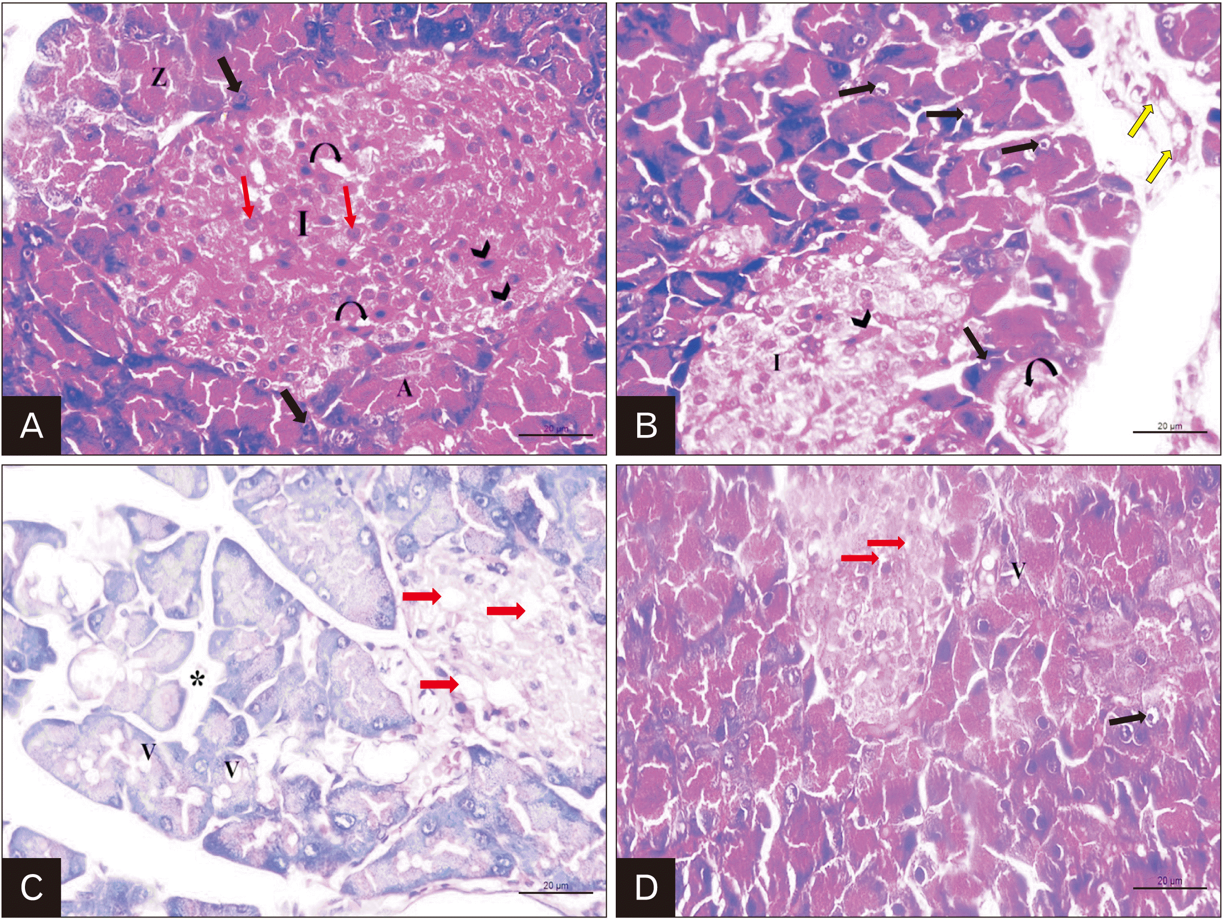 | Fig. 6Representative H&E staining of rat pancreas; (A) Control group: illustrating that the pancreatic acini (A) have basal basophilic cytoplasm containing rounded vesicular nuclei (black arrow) and apical acidophilic zymogen granules (Z). The islet of Langerhans (I) is composed of cords of secretory cells separated by blood capillaries (curved arrow). Two types of cells are recognized; central beta cells with rounded and lighter nuclei (red arrow) and peripheral alpha cells with oval darkly stained nuclei (arrow head). (B) Cipralex group: showing that many acinar cells have darkly stained pyknotic nuclei (black arrow). Distorted shape of islet cells with apparent decrease in the number (I) and congested blood capillaries (arrow head) are noticed. Thickening in the wall of pancreatic duct (curved arrow) and thickening of the interlobular septa are observed (yellow arrow). (C) Cipralex group: also demonstrating that most of pancreatic acinar cells have cytoplasmic vacuolations (V) and many β-cells of islet of Langerhans are lost and marked vacuolations (red arrow) are seen. Empty spaces (*) are found between pancreatic acini. (D) Cipralex plus silymarin group: showing few acinar cells with pyknotic nuclei (black arrow) and some cells have mild vacuolated cytoplasm (v). Few β-cells appear vacuolated (red arrow) (×400, scale bar =20 μm).
|
Masson’s trichrome stain results
Control group revealed delicate collagen fibers around the pancreatic acini, pancreatic ducts and blood vessels (Fig. 7A). Group II (cipralex group) exhibited a dense collagen fiber deposition (Fig. 7B). Cipralex plus SIL group showed moderate collagen fiber deposition (Fig. 7C).
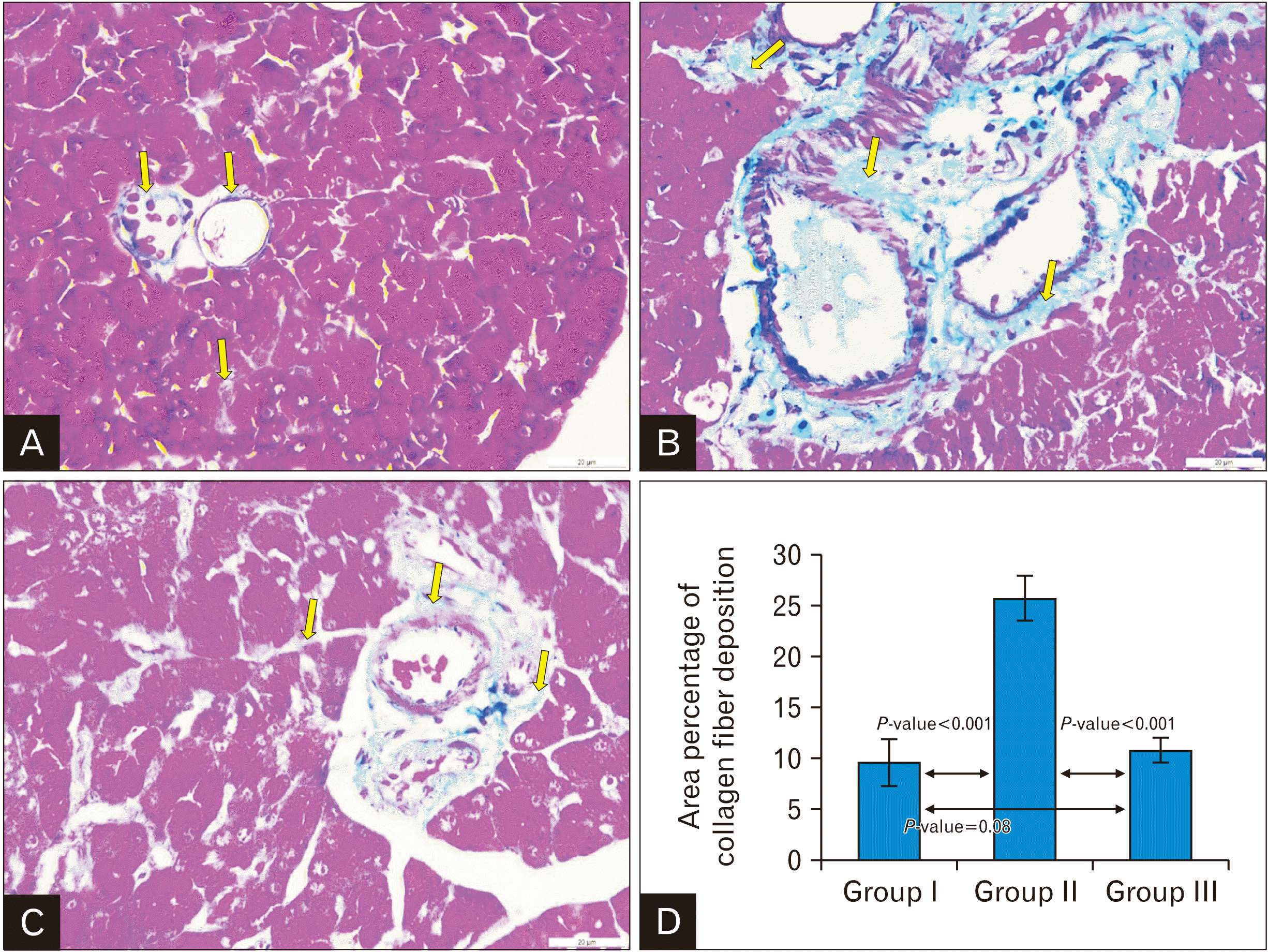 | Fig. 7Representative Masson’s Trichrome stain of rat pancreas; (A) Control group: showing delicate collagen fibers around the pancreatic acini, pancreatic ducts and blood vessels (yellow arrow). (B) Cipralex group: showing dense collagen fiber deposition (yellow arrow). (C) Cipralex plus SIL group: showing moderate collagen fiber deposition (yellow arrow) as compared with cipralex group (yellow arrow) (×400, scale bar=20 μm). (D) A histogram demonstrating a highly significant increase in the mean area percentage of collagen fibers in cipralex group compared to control group. While cipralex plus SIL group showing a highly significant decrease compared to cipralex group, with no significant difference between control and cipralex plus SIL groups. SIL, silymarin.
|
Immunohistochemical findings
Regarding iNOS immunoreaction, control group showed negative immunoreactivity in pancreatic acini and islet cells (Fig. 8A). While cipralex group showed strong cytoplasmic immunoreactivity in most pancreatic acini and most of islet cells (Fig. 8B). Cipralex plus SIL group showed mild immunoreactivity in pancreatic acini and islet cells as compared with cipralex group (Fig. 8C).
 | Fig. 8Representative iNOS immunostaining of rat pancreas; (A) Control group: illustrating negative immunoreactivity in pancreatic acini and islet cells. (B) Cipralex group: illustrating strong cytoplasmic immunoreactivity in most pancreatic acini and islet cells. (C) Cipralex plus SIL group: illustrating mild cytoplasmic immunoreactivity in pancreatic acini and islet cells as compared with cipralex group (×400, scale bar=20 μm). (D) A histogram illustrating a highly significant increase in iNOS immune expression in cipralex group compared to control group. While cipralex plus SIL group showing a highly significant decrease compared to cipralex group, with no significance between control and cipralex plus SIL groups. iNOS, inducible nitric oxide synthase; SIL, silymarin.
|
Regarding TNF-α immunoreaction, control group showed negative immunoreactivity in pancreatic acini and islet cells (Fig. 9A). While cipralex group showed strong cytoplasmic immunoreactivity in most pancreatic acini and most of islet cells (Fig. 9B). Cipralex plus SIL group showed mild immunoreactivity in pancreatic acini and islet cells as compared with cipralex group (Fig. 9C).
 | Fig. 9Representative TNF-α immunostaining of rat pancreas; (A) Control group: revealing negative immunoreactivity in pancreatic acini and islet cells. (B) Cipralex group: showing strong immunoreactivity in most of pancreatic acini and islet cells. (C) Cipralex plus SIL group: showing mild immunoreactivity in pancreatic acini and islet cells as compared with cipralex group (×400, scale bar=20 μm). (D) A histogram showing a highly significant increase in TNF-α immune expression in cipralex group compared to control group. While cipralex plus SIL group showing a highly significant decrease compared to cipralex group, with no significant difference between control and cipralex plus SIL groups. TNF-α, tumour necrosis factor-alpha; SIL, silymarin.
|
Regarding caspase-3 immunoreaction, control group showed negative immunoreactivity in pancreatic acini and islet cells (Fig. 10A). While cipralex group showed a strong cytoplasmic and nuclear immunoreactivity in most pancreatic acini and most of islet cells (Fig. 10B). Cipralex plus SIL group showed moderate immunoreactivity in pancreatic acini and islet cells as compared with cipralex group (Fig. 10C).
 | Fig. 10Representative caspase-3 immunostaining of rat pancreas; (A) Control group: demonstrating negative immunoreactivity in pancreatic acini and islet cells. (B) Cipralex group: demonstrating strong cytoplasmic and nuclear immunoreactivity for caspase-3 in most pancreatic acini and islet cells. (C) Cipralex plus SIL group: demonstrating moderate immunoreactivity in pancreatic acini and islet cells as compared with cipralex group (×400, scale bar=20 μm). (D) A histogram illustrating a highly significant increase in caspase-3 immune expression in cipralex group compared to control group. While cipralex plus SIL group demonstrating a highly significant decrease compared to cipralex group, with no significance between control and cipralex plus SIL groups. SIL, silymarin.
|
Regarding PCNA immunoreaction, control group illustrated strong nuclear immunoreactivity in most of pancreatic acini and islet cells (Fig. 11A). While cipralex group showed mild nuclear immunoreactivity in pancreatic acini and islet cells (Fig. 11B). Cipralex plus SIL group showed moderate immunoreactivity in pancreatic acini and islet cells as compared with cipralex group (Fig. 11C).
 | Fig. 11Representative PCNA immunostaining of rat pancreas; (A) Control group: illustrating strong nuclear immunoreactivity pancreatic acini and islet cells. (B) Cipralex group: showing mild immunoreactivity for PCNA in most of pancreatic acini and islet cells. (C) Cipralex plus SIL group: showing moderate immunoreactivity pancreatic acini and islet cells as compared with cipralex group (×400, scale bar=20 μm). (D) A histogram exhibiting a highly significant decrease in PCNA immune expression in cipralex group compared to control group. While cipralex plus SIL group showing a highly significant increase compared to cipralex group, with no significant difference between control and cipralex plus SIL groups. PCNA, proliferating cell nuclear antigen; SIL, silymarin.
|
Regarding anti-insulin immunoreaction, control group revealed strong cytoplasmic immunoreactivity for insulin in most of the cells of the pancreatic islet (Fig. 12A). While cipralex group showed mild immunoreactivity in a small number of islet cells (Fig. 12B). On the other hand, cipralex plus SIL group showed moderate immunoreactivity for insulin in the pancreatic islet as compared with cipralex group (Fig. 12C).
 | Fig. 12Representative insulin immunostaining of rat pancreas; (A) Control group: revealing strong cytoplasmic immunoreactivity for insulin in most of the cells of the pancreatic islet. (B) Cipralex group: showing mild immunoreactivity for insulin in a small number of islets cells. (C) Cipralex plus SIL group: showing moderate cytoplasmic immunoreactivity for insulin in most of the pancreatic islet as compared with cipralex group (×400, scale bar=20 μm). (D) A histogram exhibiting a highly significant decrease in insulin positive β-cells immune expression in cipralex group compared to control group. While cipralex plus SIL group showing a significant increase compared to cipralex group, with no significant difference between control and cipralex plus SIL groups. SIL, silymarin.
|
Morphometrical and Statistical analysis results
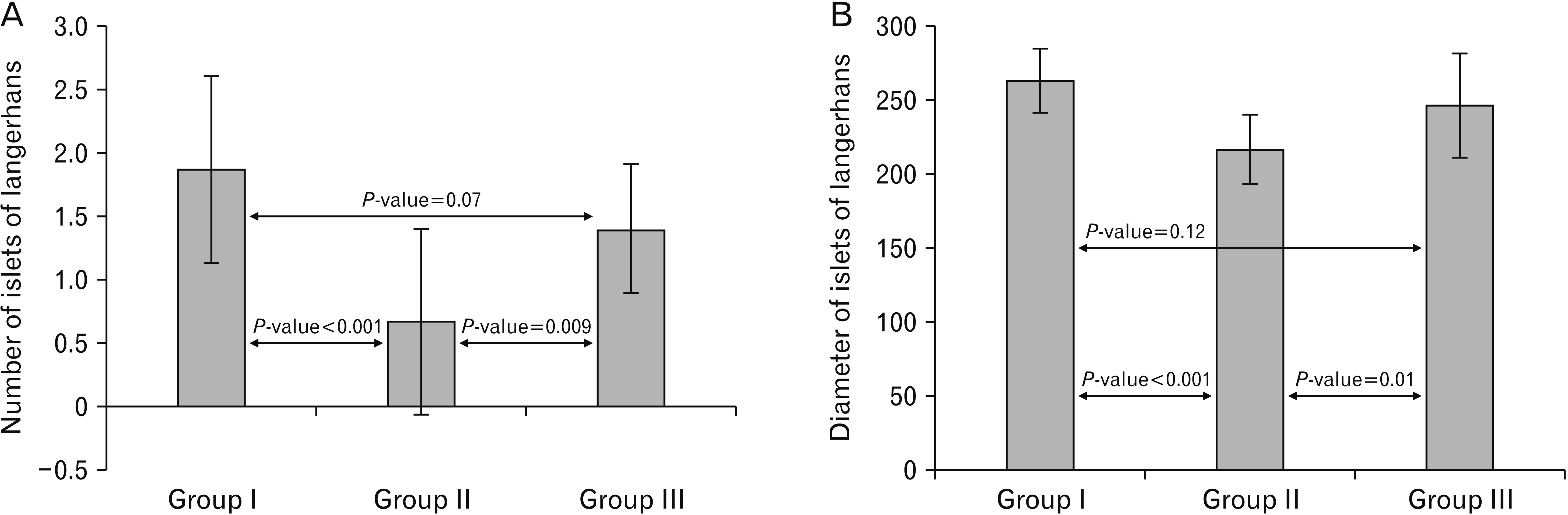
Regarding the number and diameter of islets of Langerhans, cipralex group exhibited a highly significant decrease compared to control group. While cipralex plus SIL group showed a significant increase compared to cipralex group (Table 3, Fig. 13).
 | Fig. 13A histogram showing a highly significant decrease (P<0.001) in the number and diameter of islets of Langerhans in the cipralex group compared to the control group, while the cipralex plus SIL group, illustrating a significant increase compared to the cipralex group, with no significant difference between control and cipralex plus SIL groups. SIL, silymarin.
|
Table 3
Number and diameter of islets of Langerhans among the studied groups
| Islets of Langerhans parameter |
Group I (control) n=15 |
Group II (cipralex) n=15 |
Group III (cipralex plus SIL) n=15 |
Test | P-value |
|---|---|---|---|---|---|
| Number of islets of Langerhans | 3.50* | <0.001a | |||
| 1.87±0.74 | 0.67±0.73 | 1.40±0.51 | 1.79* | 0.07b | |
| 1–3 | 0–2 | 1–2 | 2.78* | 0.009c | |
| Diameter of islets of Langerhans | 5.54** | <0.001a | |||
| 263.4±21.87 | 217.0±23.96 | 246.33±35.38 | 1.59** | 0.12b | |
| 230–290 | 180–240 | 180–320 | 2.66** | 0.01c |
![]()
Concerning the mean area percentage of collagen fibers, there were a highly significant increase in cipralex group compared to control group. While cipralex plus SIL group showed a highly significant decrease compared to cipralex group (Table 4, Fig. 7D).
Table 4
Area percentage of collagen fiber deposition among the studied groups
| Collagen fiber deposition parameter |
Group I (control) n=15 |
Group II (cipralex) n=15 |
Group III (cipralex plus SIL) n=15 |
t-test | P-value |
|---|---|---|---|---|---|
| Area percentage of collagen fiber deposition | 19.46 | <0.001a | |||
| 9.56 ±2.36 | 25.70 ±2.17 | 10.81±1.24 | 1.81 | 0.08b | |
| 6.45–14.58 | 21.69–29.34 | 9.0–13.54 | 23.04 | <0.001c |
![]()
Concerning the numbers of iNOS, TNF-α, and caspase-3 positive immunoreactive cells in the islets of Langerhans and in the pancreatic acinar cells, there were a highly significant increase in cipralex group compared to control group. While cipralex plus SIL group showed a highly significant decrease compared to cipralex group (Table 5, Figs. 8D, 9D, 10D).
Table 5
Immunohistochemical markers among the studied groups
| Immunohistochemical marker parameter |
Group I (control) n=15 |
Group II (cipralex) n=15 |
Group III (cipralex plus SIL) n=15 |
Test | P-value |
|---|---|---|---|---|---|
| iNOS | 4.67* | <0.001a | |||
| 6.11±2.84 | 25.05±7.11 | 8.38±2.99 | 1.76* | 0.08b | |
| 0.7–9.8 | 14.5–40.2 | 3.7–15.6 | 4.63* | <0.001c | |
| TNF-α | 4.67* | <0.001a | |||
| 5.30±4.24 | 30.93±6.46 | 8.19±3.47 | 1.81* | 0.07b | |
| 0.15–13.2 | 22–42 | 2.5–13.2 | 4.67* | <0.001c | |
| Caspase 3 | 4.67* | <0.001a | |||
| 5.25±2.78 | 45.87±9.11 | 7.03±2.91 | 1.64* | 0.10b | |
| 1.2–11.4 | 22.8–57.8 | 2.2–12.5 | 4.67* | <0.001c | |
| PCNA | 16.52** | <0.001a | |||
| 9.82±2.49 | 4.40±1.16 | 7.36±0.71 | 1.71** | 0.09b | |
| 4.5–14.9 | 2.5–6.2 | 6.1–8.9 | 15.73** | <0.001c | |
| Anti-insulin antibody | 5.23** | <0.001a | |||
| 43.15±7.08 | 32.68±3.14 | 38.69±5.83 | 1.88** | 0.07b | |
| 30.9–54.2 | 27.8–40.2 | 25.5–45.4 | 3.52** | 0.002c |
Values are presented as mean±SD or range. SIL, silymarin; iNOS, inducible nitric oxide synthase; TNF-α, tumour necrosis factor-alpha; PCNA, proliferating cell nuclear antigen. aComparison of group I and group II. bComparison of group I and group III. cComparison of group II and group III. *Mann Whitney U-test, **t-test.
![]()
Regarding the number of PCNA positive immunoreactive cells, cipralex group exhibited a highly significant decrease compared to control group. While cipralex plus SIL group showed a highly significant increase compared to cipralex group (Table 5, Fig. 11D).
Regarding the number of insulin positive β-cells, cipralex group exhibited a highly significant decrease compared to control group. While cipralex plus SIL group showed a significant increase compared to cipralex group (Table 5, Fig. 12D).
There wasn’t a significant difference between control and cipralex plus SIL groups in all morphometrical parameters.
Go to :
Discussion
Cipralex group, in the running study exhibited marked destruction of the normal pancreatic architecture of the acinar exocrine part, and endocrine islet of Langerhans portions of the pancreas together with remarkable inflammatory cell infiltration. Also, exhibited a dense collagen fiber deposition. These findings attributed to the progression of CP, as evidenced by many studies that defined CP as an irreversible inflammatory advanced disease of the pancreas that drives to remarkable destruction of the pancreatic acinar cells with progression of pancreatic fibrosis. Furthermore, destruction of the pancreas progresses to definitive loss of its exocrine and endocrine functions [38]. Also, the marked fibrotic changes attributed to the invigoration of pancreatic stellate cells to hyperglycaemia [39], as proofed by the highly significant fasting glucose level rise (P<0.001); together with a highly significant decrease (P<0.001) of the number and diameter of islets of Langerhans in cipralex group.
These histological results in cipralex group was confirmed by the biochemical findings including a highly significant reduction (P<0.001) in serum amylase level compared to control group, as the amylase released in the serum is an important pancreatic activity parameter [40], and highly significant increase (P<0.001) of fasting glucose level compared to control. The destruction of the β-cells is linked to decreased amylase production and release from the exocrine portion of the pancreas of diabetic rats, which is consistent with earlier studies that indicated a close relationship between the endocrine and exocrine parts of the pancreatic vascular network [41]. Additionally, it has been confirmed that the amylase enzyme significantly decreases in diabetic mouse acinar cells [42].
Moreover, there is an evident highly significant decrease (P<0.001) of PW/RW ratio in cipralex group than control group, that was synchronous with the decreased pancreatic index after the second hit of induced sever AP by L-arginine [43], that explained by serious destruction and a self-digestion of the pancreatic tissue [44].
ADs, amitriptyline, citalopram, clomipramine, and fluoxetine all cause mitochondrial dysfunction in other cell systems [45, 46]. Leading up to oxidative stress and apoptosis [47, 48]. Also, most of these ADs were all cytotoxic to pancreatic β-cells at therapeutic concentrations; inducing oxidative stress and apoptosis secondary to suppression of mitochondrial bioenergetics. These actions help to explain the diabetogenic potential of these ADs in humans [49]. The previous studies, coincident with the findings in the current study of strong immunohistochemical expression of iNOS, TNF-α, caspase-3 in cipralex group.
Additionally, prior studies demonstrated that after, the foremost injury of pancreatic acinar cells, the inflammatory cells infiltrate and cohere to the endothelium [50], releasing some variable inflammatory mediators as nitric oxide, proinflammatory cytokines, and reactive oxygen species [51]. Increased vascular permeability, activation of leukocyte, and tissue damage with marked systemic inflammation may be caused by proinflammatory cytokines particularly, IL-1β, IL-6, and TNF-α, which are present in high concentrations [51]. Also, the severity of depression may be attributed to the remarkable increase in the proinflammatory cytokines [52].
Generally, SSRIs enhances β-cell apoptosis, and prohibit insulin release [53]. The mechanism of short-term insulin inhibition by SSRIs may comprise animating insulin receptor substrate proteins-2 kinase, and promoting its phosphorylation at some inhibitory sites. In addition to suppression of glucose-stimulated insulin secretion [54]. Contrary, via biochemical and electrophysiological analyses, serotonin (5-HT) adjusts secretion of insulin by serotonylation of GTPases inside pancreatic β-cells, and SSRIs prohibit this intracellular process, leading to suppression of insulin β-cells secretion [55]. These preceding studies synchronous with the findings in cipralex group that include, mild anti-insulin antibody immunoreactivity and the remarkable highly significant diminution (P<0.001) in insulin level compared to control group.
In the present research, cipralex group revealed a highly significant decrease in expression of PCNA. These effects may be allocated to the capability of cipralex to prohibit DNA repair and cell proliferation, as similarly explained in a previous study that demonstrated significant decline in PCNA expression with reserpine due to suppression of cell proliferation and DNA repair, and on the contrary showed significant increase in the PCNA expression with the AD paroxetine, and attributed such increase to its ability to increase DNA fragmentation [56].
SIL is a common herbal production and is at most utilized to manage acute and chronic inflammatory liver diseases, as a result of its anti-oxidant, anti-inflammatory, and anti-fibrotic properties, as well as, recently, type 2 diabetes [23, 57]. Furthermore, SIL, displays significant antioxidant and membrane-stabilizing activity, therefore conserving several organs against chemical injury. Also, it can reinforce the regenerative ability of the liver [58]. Latterly, this flavonoid agent has also been illustrated as having a significant anti-neoplastic effects in a number of in vitro and in vivo cancer patterns [59].
However, the profitable effects of SIL on pancreatitis aren’t renowned. Here, for the primary time, we state that SIL mitigated the severity of cipralex-induced pancreatitis.
In this study, cipralex plus SIL group showed marked improvement in the pancreatic histological architecture with few acinar cells exhibited pyknotic nuclei, mild vacuolated cytoplasm, few beta cells appeared vacuolated, and significantly increased PW/RT ratio. Moderate collagen fiber deposition appeared. Similarly, Kim et al. [31] reported that pre- and post-treatment with SIL minimized histological damage of the pancreas, suggesting that SIL reveals protective and therapeutic effects versus AP and pulmonary damage caused by cerulein.
Also in this study, the improvement in the endocrine and exocrine components of the pancreas was confirmed by an evident high significant decrease of fasting glucose level, significant rise of the insulin level and the number of insulin positive β-cells, high significant increase of amylase level, and significant increase of the number and diameter of islets of Langerhans in cipralex plus SIL group compared to cipralex group. These results synchronous with preceding study documented that SIL was eligible to retrieve the streptozotocin-damaged pancreatic β cells, leading to marked increased insulin and decreased glucose blood levels [60].
Modification of cytokine rising is critical to the prosperous remedy of pancreatitis. Therefore, cipralex plus SIL group revealed mild iNOS and TNF-α immunoreactivity and moderate caspase-3 immunoreactivity in pancreatic acini and islet cells, suggesting that SIL inhibits pancreatic inflammation and apoptosis as supported by inhibiting the rise of TNF-α, IL-1β, and IL6 in response to SIL in the pancreas and serum [31].
Additionally, cipralex plus SIL group showed moderate PCNA immunoreactivity in pancreatic acini and islet cells as compared to cipralex group. In a relevant study, it was shown that SIL encapsulated in polymersome nanoparticles induced apoptosis, and inhibited migration and proliferation in pancreatic cancer cells and cancer stem cells [61].
Subsequently, presenting SIL as a drug or submitting foods including SIL may be advantageous in remeding cipralex induced pancreatic injury.
Eventually; publicity, availability, agreeably, safety, and low cost of herbal medicine are constantly rising. Improvements in the pancreatic histological architecture, PW loss, biochemical, and immunohistochemical analyses in the current study, confirmed that SIL portrays distinct anti-inflammatory, anti-oxidant, and anti-apoptotic qualities against the rat’s pancreas vulnerability induced by cipralex. Therefore, SIL may be utilized as a prospect candidate for managing various disorders as a complementary and alternative medicine.
Go to :
 XML Download