Journal List > Anat Cell Biol > v.56(1) > 1516081657

Patel, Dupont, Katsuta, Iwanaga, Olewnik, and Tubbs: Concomitant variations of the tibialis anterior, and extensor hallucis longus, and extensor hallucis brevis muscles

Abstract

Tibialis anterior (TA) muscle originates from the lateral surface of tibia and its tendon attaches to the medial cuneiform and base of the first metatarsal. The TA muscle is responsible for both dorsiflexion and inversion of the foot. We present a case of bilateral TA muscle variations that diverge slightly from the current classification systems of this muscle. Recognizing variations such as these may be important for anatomists, surgeons, podiatrists, and physicians. Following routine dissection, an accessory tendon of the TA muscle was found on both sides. Accessory tendons of the extensor hallucis longus and extensor hallucis brevis joined to form a common tendon on both sides. We believe that this unique case will help further the classification systems for the tendons of the TA and also be informative for clinical anatomists as well as physicians treating patients with pathology in this region.

Introduction

The anterior compartment of the leg is primarily comprised of four muscles: tibialis anterior (TA), extensor hallucis longus (EHL), and extensor digitorum longus (EDL), fibularis tertius [1]. Collectively, one of the main functions of the anterior compartment of the leg is dorsiflexion (extension) of the foot. The TA muscle originates from the lateral surface of the tibia and its tendon attaches to the medial cuneiform and base of the first metatarsal [2]. The TA muscle is responsible for both dorsiflexion (extension) and inversion of the foot. Variation of the insertion of TA is reported around 21.7% [3]. The EHL muscle originates from the medial surface of the body of the fibular and its tendon inserts on the base of the distal phalanx of the great toe [2]. The EHL muscle is involved in dorsiflexion of the foot and extension of the great toe. The dorsum of the foot is essentially made up of two muscles: extensor hallucis brevis (EHB) and extensor digitorum brevis (EDB). The EHB muscle originates from the calcaneus and attaches to the proximal phalanx of the great toe [4]. It is responsible for extension of the great toe and the EDB extends the lateral four digits. Herein, we describe a case of bilateral TA variations that diverge slightly from the current classification systems of this muscle. Recognizing such variations might help physicians understand additional functions of the muscles that cannot be understood with only normal anatomical knowledge.

Case Report

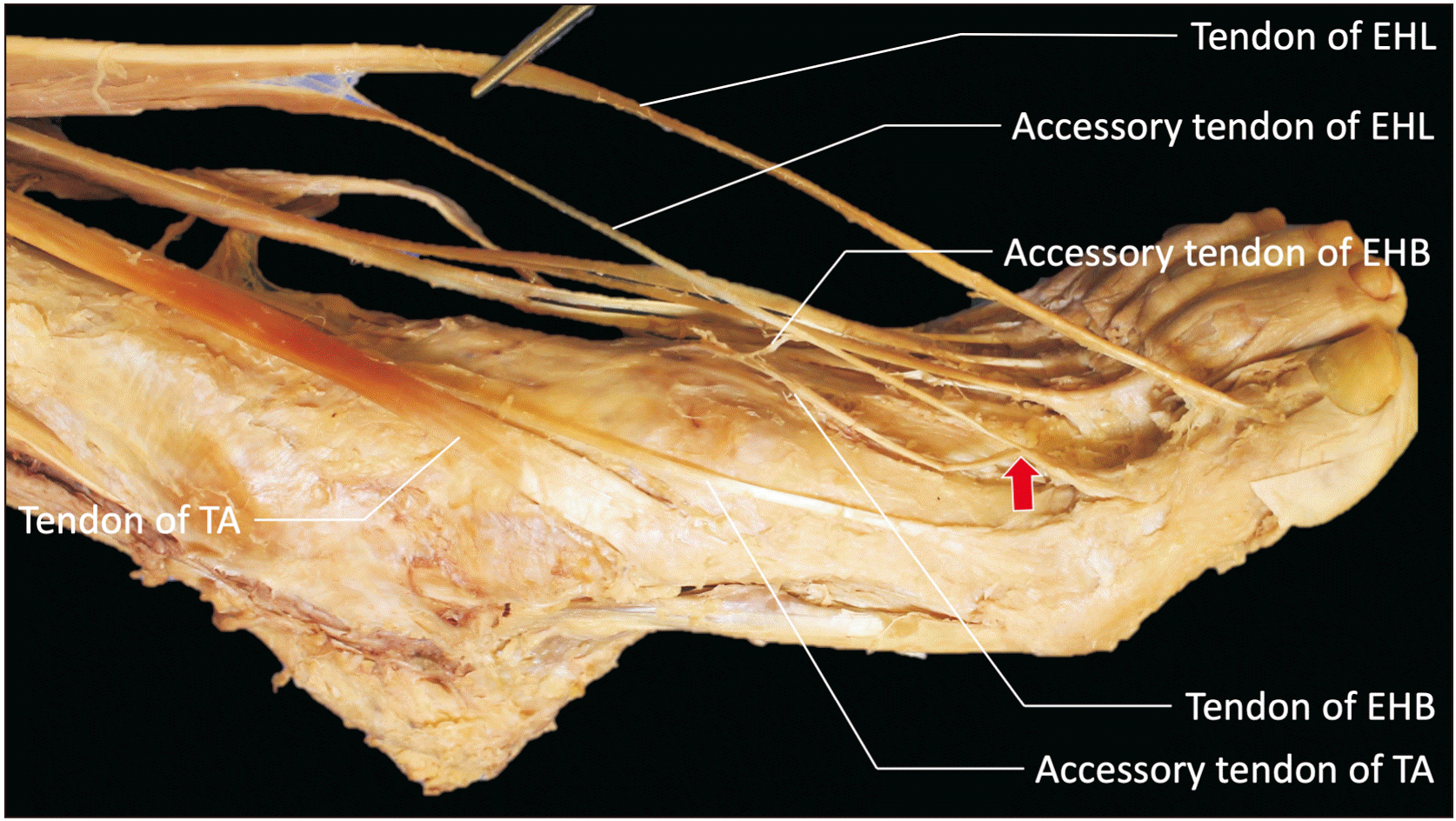
Following routine dissection, on the left side, an accessory tendon was found arising from the lateral aspect of the muscle belly of the TA. The specimen was derived from a male cadaver who was 91-years-old at death. The insertion of this accessory tendon was to the medial aspect of the first metatarsophalangeal (MTP) joint and the body of first proximal phalanx. The insertion of the TA tendon was normal (inferomedial aspect of the medial cuneiform and the base of the first metatarsal). The width of the TA tendon at the cuneiform and that of the accessory tendon at the first metatarsal was 11.9 mm and 2.4 mm, respectively. The accessory tendon arising from the lateral surface of the EHL and the accessory tendon arising from the superficial surface of the EHB joined the distal end of the EHB muscle belly and ran parallel to the main EHB tendon superficially. Then, the two tendons joined to form a common tendon which inserted into the dorsal aspect of the base of the proximal phalanx (Fig. 1).
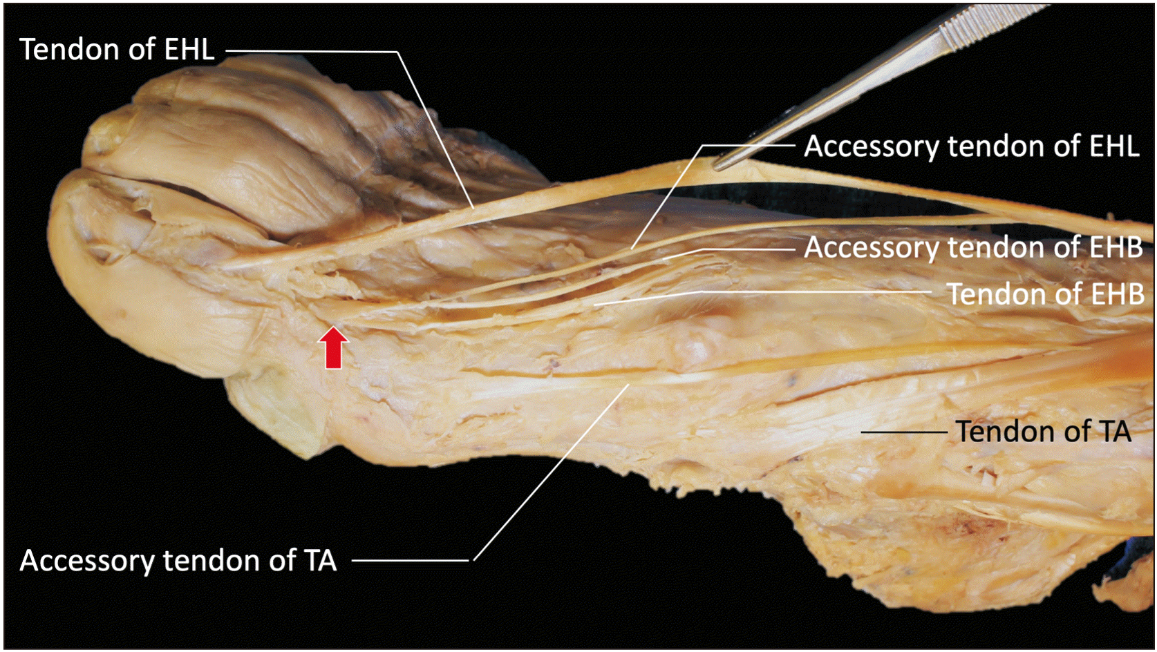
On the right side, an accessory muscle tendon was found arising from the lateral aspect of the TA tendon. The insertion of the TA was normal (inferomedial aspect of the medial cuneiform and the base of the first metatarsal). The width of the TA tendon at its insertion and that of the accessory tendon at the first metatarsal was 11.4 mm and 2.5 mm, respectively. The accessory tendon arising from the lateral surface of the EHL ran parallel to the EHL tendon. The accessory tendon arising from the superficial surface of the EHB ran parallel to the main EHB tendon superficially. The accessory EHL tendon and accessory EHB tendon joined at the first MTP joint to form a common tendon which then joined the EHB tendon and inserted into the dorsal aspect of the base of the proximal phalanx (Fig. 2).

Discussion

In the anterior compartment of the leg, accessory tendons arising/splitting from a main tendon occur in 9%–22% of the population [5]. The prevalence of TA accessory tendons range from 20%–100% compared to 24%–42.5% for EHL bands [1]. Characterization of the tendon of the TA depends on where the tendon attaches distally and characterization of the EHL depends on the morphological variations of its additional bands and their insertions [6, 7]. To date, there are four classification systems for TA tendons:

Musiał, 1963 [8]

  • - Type I: The tendon portion is divided into two distal attachments with similar width, one attaching to the medial cuneiform, and the other attaching to the base of the 1st metatarsal (occurrence=37.7% specimens).

  • - Type II: Same characteristics as type I, but the tendons are not of equal width. The wider part attaches to the medial cuneiform, and the narrower part attaches to the base of the 1st metatarsal (occurrence=56.5%).

  • - Type III: Tendon divides into a broad attachment at the medial cuneiform, with smaller strands attaching to the base of the 1st metatarsal (occurrence=4.1%).

  • - Type IV: Similar to type II, except the wide and narrow attachments are reversed.

Arthornhurasook and Gaew-Im [9]

  • - Type I, II: As described by Musial (occurrence=56.8%, 27.3%).

  • - Type III: Single strand inserting into the medial cuneiform (occurrence=15.9%).

Brenner [10]

  • - Type I, II, III: As described by Musial (occurrence=27.6%, 45.5%, 25.6%)

  • - Type IV: Identical to type III described by Arthornhurasook and Gaew-Im.

Olewnik et al. [7]

  • - Types I–IV: As described by Musial and Arthornhurasook and Gaew-Im.

  • - Type V: Identical to type IV as described by Arthornhook and Gaew-Im.

The specimen in the present case demonstrated a slight morphological variation in Type IV TA, as well as a slight variation in Type III EHL tendons, both based on Olewnik et al.’s classification [6]. According to their classification, the type IV TA tendon splits into three bands and inserts into the medial cuneiform (one accessory band) and first metatarsal (one band to the base and another band to the distal part) [6]. This specimen differs from Olewnik et al.’s type IV classification [6] as the accessory TA tendon inserts into the first MTP joint and body of the first proximal phalanx. The main TA tendon insertion was normal, inserting inferomedially to the base of the first metatarsal and medial cuneiform on both left and right sides [6]. Based on Olewnik et al.’s classification [7], the type III EHL tendon trifurcates into the main EHL tendon which attaches at the distal region of the distal phalanx; a “medial auxiliary band” attaches at the distal phalanx as well (more proximally); and a “lateral auxiliary band”, which is the strongest band, merges with the EHB and inserts at the proximal phalanx. Our case varied slighty from Olewnik et al.’s type III classification [7] in that the two accessory tendons of the EHL and EHB combined at the EHB muscle belly distally and ran parallel to the main EHB tendon (superficially). Furthermore, the two accessory tendons of the EHL and EHB coursed together into a common tendon that attached to the base of the proximal phalanx.
Generally, limb muscle development begins during the 7th week of fetal life, in tandem with reciprocal morphogen signaling of outward growth, and polarization of the limbs via the apical ectodermal ridge and the zone of polarizing activity [10]. Defects in this signaling can lead to various limb defects, but since these types of anatomical variations are not deleterious in nature, one can presume that these types of variations may or may not confer extra- or inability. Further, there are no biochemical tests in the literature to determine whether different muscle and tendon configurations of the TA confer a mechanical advantage. As the limb buds splits into flexor and extensor components, variations in signaling (extensive migration and rearrangement of the muscles) may take place and lead to different configurations of the TA tendons. TA variations involving thinner tendons may be more susceptible to traumatic tendon ruptures due to overloading a thin tendon with a maladaptive force vector, as well as ruptures originating from diabetes, and rheumatic diseases such as gout and rheumatoid arthritis [11-13].
We believe that this case report will help further the classification systems for the tendons of the TA and also be informative for clinical anatomists as well as physicians treating patients with pathology in this region.

Acknowledgements

The authors sincerely thank those who donated their bodies to science so that anatomical research could be performed. Results from such research can potentially increase mankind’s overall knowledge that can then improve patient care. Therefore, these donors and their families deserve our highest gratitude [14].

Notes

Author Contributions

Conceptualization: JI, ŁO, RST. Data acquisition: RST, JI. Data analysis or interpretation: JP. Drafting of the manuscript: JP, GD, JK. Critical revision of the manuscript: JK, JI, ŁO, RST. Approval of the final version of the manuscript: all authors.

Conflicts of Interest

No potential conflict of interest relevant to this article was reported.

References

1. Karauda P, Podgórski M, Paulsen F, Polguj M, Olewnik Ł. 2021; Anatomical variations of the tibialis anterior tendon. Clin Anat. 34:397–404. DOI: 10.1002/ca.23663. PMID: 32713016.
crossref
2. Drake RL, Vogl AW, Mitchell AWM. Drake RL, Vogl AW, Mitchell AWM, editors. 2020. Lower limb/leg. Gray's Anatomy for Students. 4th ed. Elsevier;Philadelphia: p. 612–27.
3. Htwe O, Swarhib M, Pei TS, Naicker AS, Das S. 2012; Congenital bilateral agenesis of the tibialis anterior muscles: a rare case report. Rom J Morphol Embryol. 53:657–9. PMID: 22990563.
4. Drake RL, Vogl AW, Mitchell AWM. Drake RL, Vogl AW, Mitchell AWM, editors. 2020. Lower limb/foot. Gray's Anatomy for Students. 4th ed. Elsevier;Philadelphia: p. 627–59.
5. DeLuca M, Boucher L. 2019; Morphological variations and accessory ossicles in the peroneal and tibialis muscles. Anat Cell Biol. 52:344–8. DOI: 10.5115/acb.19.030. PMID: 31598366. PMCID: PMC6773904.
crossref
6. Olewnik Ł, Podgórski M, Polguj M, Topol M. 2019; A cadaveric and sonographic study of the morphology of the tibialis anterior tendon - a proposal for a new classification. J Foot Ankle Res. 12:9. DOI: 10.1186/s13047-019-0319-0. PMID: 30733832. PMCID: PMC6359855. PMID: b635b5eda22148dc836d413a2ea51a35.
crossref
7. Olewnik Ł, Podgórski M, Polguj M, Ruzik K, Topol M. 2019; A cadaveric study of the morphology of the extensor hallucis longus - a proposal for a new classification. BMC Musculoskelet Disord. 20:310. DOI: 10.1186/s12891-019-2688-8. PMID: 31266496. PMCID: PMC6607556. PMID: 303dc2ed5e6c4623b63768e75a71ace8.
crossref
8. Musiał W. 1963; Variations of the terminal insertions of the anterior and posterior tibial muscles in man. Folia Morphol. 26:237–47.
9. Arthornthurasook A, Gaew Im K. 1990; Anterior tibial tendon insertion: an anatomical study. J Med Assoc Thai. 73:692–6. PMID: 2086718.
10. Brenner E. 2002; Insertion of the tendon of the tibialis anterior muscle in feet with and without hallux valgus. Clin Anat. 15:217–23. DOI: 10.1002/ca.10021. PMID: 11948958.
crossref
11. DiDomenico LA, Williams K, Petrolla AF. 2008; Spontaneous rupture of the anterior tibial tendon in a diabetic patient: results of operative treatment. J Foot Ankle Surg. 47:463–7. DOI: 10.1053/j.jfas.2008.05.007. PMID: 18725129.
crossref
12. Constantinou M, Wilson A. 2004; Traumatic tear of tibialis anterior during a Gaelic football game: a case report. Br J Sports Med. 38:e30. DOI: 10.1136/bjsm.2003.007625. PMID: 15562145. PMCID: PMC1724965.
crossref
13. Jerome JT, Varghese M, Sankaran B, Thomas S, Thirumagal SK. 2008; Tibialis anterior tendon rupture in gout--case report and literature review. Foot Ankle Surg. 14:166–9. DOI: 10.1016/j.fas.2007.12.001. PMID: 19083637.
crossref
14. Iwanaga J, Singh V, Ohtsuka A, Hwang Y, Kim HJ, Moryś J, Ravi KS, Ribatti D, Trainor PA, Sañudo JR, Apaydin N, Şengül G, Albertine KH, Walocha JA, Loukas M, Duparc F, Paulsen F, Del Sol M, Adds P, Hegazy A, Tubbs RS. 2021; Acknowledging the use of human cadaveric tissues in research papers: recommendations from anatomical journal editors. Clin Anat. 34:2–4. DOI: 10.1002/ca.23671. PMID: 32808702.
crossref

Fig. 1
Variant anatomy on the left foot. EHB, extensor hallucis brevis; EHL, extensor hallucis longus; TA, tibialis anterior.
acb-56-1-137-f1.tif
Fig. 2
Variant anatomy on the right foot. EHB, extensor hallucis brevis; EHL, extensor hallucis longus; TA, tibialis anterior.
acb-56-1-137-f2.tif
TOOLS
Similar articles