

 PDF
PDF Citation
 Print
Print


INTRODUCTION
Acute appendicitis accounts for the largest proportion of emergency gastrointestinal surgeries, and establishing its diagnosis is usually straightforward.1-3 The diagnostic uncertainty caused by the atypical presentation of acute appendicitis can be attributed to the malposition of the appendix.4 The appendix is a vestigial organ located 2.5 cm below the ileocecal plate and on the posteromedial side of the cecum. This organ is one of the few where the anatomical position is not uniform. The location of the appendix varies and can be found in the retrocecal, pelvic, subcecal, preileal, and postileal regions. The appendix is rarely located in the subhepatic, mesoceliac, intrahernial, and left-side regions. Appendicitis causing pain in the lower left quadrant (LLQ) is remarkably rare and can occur as a congenital anomaly, including true left-sided appendicitis, or as an atypical representation of the right, elongated appendix projecting into the LLQ. Left-sided appendicitis is associated with two types of congenital anomalies: situs inversus (SI) and midgut malrotation (MM).5-7
This paper reports the process of performing appropriate surgery in a case of left-sided appendicitis with an intestinal rotation abnormality mistaken for diverticulitis of the left sigmoid colon. Through this case report, the diseases to be differentiated from left-sided appendicitis are investigated, and the diagnostic process conducted at the authors’ hospital is introduced.
The Institutional Review Board of Dankook University Hospital reviewed and approved this study (IRB No. 2022-10-008).
CASE REPORT
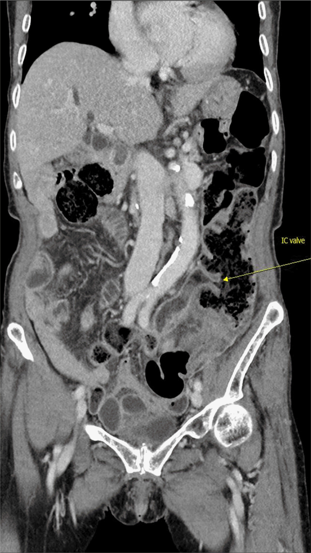
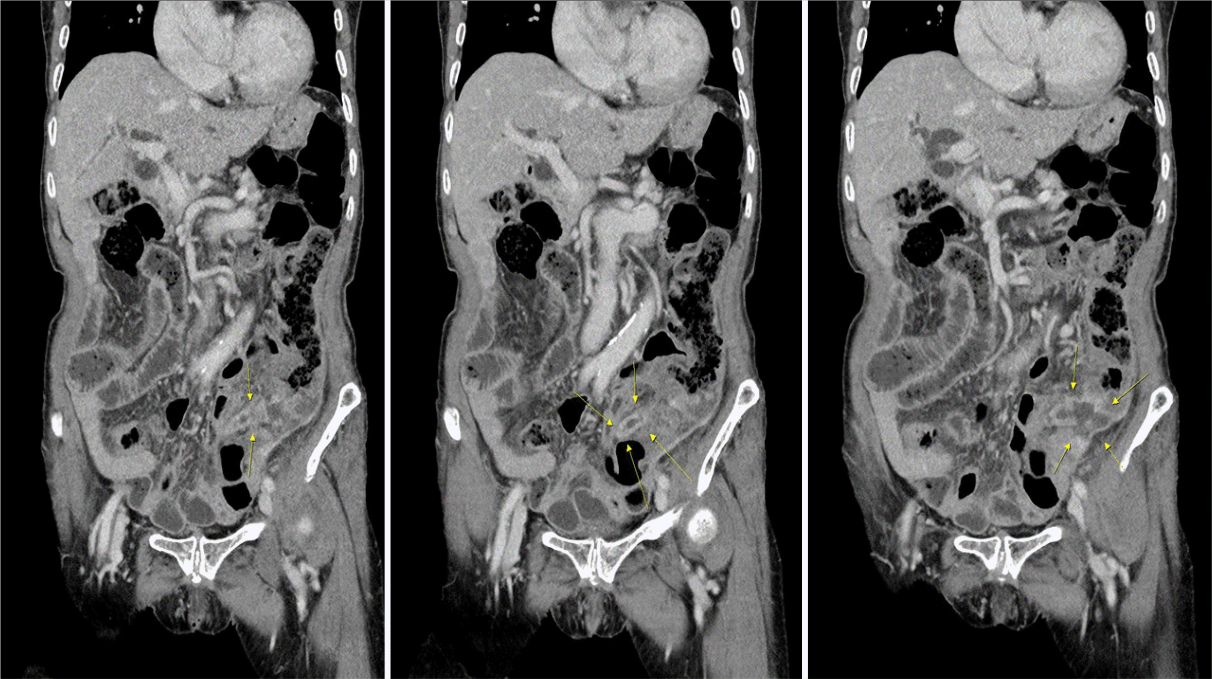
The current case report presents the case of an 86-year-old woman who had continuous abdominal pain two days before the visit to the emergency room. Thus, a medical examination was conducted. The patient had no specific present illness other than taking medicine for hypertension. The patient underwent cholecystectomy 20 years prior, had a history of recurring common bile duct (CBD) stones and cholangitis due to a distal CBD deformity (acute angulation), and had undergone endoscopic retrograde cholangiopancreatography, percutaneous transhepatic bile drainage, and endoscopic retrograde biliary drainage insertion several times at the authors’ hospital. The patient's examination revealed tenderness in the LLQ area but no rebound tenderness. The white blood cell was 13,210/μL and showed leukocytosis. CRP was also increased at 7.86 mg/dL. No specific abnormalities were observed on the simple plain abdominal X-ray. Abdominopelvic computed tomography revealed MM (Fig. 1), and uneven concentric wall thickening from the distal descending colon to the proximal sigmoid colon with surrounding fatty infiltration was confirmed in the left lower abdomen (Fig. 2). No evidence of intestinal free perforation was found in the abdominal cavity, but an abscess, approximately 2 cm in size, was suspected around the sigmoid colon. The patient decided to proceed with the antibiotic and fasting treatments, rather than surgery because of her old age.
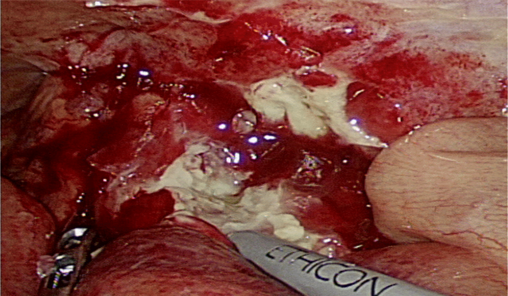
The next day, the patient had increased abdominal pain and increased tenderness in the LLQ region on the physical examination. Therefore, the various possibilities of colonic diverticulitis, left-sided appendicitis, malignant diseases, and colon perforation originating from those diseases were explained to the patient, and emergency surgery was then performed. The operation performed was laparoscopic surgery. The lesion was located on the left side. A 12-mm camera trocar was inserted into the umbilicus. Subsequently, a 5-mm trocar was inserted into the right lower quadrant and middle lower areas. According to the surgical findings, an abscess formed due to perforation caused by inflammation of the proximal part of the appendix (Fig. 3). The sigmoid colon was located just behind the abscess sac around the appendix with mild adhesion.
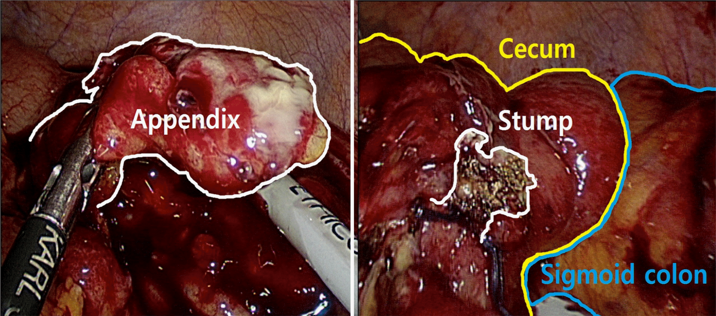
In conclusion, the cause of the patient’s symptoms was a perforation from left appendicitis with an abscess, not a disease of the colon. The base of the appendix was intact, and only an appendectomy was performed (Fig. 4). After intra- abdominal irrigation, a drain was placed, and the operation was terminated. A malrotated cecum, descending colon, and sigmoid colon were observed during surgery, and there were no specific abnormalities. This patient belongs to Stringer classification ⅢC, in which both the cecum and mesentery are not fixed among the midgut malrotation.
The patient proceeded on a soft diet from the second day after surgery and was discharged without complications. No surgical site infection or specific complications were observed on follow-up at the outpatient clinic one week after discharge. Based on postoperative biopsy findings, the patient was diagnosed with acute perforated gangrenous appendicitis. The patient was informed about the need for a colonoscopy and follow-up CT examination in the future for diverticulitis and malignant disease.
DISCUSSION
Abdominal pain is one of the most common symptoms in need of surgery. Among the diagnoses due to abdominal pain, appendicitis is the most common surgical disease.8 A diagn osis of appendicitis is possible based on the identification of well-established clinical symptoms, physical examination, and surgeon’s experience.9,10 In approximately one-third of appendicitis cases, the location of the appendix can be significantly different, and the pain is usually localized outside the lower right quadrant. On the other hand, appendicitis expressed as LLQ pain is relatively rare and often misdiagnosed. In adults, the differential diagnoses caused by pain in the LLQ include diverticulitis, renal colic, ovarian cyst rupture, epididymitis, hernia, focal enteritis, and psoas abscess.11
Left-sided acute appendicitis has two major anatomical causes: MM and a less common variant reported as SI. MM is a congenital positional malformation of the intestine resulting from non-rotation or incomplete rotation of the primordial loop on the superior mesenteric artery axis during fetal life. According to the literature, the incidence of midgut abnormal rotational malformations varies from 0.03% to 0.5% at birth. Most cases of MM show biliary vomiting in the first months after birth, but this symptom is rarely observed in adulthood.12,13
SI is a rare autosomal recessive birth defect characterized by the transposition of abdominal and thoracic organs. Fabricius first reported SI in 1600. The transposition of the organs due to this disease may affect the entire lung and abdomen, or only one of the two areas may be partially affected. The incidence of site anomalies reported in the literature varies from 0.001% to 0.01% in the general population.14-16
Diagnosis of acute appendicitis in patients with MM or SI is possible based on a physical examination, electrocardiogram, chest X-ray, barium study, ultrasound-guided (USG) scan, CT scan, and diagnostic laparoscopy. The use of imaging modalities for diagnosing acute appendicitis, such as USG scans and CT, has increased over the past two decades. The USG scan is a widely used test for appendicitis, but it has limitations. The results vary depending on the operator and may be inaccurate because of patients with a relatively large lower quadrant or gas in the gastrointestinal tract. The value of CT in the diagnosis of acute appendicitis has been reported, with an accuracy of 80-90%. CT may also help detect SI and MM.17
In the present case, it was difficult to differentiate left-sided appendicitis from diverticulitis of the sigmoid colon in the preoperative imaging examination. Therefore, it was necessary to discriminate the possibility of chronic diverticulitis due to chronic inflammation and possible micro-perforation caused by malignant lesions of the cecum. Moreover, the possibility of left-sided appendicitis due to MM could not be excluded. Several occurrences of diverticulitis in the sigmoid colon have been reported. Moreover, it is essential to differentiate diverticulitis without complications from acute appendicitis that requires surgery because nonsurgical treatment through fasting and antibiotic administration is preferred over surgical treatment.18
In the present case, the suggestions of the radiology department were important in diagnosing the patient’s MM and differentiating between diverticulitis and left-sided appendicitis. Moreover, it is also essential for the surgeon to decide based on sufficient experience. Although diagnostic laparoscopic surgery is a relatively accurate means of establishing a diagnosis, it must be accompanied by the induction of general anesthesia, and this procedure should be performed carefully in elderly patients and patients with several underlying diseases. Nevertheless, surgeons, who should be equipped with sufficient experience and training, have an important role when deciding on surgery and treating the patient before the disease worsens. In addition, good cooperation with a gastroenterologist is vital for continuous management after surgery. In the present case, the presence of diverticulum and the possibility of malignant disease had to be considered; thus, an endoscopy after surgery was necessary. In patients with MM, the role of a gastroenterologist is important because patients with MM are different from general patients when performing a colonoscopy.19,20
Left-sided appendicitis due to MM or SI is a rare disease, but a careful diagnosis is required. Diagnostic laparoscopic surgery through a physical examination of patients based on the surgeon’s experience is essential for patient management.
Multidisciplinary collaboration with radiologists and gastroenterologists is essential because left-sided appendicitis is challenging to diagnose and differentiate from diverticulitis of the sigmoid colon.
 XML Download