Journal List > Korean J Gastroenterol > v.81(2) > 1516081650

자가면역간염의 진단

Abstract

Autoimmune hepatitis (AIH) is an immune-mediated inflammatory liver disease with an uncertain cause. The diagnosis of AIH is based on the characteristic clinical and laboratory findings (elevated liver enzyme and hypergammaglobulinemia), the presence of characteristic autoantibodies, and compatible histological abnormalities. AIH lacks a signature diagnostic marker, and the diagnosis requires the exclusion of other diseases (viral hepatitis, alcoholic liver disease, non-alcoholic steatohepatitis, drug-induced liver injury, Wilson’s disease, and hereditary hemochromatosis). Therefore, collaboration between the clinical physician, laboratory medicine experts, and pathologists is important for a diagnosis. In December 2022, the Korean Association for the Study of the Liver (KASL) clinical practice guidelines were established. This review article summarizes the diagnosis part of these guidelines.

서 론

자가면역간염(autoimmune hepatitis, AIH)은 자가면역기전에 의해 발생하는 원인 미상의 염증성 간질환으로 희귀난치성 질환으로 분류된다. 모든 연령층에서 발생할 수 있고, 무증상, 급성 간염, 급성 간부전, 만성간염, 간경변증 등 모든 유형의 간질환으로 발현할 수 있다. 자가면역간염의 진단은 근거기반(evidence based)이라기 보다는 전문가 합의(expert consensus)의 영역으로 이는 자가면역간염만의 증상이 존재하지 않고 임상양상과 검사소견, 병리소견 및 다른 질환과의 감별을 통해서 이루어지기 때문이다.1 2022년 12월, 대한간학회 자가면역간염 진료 가이드라인이 제정되었으며 이 중 진단 부분을 요약 정리하고자 한다.

본 론

1. 진단 기준

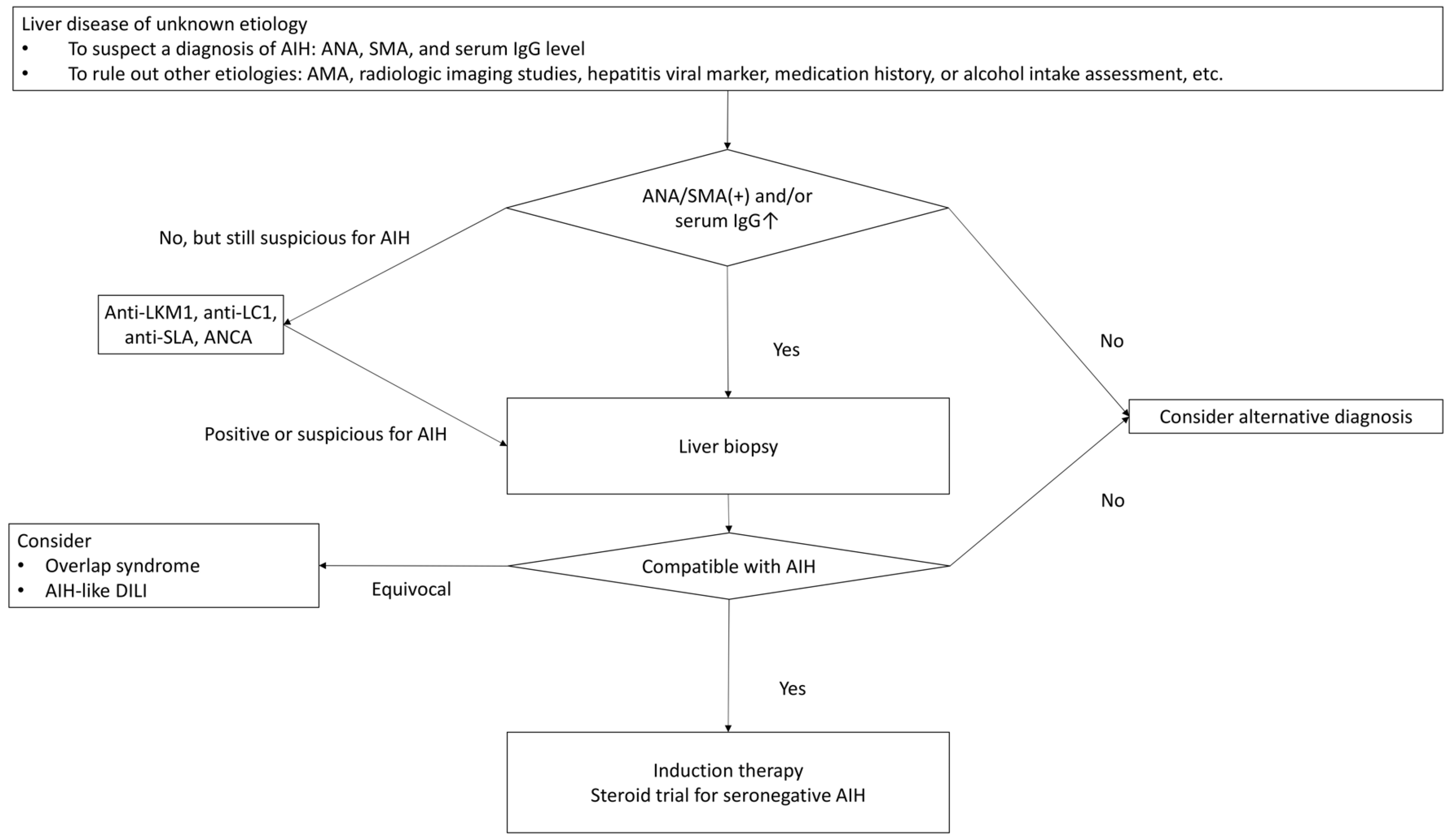
자가면역간염은 특징적인 임상 및 검사 소견(혈청 AST, ALT 및 면역글로불린 G [IgG] 상승), 특징적인 자가항체의 존재 및 합당한 간조직 소견을 근거로 진단한다(Fig. 1). 자가면역간염은 특징적인 진단 표지자가 없어, 일반적으로 다른 원인의 만성 간질환(바이러스 간염, 알코올 간질환, 비알코올 지방간염, 약인성간손상, 윌슨병, 유전성 혈색소 침착증 등)을 배제해야 진단할 수 있다.1-3
혈청 빌리루빈과 아미노전이효소(aminotransferase)가 매우 증가되더라도 담즙정체효소는 정상 또는 미세한 상승 정도만 보이는 것이 자가면역간염의 특징적 소견이다. 자가면역간염에서 혈청 IgG 상승은 흔하지만, 면역글로불린 A (IgA) 및 면역글로불린 M (IgM)은 일반적으로 정상이다.3-6 전체 약 10%의 환자에서, 그리고 급성 발현한 자가면역간염의 25-39%에서 IgG가 정상이며 이러한 환자에서도 임상 양상은 전형적인 자가면역간염과 유사하다.7-10
다른 질환과의 감별, 중복증후군의 판별 등을 고려할 때 조직검사는 금기가 없는 한 자가면역간염의 진단에 필수적이라는 것이 일반적인 견해이다.1-3,7

2. 자가항체(Autoantobidies)

자가면역간염이 의심되는 경우 자가항체(autoantibody)인 항핵항체(ANA), 항평활근항체(anti-smooth muscle antibody, SMA)를 선별 검사로 시행한다(Table 1).11 이들 검사에서 음성을 보이는 경우, 항 간-콩팥-미소솜체 항체 1 (antibody to liver kidney microsome type 1, anti-LKM1), 항 간-세포액 -1 항체(antibody to liver cytosol type 1, anti-LC1), 항수용성간항원항체(antibody to soluble liver antigen, anti-SLA),핵주위 항호중구핵항체(perinuclear antineutrophil cytoplasmic antibody, p-ANCA) 검사를 추가로 실시할 수 있다.
ANA와 SMA가 동반된 경우 진단적 가치가 높으며 Anti-SLA는 자가면역간염에 유일한 질병 특이 자가항체이므로 진단 가치가 높다.12,13 Anti-LKM1 및 anti-LC1은 2형 자가면역간염의 혈청학적 표지자이지만, 만성 C형간염 바이러스 감염이 있는 성인 및 소아 환자의 5-10%에서 검출될 수 있기 때문에 이를 배제해야 한다. 핵주위 항호중구핵항체(perinuclear anti-neutrophil nuclear antibody, p-ANNA)나 p-ANCA가 1형 AIH의 일부 환자에서 유일한 혈청학적표지자일 수 있다.14,15 원발담도담관염(primary biliary cholangitis, PBC) 특이 혈청학적 표지자인 항미토콘드리아 항체(AMA)는 중복증후군 등의 감별진단을 위해서 검사를 시행하며, 중복중후군(overlap syndrome)이 아닌 전형적인 자가면역간염 표현형을 가진 환자의 8-12%에서도 검출될 수 있다.16,17 약 19-34%의 환자가 자가면역간염의 임상 특징을 가지지만, ANA, SMA, anti-LKM1 자가항체가 검출되지 않는자가항체음성 자가면역간염(seronegative AIH)의 형태로 발현하여 원인불명의 간염으로 진단되기 쉽다.1 자가항체음성자가면역간염은 진단 점수 체계에 근거한 임상적인 의심과 글루코코티코이드 치료에 대한 반응 여부를 통해 진단한다.18
혈청 검사실과 임상의는 환자에게 최대한의 이점을 제공하기 위해 자가면역간질환 혈청 검사의 해석에 대한 전문성을 높이고 긴밀히 의사소통해야 한다. 진단이 불확실한 경우 완전한 평가를 위해 전문 검사기관에 혈청 검사를 의뢰할 필요가 있다.

3. 병리(Histological findings)

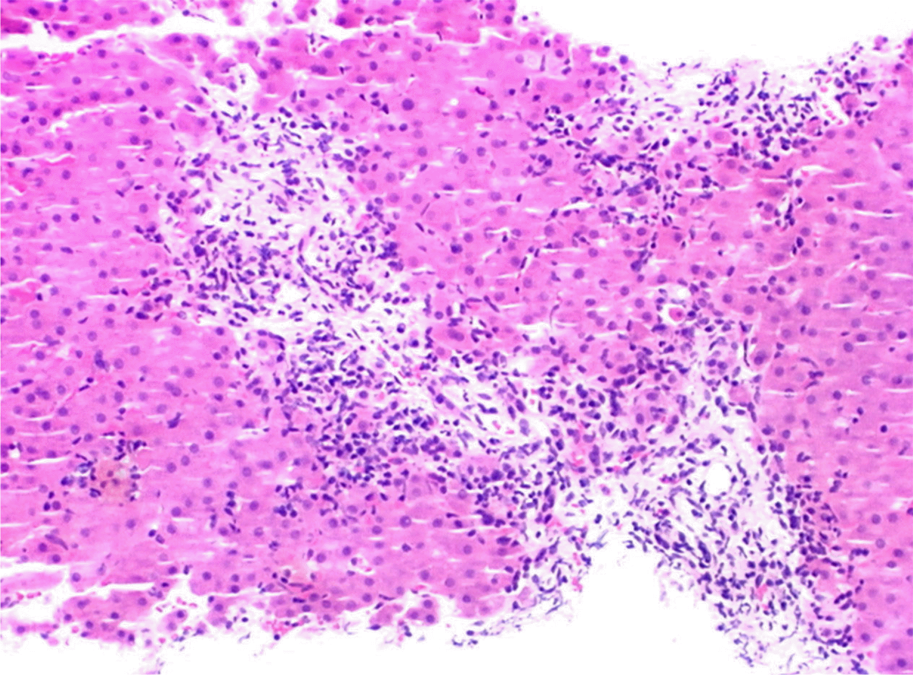
전형적인 자가면역간염의 조직소견은 다음과 같다. 문맥역(portal tract)에 단핵구 세포(mononuclear cell) 침윤이 있으며 경계면 간염을 보인다(Fig. 2).6,19-21 단핵구 세포 중 형질세포(plasma cell)가 뚜렷하게 많으며 간소엽(lobule)에 다양한 정도의 간세포 손상이 관찰된다. 섬유화는 만성바이러스간염과 유사한 양상으로 문맥역을 중심으로 발생하며, 간경변증으로 점차 진행한다. 자가면역간염의 진단에 사용되는 1999년 개정 진단 점수 체계와 2008년 간소화 진단 점수 체계에 제시된 내용에 따르면 림프구 및 형질세포의 침윤, 경계면 간염, 간세포 로제트와 엠페리포레시스가 자가면역간염의 병리학적 진단에 중요한 요소인 것으로 강조되어 있다.6,19 그러나 간세포 로제트와 엠페리포레시스는 심한 염증과 재생에 따른 비특이적인 소견들이고, 급성 간염으로 발병하는 자가면역간염의 경우에는 현 점수 체계를 적용하기가 현실적으로 어렵다는 점이 지적되고 있다. 이러한 점들을 고려하여, 문맥역의 변화는경미하거나 없더라도 중등도 이상의 간소엽 염증이 관찰되는경우에도 임상적으로 다른 원인을 배제할 수 있다면 자가면역간염의 가능성을 제시할 수 있는 방향으로 진단 점수 체계가변경될 것으로 전망된다.20,21
담관 손상은 자가면역간염의 전형적인 특징은 아니며, 전형적인 자가면역간염의 조직소견이 보이는데 심한 담관 손상이 동반된 경우에는 원발담도담관염이나 원발경화담관염(primary sclerosing cholangitis, PSC)과 겹치는 중복증후군의 가능성을 고려해 볼 수 있다.

4. 진단 점수 체계(Diagnostic scoring systems)

자가면역간염의 전형적인 사례 외에 비전형적인 사례의 진단을 돕고, 진단을 정량화하여 객관적인 비교가 가능하도록 1993년 국제 자가면역간염 그룹(International Autoimmune Hepatitis Group, IAIHG)에서 진단 점수 체계(diagnostic scoring system)가 고안되었고 1999년 개정 진단 점수 체계(revised original scoring system)가 발표되었으며 2008년 간소화 진단 점수 체계(simplified scoring system)도 개발되었다(Tables 2, 3).6,19,22 국내에서도 개정 진단 점수 체계와 간소화 진단 점수 체계를 진단에 사용하고 있다.23 개정 진단 점수 체계는 복잡하거나 비전형적인 특징을 가진 환자의 진단에 도움이 되고, 간소화 진단 점수 체계는 전형적인 환자에서 더 정확하다고 알려져 있다.24 일본 연구에서는 개정 진단 점수 체계로 100%의 민감도와 93%의 특이도, 간소화 진단 점수 체계로 85%의 민감도와 99%의 특이도를 보였다.25 국내 연구에서는 간소화 진단 점수 체계에서 69.9%의 민감도와 86.4%의 양성 예측도로 비전형적인 환자의 진단에 제한이 있고 중등도 민감도를 보이는 결과를 나타낸 바 있다.26,27 따라서 개정 진단 점수 체계는 민감도가 높고 간소화 진단 점수 체계는 특이도가 높은 점을 고려하여 간소화 진단 점수 체계로 산정된 점수가 낮으면, 개정 진단 점수 체계로 재평가를 고려해야 한다.24
소아 환자에게 개정 진단 점수 체계를 적용할 수 있으나 자가항체 역가가 성인보다 낮은 점에 유의하여야 한다.19

5. 중복증후군(Overlap Syndromes)

1) 자가면역간염-원발담도담관염 중복증후군(AIH-PBC Overlap Syndrome)

자가면역간염-원발담도담관염 중복증후군의 진단은 파리 기준(Paris criteria)이 가장 흔히 사용되고 효과적인 방법으로 알려져 있다.28 다음 항목 중 최소 2개 이상의 기준을 충족하여야 한다. 원발담도담관염 진단을 위해 (1) 혈청 ALP 정상 상한치의 2배 이상 또는 혈청 글루타밀전이효소(GGT) 정상 상한치의 5배 이상; (2) AMA 양성; (3) 합당한 간 조직검사 소견(소엽간 담관[interlobular bile duct])의 비화농성 파괴성 담관염(non-suppurative destructive cholangitis) 중 2개 이상, AIH 진단을 위해 (1) ALT 정상 상한치의 5배 이상; (2) IgG 정상 상한치의 2배 이상 그리고/또는 SMA 양성; (3) 간 조직검사 상 중등도 또는 중증 경계면 간염 중 2개 이상 각각 만족하여야 한다.29

2) 자가면역간염-원발경화담관염 중복증후군(AIH-PSC Overlap Syndrome)

자가면역간염-원발경화담관염 중복증후군은 자가면역간염의 진단 기준을 만족시키고 AMA가 음성인 환자에서, MRCP 또는 ERCP에서 특징적인 담관의 국소 협착과 확장으로 염주 모양의 담도상을 보이는 경우, 대담관형의 원발경화담관염(large-duct PSC)으로, 혹은 담관 영상검사에서 이상은 없으나 간 조직검사에서 특징적인 섬유폐쇄담관염(fibrous obliterative cholangitis)를 보이는 경우, 소담관형의 원발경화담관염(small-duct PSC)으로 진단할 수 있다.30

6. 자가면역간염 유사 약인성 간손상과의 감별진단

약인성 간손상이 혈청 자가항체 생성과 감마글로불린혈증을 유도하여 마치 자가면역간염의 임상소견을 보이는 경우가 있다. 간 조직검사로 감별 진단해 볼 수 있으나, 약인성 간손상이 경계면 간염과 형질세포의 침윤이 관찰되는 조직소견을 보여 자가면역간염과 감별하기 어려운 경우도 있다.31 따라서, 질병 발생 이전에 노출된 약제와 보조제를 잘 파악하여야 한다. 약제 노출 후 자가면역간염 유사 약인성 간손상 발생까지의 잠복기는 1-8주부터 3-12개월까지 범위가 넓다.1,32 또한, 글루코코티코이드 치료 반응과 치료 종료 후 재발 여부 평가
가 두 질환을 감별하는데 도움이 된다.33 약제를 중단해도 호전되지 않고 Hy’s law의 기준(혈청 AST 또는 ALT가 정상상한치의 3배를 초과하거나, 혈청 빌리루빈이 2배를 초과할경우)에 부합하면 글루코코티코이드 사용을 고려한다. 글루코코티코이드 치료 중단 후 혈액검사가 지속적으로 정상으로 유지되면 약인성 간손상으로 진단이 가능하지만, 반복적인 간수치의 상승은 자가면역간염을 시사한다.

결 론

자가면역간염의 진단은 다른 질환을 배제하고 임상양상과 검사소견, 조직소견을 종합하여야 한다. 이를 위해서는 임상진료의사, 진단검사의학 전문가, 병리전문가 등의 협업이 중요하다. 특히 국내 진단 환자의 54-75%에서 조직검사가 시행되었다는 보고가 있어 이에 대한 개선의 필요성이 있다 하겠다.19 또한, 진단 점수 체계는 우리나라를 포함한 아시아인에서 고안된 것이 아니어서 각 항목의 진단과의 연관성 및 가중치에 대한 연구 및 개선이 필요하다.
자가면역간염은 드문 질환이고 국내 연구 자료가 상대적으로 적어 그동안 미국이나 유럽 가이드라인에 의존하여 진단 및 치료가 이루어져 왔다. 2022년 12월 제정된 대한간학회 가이드라인도 많은 부분 외국 자료를 기반으로 하고 있으나 최대한 국내 실정 및 자료를 반영하려고 노력하였고 이를 토대로 향후 자가면역간염의 진단과 치료가 보다 체계적으로 이루어져야 할 것이다. 앞으로 추가적인 국내 자료가 축적된다면 우리나라 자가면역간염 환자에 더욱 적합한 진단 기준 개발도 가능할 것이다.

Notes

Financial support

None.

Conflict of interest

None.

REFERENCES

1. Mack CL, Adams D, Assis DN, et al. 2020; Diagnosis and management of autoimmune hepatitis in adults and children: 2019 Practice Guidance and Guidelines From the American Association for the Study of Liver Diseases. Hepatology. 72:671–722. DOI: 10.1002/hep.31065. PMID: 31863477.
crossref
2. Wang G, Tanaka A, Zhao H, et al. 2021; The Asian Pacific Association for the Study of the Liver clinical practice guidance: the diagnosis and management of patients with autoimmune hepatitis. Hepatol Int. 15:223–257. DOI: 10.1007/s12072-021-10170-1. PMID: 33942203. PMCID: PMC8144150.
crossref
3. Lim K, Park JG. 2022; Diagnostic approach to autoimmune hepatitis. Korean J Med. 97:33–41. DOI: 10.3904/kjm.2022.97.1.33.
crossref
4. Tanaka A. 2020; Autoimmune hepatitis: 2019 update. Gut Liver. 14:430–438. DOI: 10.5009/gnl19261. PMID: 32301319. PMCID: PMC7366136.
crossref
5. Zachou K, Muratori P, Koukoulis GK, et al. 2013; Review article: autoimmune hepatitis -- current management and challenges. Aliment Pharmacol Ther. 38:887–913. DOI: 10.1111/apt.12470. PMID: 24010812.
crossref
6. Hennes EM, Zeniya M, Czaja AJ, et al. International Autoimmune Hepatitis Group. Simplified criteria for the diagnosis of autoimmune hepatitis. Hepatology. 2008; 48:169–176. DOI: 10.1002/hep.22322. PMID: 18537184.
crossref
7. Fujiwara K, Fukuda Y, Yokosuka O. 2008; Precise histological evaluation of liver biopsy specimen is indispensable for diagnosis and treatment of acute-onset autoimmune hepatitis. J Gastroenterol. 43:951–958. DOI: 10.1007/s00535-008-2254-x. PMID: 19107339.
crossref
8. Yasui S, Fujiwara K, Yonemitsu Y, Oda S, Nakano M, Yokosuka O. 2011; Clinicopathological features of severe and fulminant forms of autoimmune hepatitis. J Gastroenterol. 46:378–390. DOI: 10.1007/s00535-010-0316-3. PMID: 20821236.
crossref
9. Laschtowitz A, Zachou K, Lygoura V, et al. 2021; Histological activity despite normal ALT and IgG serum levels in patients with autoimmune hepatitis and cirrhosis. JHEP Rep. 3:100321. DOI: 10.1016/j.jhepr.2021.100321. PMID: 34381983. PMCID: PMC8333110.
crossref
10. Joshita S, Yoshizawa K, Umemura T, et al. Japan Autoimmune Hepatitis Study Group (JAIHSG). Clinical features of autoimmune hepatitis with acute presentation: a Japanese nationwide survey. J Gastroenterol. 2018; 53:1079–1088. DOI: 10.1007/s00535-018-1444-4. PMID: 29476251.
crossref
11. Terziroli Beretta-Piccoli B, Mieli-Vergani G, Vergani D. 2022; Autoimmune hepatitis: serum autoantibodies in clinical practice. Clin Rev Allergy Immunol. 63:124–137. DOI: 10.1007/s12016-021-08888-9. PMID: 34491531. PMCID: PMC9464171.
crossref
12. Wies I, Brunner S, Henninger J, et al. 2000; Identification of target antigen for SLA/LP autoantibodies in autoimmune hepatitis. Lancet. 355:1510–1515. DOI: 10.1016/S0140-6736(00)02166-8. PMID: 10801173.
crossref
13. European Association for the Study of the Liver. 2015; EASL Clinical Practice Guidelines: Autoimmune hepatitis. J Hepatol. 63:971–1004. Erratum in: J Hepatol 2015;63:1543-1544. DOI: 10.1016/j.jhep.2015.09.016.
14. Zachou K, Rigopoulou E, Dalekos GN. 2004; Autoantibodies and autoantigens in autoimmune hepatitis: important tools in clinical practice and to study pathogenesis of the disease. J Autoimmune Dis. 1:2. DOI: 10.1186/1740-2557-1-2. PMID: 15679907. PMCID: PMC544946.
15. Targan SR, Landers C, Vidrich A, Czaja AJ. 1995; High-titer antineutrophil cytoplasmic antibodies in type-1 autoimmune hepatitis. Gastroenterology. 108:1159–1166. DOI: 10.1016/0016-5085(95)90215-5. PMID: 7698584.
crossref
16. O'Brien C, Joshi S, Feld JJ, Guindi M, Dienes HP, Heathcote EJ. 2008; Long-term follow-up of antimitochondrial antibody-positive autoimmune hepatitis. Hepatology. 48:550–556. DOI: 10.1002/hep.22380. PMID: 18666262.
17. Farias AQ, Gonçalves LL, Bittencourt PL, et al. 2006; Applicability of the IAIHG scoring system to the diagnosis of antimitochondrial/anti-M2 seropositive variant form of autoimmune hepatitis. J Gastroenterol Hepatol. 21:887–893. DOI: 10.1111/j.1440-1746.2006.04130.x. PMID: 16704541.
crossref
18. Gassert DJ, Garcia H, Tanaka K, Reinus JF. 2007; Corticosteroid-responsive cryptogenic chronic hepatitis: evidence for seronegative autoimmune hepatitis. Dig Dis Sci. 52:2433–2437. DOI: 10.1007/s10620-006-9665-4. PMID: 17429719.
crossref
19. Alvarez F, Berg PA, Bianchi FB, et al. 1999; International Autoimmune Hepatitis Group Report: review of criteria for diagnosis of autoimmune hepatitis. J Hepatol. 31:929–938. DOI: 10.1016/S0168-8278(99)80297-9. PMID: 10580593.
crossref
20. Balitzer D, Shafizadeh N, Peters MG, Ferrell LD, Alshak N, Kakar S. 2017; Autoimmune hepatitis: review of histologic features included in the simplified criteria proposed by the international autoimmune hepatitis group and proposal for new histologic criteria. Mod Pathol. 30:773–783. DOI: 10.1038/modpathol.2016.267. PMID: 28106105.
crossref
21. Lohse AW, Sebode M, Bhathal PS, et al. 2022; Consensus recommendations for histological criteria of autoimmune hepatitis from the International AIH Pathology Group: Results of a workshop on AIH histology hosted by the European Reference Network on Hepatological Diseases and the European Society of Pathology: Results of a workshop on AIH histology hosted by the European Reference Network on Hepatological Diseases and the European Society of Pathology. Liver Int. 42:1058–1069. DOI: 10.1111/liv.15217. PMID: 35230735.
crossref
22. Johnson PJ, McFarlane IG. 1993; Meeting report: International Autoimmune Hepatitis Group. Hepatology. 18:998–1005. DOI: 10.1002/hep.1840180435. PMID: 8406375.
crossref
23. Jeong SH. 2018; Current epidemiology and clinical characteristics of autoimmune liver diseases in South Korea. Clin Mol Hepatol. 24:10–19. DOI: 10.3350/cmh.2017.0066. PMID: 29307132. PMCID: PMC5875193.
crossref
24. Czaja AJ. 2008; Performance parameters of the diagnostic scoring systems for autoimmune hepatitis. Hepatology. 48:1540–1548. DOI: 10.1002/hep.22513. PMID: 18924244.
crossref
25. Miyake Y, Iwasaki Y, Kobashi H, et al. 2010; Clinical features of autoimmune hepatitis diagnosed based on simplified criteria of the International Autoimmune Hepatitis Group. Dig Liver Dis. 42:210–215. DOI: 10.1016/j.dld.2009.06.013. PMID: 19632907.
crossref
26. Kim BH, Kim YJ, Jeong SH, et al. 2013; Clinical features of autoimmune hepatitis and comparison of two diagnostic criteria in Korea: a nationwide, multicenter study. J Gastroenterol Hepatol. 28:128–134. DOI: 10.1111/j.1440-1746.2012.07292.x. PMID: 23033899.
crossref
27. Lee YN, Kim YS, Kim SG, et al. 2011; Diagnostic Value and Utility of the Simplified International Autoimmune Hepatitis Group (IAIHG) criteria for autoimmune hepatitis in Korea. Korean J Med. 81:340–350. DOI: 10.3904/kjim.2011.26.3.340. PMID: 22016595. PMCID: PMC3192207.
28. Kuiper EM, Zondervan PE, van Buuren HR. 2010; Paris criteria are effective in diagnosis of primary biliary cirrhosis and autoimmune hepatitis overlap syndrome. Clin Gastroenterol Hepatol. 8:530–534. DOI: 10.1016/j.cgh.2010.03.004. PMID: 20304098.
crossref
29. Chazouillères O, Wendum D, Serfaty L, Montembault S, Rosmorduc O, Poupon R. 1998; Primary biliary cirrhosis-autoimmune hepatitis overlap syndrome: clinical features and response to therapy. Hepatology. 28:296–301. DOI: 10.1002/hep.510280203. PMID: 9695990.
crossref
30. Czaja AJ. 2014; Cholestatic phenotypes of autoimmune hepatitis. Clin Gastroenterol Hepatol. 12:1430–1438. DOI: 10.1016/j.cgh.2013.08.039. PMID: 24013108.
crossref
31. Hisamochi A, Kage M, Ide T, et al. 2016; An analysis of drug-induced liver injury, which showed histological findings similar to autoimmune hepatitis. J Gastroenterol. 51:597–607. DOI: 10.1007/s00535-015-1131-7. PMID: 26519284.
crossref
32. Liu ZX, Kaplowitz N. 2002; Immune-mediated drug-induced liver disease. Clin Liver Dis. 6:755–774. DOI: 10.1016/S1089-3261(02)00025-9. PMID: 12362579.
crossref
33. deLemos AS, Foureau DM, Jacobs C, Ahrens W, Russo MW, Bonkovsky HL. 2014; Drug-induced liver injury with autoimmune features. Semin Liver Dis. 34:194–204. DOI: 10.1055/s-0034-1375959. PMID: 24879983.
crossref

Fig. 1
Diagnostic algorithm of autoimmune hepatitis. AIH, autoimmune hepatitis; ANCA, antineutrophil cytoplasmic antibody; anti-LC1, antibody to the liver cytosol type 1; anti-LKM1, antibody to the liver kidney microsome type 1; anti-SLA, antibody to soluble liver antigen; DILI, drug-induced liver injury; IgG, immunoglobulin G; SMA, smooth muscle antibody.
kjg-81-2-66-f1.tif
Fig. 2
Histopathology of autoimmune hepatitis. Microscopic features of autoimmune hepatitis. Chronic hepatitis with moderate porto-periportal activity, hepatic rosette formation, and lymphohistiocytic infiltration around the interlobular bile duct is seen (H&E stain, x400).
kjg-81-2-66-f2.tif
Table 1
Characteristics of Autoantibodies in Autoimmune Hepatitis
Autoantibody Method of detection Frequency Characteristics
ANA IFA
ELISA/Immunoblot
75-95% AIH-1
Homogenous pattern in 2/3; speckled or nucleolar in 1/3 on HEp-2 cell using IFA
SMA IFA
ELISA/Immunoblot (Actin)
~75% Strongly favors AIH-1, particularly when combined with ANA at high titers
Anti-LKM1 IFA
ELISA/Immunoblot
70% in AIH-2 AIH-2 after excluding hepatitis C
Titer correlates with disease activity.
Anti-LC1 IFA
ELISA/Immunoblot
30% in AIH-2 AIH-2 after excluding hepatitis C
Anti-SLA ELISA/Immunoblot 20-30% in AIH-1 and AIH-2 Specific for AIH
Associated with more severe disease
p-ANNA IFA 20-96% in AIH-1 Detected as the only serological marker in suspected AIH-1 with negative ANA, SMA, and anti-SLA

AIH-1, autoimmune hepatitis type 1; AIH-2, autoimmune hepatitis type 2; anti-LC1, antibody to liver cytosol type 1; anti-LKM1, antibody to liver kidney microsome type 1; anti-SLA, antibody to soluble liver antigen; IFA, indirect immunofluorescence assay; p-ANNA, perinuclear antineutrophil nuclear antibody; SMA, smooth muscle antibody.

Table 2
Revised Original Diagnostic Scoring System
Feature Score Feature Score
Sex Drug history
Female 2 Positive -4
Male 0 Negative 1
ALP/AST (or ALT) ratio Average alcohol intake
<1.5 2 <25 g/day 2
1.5-3.0 0 >60 g/day -2
>3.0 -2 Liver histology
Serum globulin or IgG>ULN Interface hepatitis 3
>2.0 3 Lymphoplasmacytic infiltrate 1
1.5-2.0 2 Rosetting of hepatocytes 1
1.0-1.5 1 None of the above -5
<1.0 0 Biliary changes -3
ANA, SMA or LKM-1 (titers) Other changes -3
>1:80 3 Concurrent autoimmune diseases 2
1:80 2 Additional findings
1:40 1 Other defined autoantibodies 2
<1:40 0 HLA DRB1*03 or DRB1*04 1
AMA (+) -4 Response to therapy
Hepatitis viral markers Complete 2
Positive -3 Relapse 3
Negative 3

Diagnostic scores

Pre-treatment score Post-treatment score
Definite AIH >15 Definite AIH >17
Probable AIH 10-15 Probable AIH 12-17

AIH, autoimmune hepatitis; HLA, human leukocyte antigen; IgG, immunoglobulin G; LKM-1, antibody to liver kidney microsome type 1; SMA, smooth muscle antibody; ULN, upper limit of normal range.

Table 3
Simplified Diagnostic Scoring System
Feature Value Score
ANA or SMA ≥1:40 1
ANA or SMA ≥1:80 +2a
or LKM-1 ≥1:40 +2a
or SLA Positive +2a
IgG >ULN 1
>1.1×ULN 2
Liver histology (evidence of hepatitis is a necessary condition)b Compatible
Typical
1
2
Negative viral markers Yes 2

Diagnostic scores

≥6 Probable AIH
≥7 Definite AIH

AIH, autoimmune hepatitis; IgG, immunoglobulin G; LKM1, antibody to liver kidney microsome type 1; SLA, antibody to soluble liver antigen; SMA, smooth muscle antibody; ULN, upper limit of normal range.

aAddition of points achieved for all autoantibodies (maximum, 2 points).

bInterface hepatitis, lymphocytic/lymphoplasmacytic infiltrates in portal tracts and extending into the lobule, emperipolesis, and hepatic rosette formation were regarded as typical for diagnosis of AIH. To be considered typical, each of the three features of typical AIH histology have to be present. Compatible features are a picture of chronic hepatitis with lymphocytic infiltration without all the features considered typical.

TOOLS
Similar articles