

 PDF
PDF Citation
 Print
Print


Introduction
Cell-based regenerative therapy is being extensively evaluated as an alternative treatment option for chronic kidney disease (CKD) patients. Targeting inflammatory cellular changes and stimulating the endogenous repair system could temporarily halt disease progression, allowing structural and functional repair of the damaged kidney. Mesenchymal stem cells (MSC) are ideal candidates for cell-based therapy aimed at restoration of the human kidney (1). However, it has not been standardized to date the injection routes of these MSC.
In general, most cells are trapped in the lungs without reaching the target organ when MSC are administered intravenously, and this phenomenon is called mechanical entrapment (2). In addition, intravenous injection of human bone marrow-derived mesenchymal stem cells (hBMSC) can cause unwanted adverse reactions in other parts of the body (3). For example, cases of peripheral venous thromboembolism have been reported in renal transplant patients who received umbilical cord-derived MSC intravenously, as well as in patients with CKD (4); this is due to a hyper-acute blood-mediated inflammatory reaction. Similarly, no standard dose for MSC injection has yet been established. In 2014, Jieru et al. compared the efficacy of injection of 1×10⁵ and 1×10⁶ stem cells in a rat model of renal injury, and found that the lower dose was more efficacious (5). And Peired et al. also reported the feasibility of injecting 5×10⁴ MSC in 2016. To ensure optimal outcomes, it is essential to standardize the injection route and dose of stem cells. In this study, we compared the efficacy and safety of hBMSC injection at different doses and via different routes in an animal model of CKD.
Materials and Methods
Isolation and culture of hBMSC
The study protocol was approved by the Institutional Review Board of the Public Institutional Bioethics Committee (Ministry of Health and Welfare, Seoul, Republic of Korea (P01-201901-31-002) and was in accordance with the Declaration of Helsinki. Bone marrow was acquired from healthy donors. All the manufacture process and tests for hBMSC generation were achieved using proper manufacturing practices (Pharmicell, Seongnam, Gyeonggi, Republic of Korea). Approximately 10 ml of bone marrow was acquired from each healthy donor’s posterior superior iliac crest. Mononuclear cells were isolated from the bone marrow by density gradient centrifugation and irrigated with phosphate-buffered saline (PBS). Cells were resuspended in Dulbecco’s Modified Eagle’s Medium (DMEM, low glucose; Gibco) supplemented with 10% PLUSTM human platelet lysate (hPL) (Compass Biomedical) and 20 μg/ml gentamicin (Gibco), and plated at a concentration of 2.0–3.0×10⁶ cells/cm2 in T-75 flasks. Cells were cultured at 37℃ in a humidified atmosphere containing 5% CO2. After 5 days, nonadherent cells were eliminated by replacing the medium, and the adherent cells were cultured for another 3 days (passage 1). After achieving 70∼80% confluence, the adherent cells were isolated with trypsin/EDTA (Gibco) and replated at a density of 4∼5×10³ cells/cm2 in 175 cm2 flasks (passage 2). The cultured cells were collected and frozen using the cryostabilizer (CELLBANKERⓇ2 (ZENOAQ), and stored in a liquid nitrogen tank to prepare a Master Cell Bank (MCB). The Working Cell Bank (WCB) was prepared by thawing the cells from MCB and culturing them for 3 to 5 days (process 3, 4, and 5), then collecting the cells of passage 5, which were subcultured twice, and stored frozen. Then, the WCB cells were thawed and suspended in DMEM culture medium containing 10% PLUSTM, incubated for 3 to 5 days (process 6 and 7). The cells in passage 7 that had been subcultured once were collected and irrigated with PBS, and irrigated again with Plasma Solution A (HK inno.N Corp) were used for nonclinical tests. hBMSC were resuspended to a final concentration of 0.5∼2.0×10⁶ cells in 100 μl plasma solution A. Precise criteria for release of hBMSC for preclinical use are as follows: absence of microbial contamination (bacteria, fungus, mycoplasma, or endotoxin); viability greater than 70%, as determined by Trypan blue exclusion assay; and immune phenotyping by flow cytometry demonstrating expression of CD73 and CD105 surface molecules (>85%) and the absence of CD14, CD34, and CD45 (<3%) (Fig. 1A). The osteogenic and adipogenic differentiation ability of MSC were confirmed by alkaline phosphatase and Oil red-O staining (Fig. 1B).
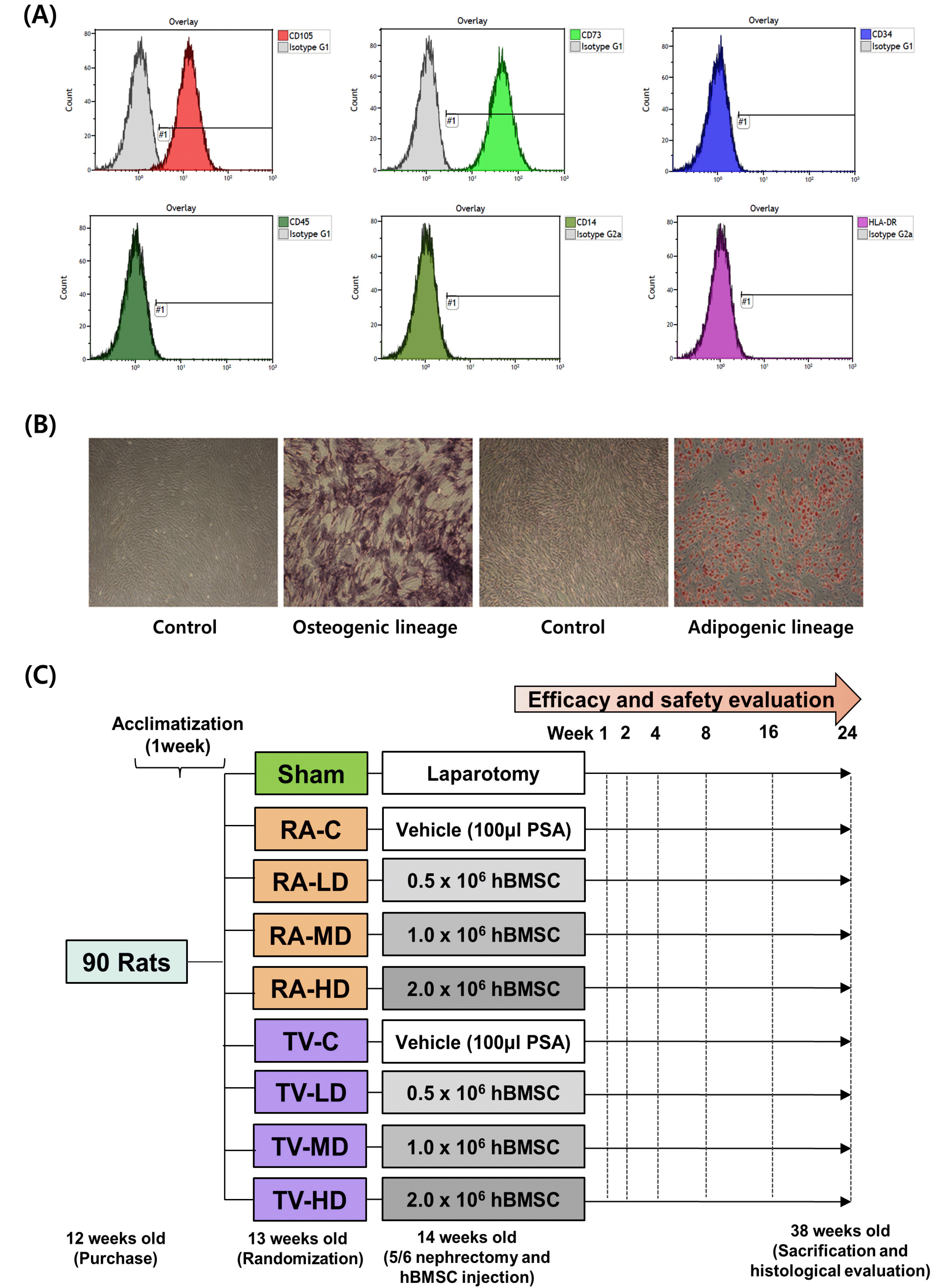
Fig. 1
Flow cytometric histograms, morphology of hBMSC and efficacy and safety evaluation schedule. (A) FACS data of hBMSC. (B) Morphology of hBMSC and osteogenic, adipogenic differentiation lineage. (C) Experimental schedule. RA: renal artery, RA-C: renal artery control, RA-LD: renal artery low dose, RA-MD: renal artery moderate dose, RA-HD: renal artery high dose, TV-C: tail vein control, TV-LD: tail vein low dose, TV-MD: tail vein moderate dose, TV-HD: tail vein high dose, PSA: plasma solution A, hBMSC: human bone marrow-derived mesenchymal stem cells.
![]()
Animal care
All aspects of animal care and animal experimentation followed the eighth edition of the Guide for the Care and Use of Laboratory Animals (6). The protocols for performing animal experiments were approved by the Institutional Animal Care and Use Committee of Asan Medical Center (2018-12-152). Ninety 12-week-old male Sprague-Dawley rats were purchased from Orient Bio (Seongnam, Gyeonggi, Republic of Korea) and acclimatized to their cages for 1 week. During the experiments, one dark cycle of 12 hour light (lights on at 08:00 AM and lights off at 20:00 PM) were maintained for all rats. Temperature was held at 22±2℃ and humidity at 50∼55%. Sufficient access to food and water was provided for all rats.
Establishment of an animal model of CKD
We ligated the left renal artery using a bulldog clamp. The upper and lower poles of the left kidney were marked using a pincette and resected with a blade. TachoSil (BBF sterilisation service GmbH, Kernen, Germany) was applied to stop bleeding at the resection surface. The bulldog clamp was removed after 10 minutes. Right nephrectomy was performed the same time. We observed no significant difference in the weight of the resected left kidney, except in the sham surgery group.
Optimization of the dose and injection route
We purchased ninety 12-week-old rats and randomized into nine equal groups (10 rats per group) after one week of acclimatization: sham (SH), renal artery control (RA-C), tail vein control (TV-C), renal artery low dose (RA-LD) (0.5×10⁶ cells), renal artery moderate dose (RA-MD) (1.0×10⁶ cells), renal artery high dose (RA-HD) (2.0×10⁶ cells), tail vein low dose (TV-LD) (0.5×10⁶ cells), tail vein moderate dose (TV-MD) (1.0×10⁶ cells), and tail vein high dose (TV-HD) (2.0×10⁶ cells). After 2 weeks, we intramuscularly anesthetized each rat with 0.3 ml of tiletamine (Zoletil; Virbac Laboratories, Carros, France) and xylazine hydrochloride (Rompun, Bayer, Germany) in a 4:1 mixture. In the sham group, only laparotomy was performed, whereas a 5/6 nephrectomy was completed in all other groups. hBMSC injection and 5/6 nephrectomy were performed during the same surgery. In the renal artery injection group, the indicated doses of cells were administered via a 33-gauge Hamilton syringe (Hamilton, Zurich, Switzerland) into the distal abdominal aorta after the aorta was clamped above and below the left renal artery. The hBMSC injection site was sealed using TachoSil, and the clamps were removed to restore abdominal aortic and left renal blood flow. In the tail vein injection group, the indicated doses of hBMSC were administrated via the tail vein through a 26-gauge Kovax syringe (Koreavaccine, Gyeonggi, Republic of Korea). In the RA-C and TV-C groups, only vehicle (100 μl plasma solution A) was injected.
Measurement of renal function and efficacy evaluation
Serum creatinine, urine creatinine, and BUN were measured at each time point in all rats. To evaluate the daily urine volume and urine total protein, urine was collected in a metabolic cage at 24 hours. The creatinine clearance (Ccr) was calculated using the following formula for: Ccr (ml/min/100 g)=(urine creatinine×24 hours urine volume×100)/(serum creatinine×1440×body weight). BUN, creatinine, and urine total protein (UTP) levels were determined using the standard laboratory kit at our hospital. BUN, serum creatinine, Ccr, urine total protein, and blood pressure were measured in each group 1 week before and 1, 2, 4, 8, 12, 16, 20, and 24 weeks after hBMSC injection (Fig. 1C). Blood pressure was measured by tail plethysmography.
Safety evaluation
During the study period, we evaluated all rats for behavioral changes and mortality at any time. Body weight and food consumption were regularly measured in each group 1 week before and 1, 2, 4, 8, 12, 16, 20, and 24 weeks after hBMSC injection.
Histopathological analysis of kidney tissue
All nephrectomy specimens were sectioned at 5 μm and stained with Periodic Acid–Schiff for light microscopy. Histopathological scoring was performed and tubular injury score was estimated based on the proportion of destroyed tubular structures in the cortex and medulla that exhibited glomerulus degeneration, interstitial fibrosis, interstitial inflammation, tubular dilatation, tubular basophilia, and intratubular eosinophilic granule in ten randomly chosen, non-overlapping fields (100×), as follows: 0 (normal histology), 1 (minimal: 0∼20%), 2 (slight: 20∼40%), 3 (moderate: 40∼60%), 4 (marked: 60∼80%), or 5 (severe: 80∼100%).
Sirius red staining
Slides were dipped in xylene I, II, and III; xylene was removed using ethanol series (100%, 90%, 80%, and 70%). After the slides were dipped in distilled water three times, they were dipped in Harris hematoxylin solution (Sigma, St. Louis, MO, USA; cat no: HHS-16). Sirius red reagent (Abcam, Cambridge, MA, USA) was dropped onto the slides, and the specimens were incubated. After the slides were dipped twice in acid solution, they were washed three times using distilled water, and then dipped again in the ethanol series. The slides were again dipped in xylene I, II, and III, and covered with coverslips using permanent mounting media.
TUNEL assay
The TUNEL assay kit (Roche Molecular Biochemicals, Mannheim, Germany) was used for microscopic evaluation. Each sectioned nephrectomy specimen was deparaffinized in xylene I, II, and III, and then rehydrated using ethanol series (100%, 90%, 80%, and 70%). After the slides were irrigating in distilled water, they were placed in a pretreated solution (a heat-induced epitope retrieval solution/a target retrieval solution). After irrigation, we added Protein Block, Serum-free, Ready-to-Use (Dako, Glostrup, Denmark) containing 20% FBS to the sections, and incubated them in a humid chamber. The TUNEL reaction mixture was distributed onto the slides, which were then incubated in a 37℃ humid chamber. Converter-Peroxidase was added, and the slides were incubated in the same manner. After irrigation, xxxx (DAB) working solution was added to the specimens.
Immunohistochemistry
Tissue sections mounted on microslides were deparaffinized in xylene, hydrated through ethanol series, and then dipped in 3% H2O2 to eliminate endogenous peroxidase activity. For antigen retrieval, we microwaved all slides for 15 minutes in Tris-EDTA buffer (pH 9.0) prepared in distilled water. Next, the slides were incubated for 2 hours at 37℃ with antibodies against α-smooth muscle actin (α-SMA) (eBioscience, Tokyo, Japan) and ED-1, a marker of macrophages (1:500, AbD Serotec, Oxford, UK). After irrigation, the sections were incubated at ambient temperature for 30 minutes using a biotin-free polymeric horseradish peroxidase–linker antibody conjugate system (Dako, Glostrup, Denmark). After the slides were washed, chromogen development was performed for 10 minutes. The slides were counterstained with Meyer’s hematoxylin and mounted using Immu-mount (Fisher Scientific, Geel, Belgium). For imaging analysis, we photographed four randomly selected kidney fields per rat in both the cortex and the medulla (40 kidney fields per group). Images were recorded at 40×magnification using the Panoramic Viewer software (3DHISTECH, Budapest, Hungary). All images were evaluated using Adobe Photoshop CS2 for quantification of signals. Outcomes of proliferation are presented as ED-1–positive area and α-SMA area as a percentage of the total area in each field (the cortex and the medulla).
Statistical analysis
All results of this study were expressed as means±standard errors. Statistical differences among groups were analyzed by one-way analysis of variance (ANOVA) followed by the Tukey’s HSD (honest significant difference) test for post-hoc comparisons. All statistical results were two-sided, and statistical significance was defined as p<0.05, p<0.01, or p<0.001. The data were analyzed by using IBM SPSS Statistics Version 21 (IBM Corporation, Armonk, NY, US).
For longitudinally measured Ccr, creatinine, and BUN, a linear mixed model (LMM) was applied to evaluate the effects of group (control, low, moderate, high), time, and the interaction of group and time. The data were summarized as least-squared means and standard errors. Statistical analyses were performed using SAS version 9 and 4 (Cary, NC, USA).
Results
Optimization of the dose and injection route
Serum BUN analysis: Serum BUN was significantly lower in the TV-HD group at 2 weeks (p<0.01), 16 weeks (p<0.05), and 24 weeks (p<0.01) than in the TV-C group at the same time points, as determined by one-way ANOVA. However, BUN levels were significantly higher in the RA-MD group at 12 weeks (p<0.05), 20 weeks (p<0.01), and 24 weeks (p<0.05), and in the RA-HD group at 16 weeks, than in the RA-C group in the same time points (p<0.05) (Fig. 2A and 2B). In the analysis of LMM for longitudinally measured data, the increase in BUN levels was significantly smaller in the TV-HD group than in the TV-C group (p<0.05).
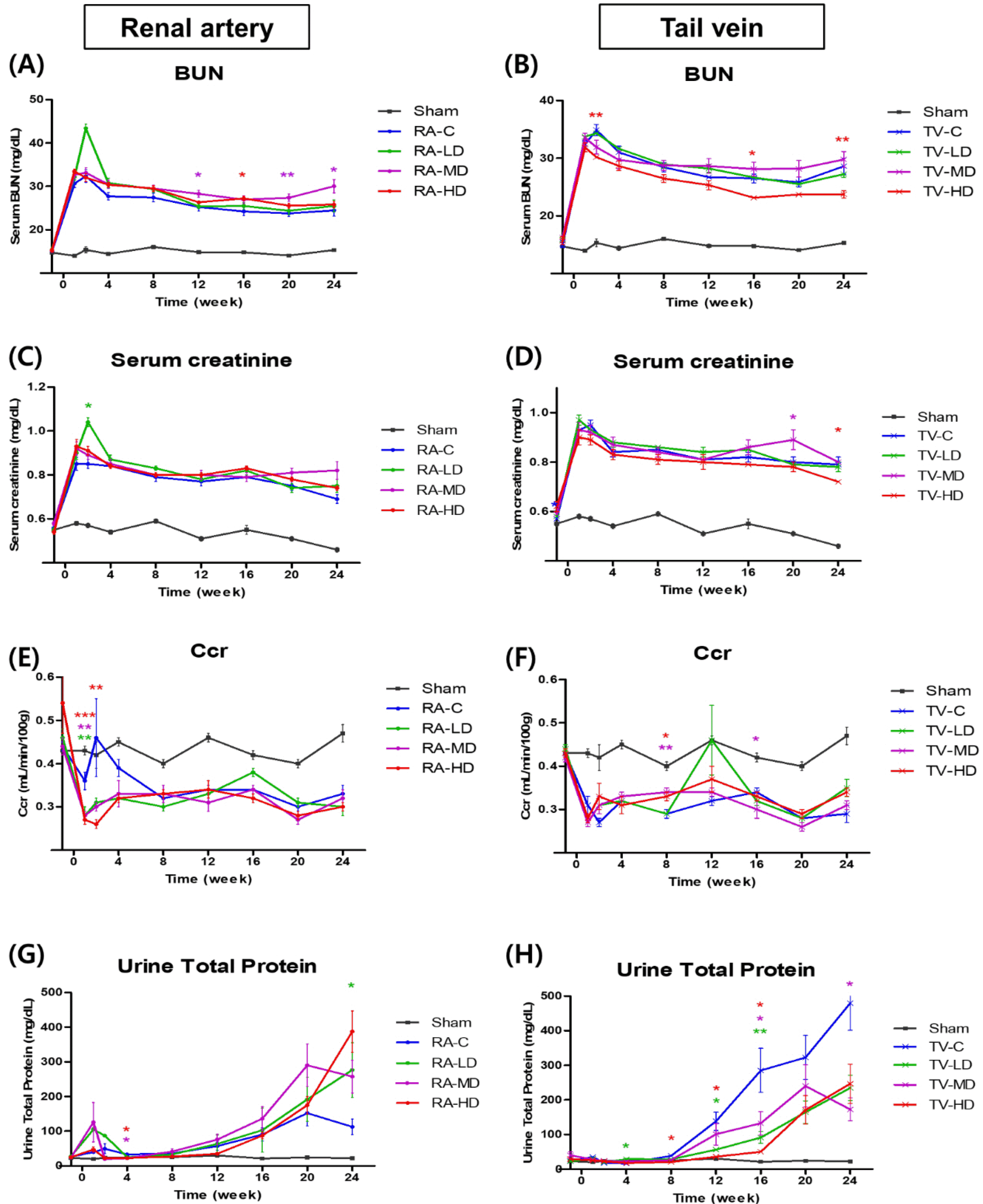
Fig. 2
Efficacy of hBMSC in CKD rat model according to injection route and hBMSC dose. BUN: blood urea nitrogen, Ccr: Creatinine clearance, RA-C: renal artery control, RA-LD: renal artery low dose, RA-MD: renal artery moderate dose, RA-HD: renal artery high dose, TV-C: tail vein control, TV-LD: tail vein low dose, TV-MD: tail vein moderate dose, TV-HD: tail vein high dose. *p<0.05; **p<0.01; ***p<0.001, vs. the RA-C group; *p<0.05; **p<0.01; ***p<0.001, vs. the TV-C group; Red colored stars indicate statistically significant differences in RA-LD, RA-MD, RA-HD, groups compared to the RA-C group, respectively. Blue colored stars indicate statistically significant differences in TV-LD, TV-MD, TV-HD, groups compared to the TV-C group, respectively. (A∼H) 3 times per rat, 10 rats per group, but 9 rats in the RA-C, TV-LD, TV-HD groups and 8 rats in the TV-MD groups.
![]()
Serum creatinine analysis: Relative to the corresponding control groups, serum creatinine was significantly lower in the TV-HD group at 24 weeks (p<0.05), but significantly higher in the preoperative TV-HD group (p<0.05), the RA-LD group at 2 weeks (p<0.05), and the TV-MD group at 20 weeks (p<0.05) (Fig. 2C and 2D). In the analysis of LMM for longitudinally measured data, the increase in serum creatinine relative to the baseline value was significantly lower in the TV-HD group than in the TV-C group (p<0.01).
Ccr analysis: At 8 weeks, Ccr was significantly higher in the TV-MD and TV-HD groups (p<0.01 and p<0.05) than in the TV-C group. At 1 week, Ccr was significantly lowerf in the RA-LD, RA-MD, and RA-HD groups than in the RA-C group (p<0.01, p<0.01, and p<0.001) (Fig. 2E and 2F). LMM analysis revealed no difference between the TV-HD and TV-C groups.
UTP analysis: In one-way ANOVA analysis, UTP levels in the RA-MD and RA-HD groups were significantly lower at 4 weeks (p<0.05 and p<0.05) than RA-C group in the same time points. Also, UTP levels were significantly higher in the TV-LD group at 12 and 16 weeks, the TV-MD group at 16 and 24 weeks, and the TV-HD group at 8, 12, and 16 weeks than in the TV-C group. However, UTP levels were significantly lower in the RA-LD group at 24 weeks and the TV-LD group at 4 weeks than in the control group (Fig. 2G and 2H).
Blood pressure: At 12 weeks, systolic blood pressure was significantly higher in the RA-LD and RA-HD groups than in the RA-C group (p<0.01, p<0.05), but no significant difference was observed in the tail vein injection group (Supplementary Fig. S1A∼D).
Safety evaluation: One case of mortality was observed in the RA-C group 1 week after nephrectomy. In the tail vein group, one case of mortality was observed in the TV-LD group, two in the TV-MD group and one in the TV-HD group. However, the survival rate did not differ significantly between groups (p=0.436).
Food consumption before and after hBMSC injection was observed in each group, and no significant difference was observed among groups (Supplementary Fig. S1E and S1F). Likewise, body weight before study initiation did not differ significantly among groups. During the study period, all the rats gradually gained weight. At the end of the study, absolute body weight did not significantly differ between normal control, and hBMSC injection groups (Supplementary Fig. S1G and S1H).
Histological analysis: We calculated the degree of glomerulus degeneration and tubular injury by histopathological scoring. In the renal artery injection group, the degree of glomerulus degeneration in the cortex and tubular injury in medulla became more severe at higher doses And the degree of tubular injury was significantly higher in the RA-HD group than in the RA-C group [p<0.05 (cortex), p<0.05 (medulla)] (Fig. 3A and 3B). However, in the tail vein injection group, we observed no significant difference between the TV-C group and the other groups (Fig. 4A and 4B). Sirius red staining in the cortex and the medulla revealed that fibrotic changes were significantly less extensive in the hBMSC injection groups than in the control group (Fig. 3C and 3D and Fig. 4C and 4D). The TUNEL assay revealed that the rate of apoptosis in the cortex and the medulla was significantly lower in all hBMSC injection groups than in the control groups (RA-C and TV-C) (Fig. 3E and 3F and Fig. 4E and 4F). Immuno-histochemical staining for α-SMA in the cortex and the medulla revealed that all injection groups showed also significantly less myofibroblast differentiation than the control groups (Fig. 3G and 3H and Fig. 4G and 4H). ED-1 immunohistochemical staining in the cortex and the medulla revealed that macrophage activation was lower in all injection groups than in the control group (Fig. 3I and 3J and Fig. 4I and 4J).


Fig. 3
Histopathological analysis of the renal artery injection group. (A) Histopathological scoring, cortex. (B) Histopathological scoring, medulla. (C) Sirius red, cortex. (D) Sirius red, medulla. (E) TUNEL, cortex. (F) TUNEL, medulla. (G) α-SMA, cortex. (H) α-SMA, medulla. (I) ED-1, cortex. (J) ED-1, medulla. Non-overlapping fields (400×). *p<0.05; **p<0.01; ***p<0.001, vs. the RA-C group; $$p<0.01, vs. the RA-LD group; RA-C: renal artery control, RA-LD: renal artery low dose, RA-MD: renal artery moderate dose, RA-HD: renal artery high dose. (A, B) 10 fields per rat, 10 rats per group, but 9 rats in the RA-C, TV-LD, TV-HD groups and 8 rats in the TV-MD groups. (C∼J) 4 fields per rat, 10 rats per group, but 9 rats in the RA-C, TV-LD, TV-HD groups and 8 rats in the TV-MD groups.
![]()
Fig. 4
Histopathological analysis of the tail vein injection group. (A) Histopathological scoring, cortex. (B) Histopathological scoring, medulla. (C) Sirius red, cortex. (D) Sirius red, medulla. (E) TUNEL, cortex. (F) TUNEL, medulla. (G) α-SMA, cortex. (H) α-SMA, medulla. (I) ED-1, cortex. (J) ED-1, medulla. Non-overlapping fields (400×). NS, p>0.05; *p<0.05; **p<0.01; ***p<0.001 vs. the TV-C group; $$p<0.01, vs. the TV-LD group; TV-C: tail vein control, TV-LD: tail vein low dose, TV-MD: tail vein moderate dose, TV-HD: tail vein high dose. (A, B) 10 fields per rat, 10 rats per group, but 9 rats in the RA-C, TV-LD, TV-HD groups and 8 rats in the TV-MD groups. (C∼J) 4 fields per rat, 10 rats per group, but 9 rats in the RA-C, TV-LD, TV-HD groups and 8 rats in the TV-MD groups.
![]()
Discussion
The ability of stem cells to restore normal kidney function can be explained by their ability to undergo selective migration into damaged tissues and their ability to express growth factor receptors and chemokines released from organs with inflammatory change (7). In damaged organs, stem cells have the potential to differentiate into various tissues, such as tubular epithelial cells, mesangial cells of nephrons, podocytes, or glomerular endothelial cells (8).
Many studies have reported the regenerative effects of cell-based treatments in animal models of CKD. However, it remains uncertain which cell types or cell-based products increase renal function most successfully in experimental models of CKD (9). Due to the diversity among preclinical trials in terms of animal models, intervention timing, cell type or cell product, number of cells, injection route, and read-out of kidney function and morphology, proper translation of preclinical studies into the clinical field is extremely complex.
The currently known mode of action of MSC is due to paracrine mechanisms through soluble mediators released constitutively or through crosstalk with target cells (10). Adenosine produced by crosstalk between MSC and T cells acts as a mediator to promote enzyme expression (CD39 and CD73) in MSC and T cells, resulting in downregulation of T cell proliferation (11). Other paracrine mechanisms, containing stem cell–derived extracellular vesicles, also play a significant role in the overall effects that MSC exerts on the immune response (12).
MSC represent 0.001∼0.01% of all nucleated cells in bone marrow (13). MSC retain their mesenchymal differentiation capacity even after repeated subculture in vitro (14). Although bone marrow has been described as the best source of available MSC (13), the use of hBMSC is not always acceptable due to the high rate of viral exposure and the substantial reduction in cell number and the proliferative/differentiation capability with donor age (15).
Despite these difficulties, we chose hBMSC for this study based on previous reports that they are superior to other stem cells in terms of efficacy and safety. In addition, we have accumulated a great deal of research experiences with these cells. In our previous study at 2018 (16), we conducted a study in which we used BMSC to treat a rat model of ischemia–reperfusion injury (IRI). In that study, we sought to determine the optimal injection route (renal arterial, renal parenchymal, or tail vein injection), dose (low dose: 1×10⁶ cells, moderate dose: 2×10⁶ cells, or high dose: 4×10⁶ cells), and injection period (pre-, concurrent-, or post-IRI). Based on these prior experiences, we were able to effectively conduct research using hBMSC in the CKD rat model.
A frequent argument in favor of the intra-arterial injection route is that stem cells are primarily entrapped inthe pulmonary microvasculature after intravenous injection (17). It is known that the degree of entrapment in the lungs increases as the number and size of injected MSC and the number of injections increase (18). A recent meta-analysis of the effects of MSC injection provided strong evidence in favor of the renal arterial delivery route for kidney regeneration in animal models with acute or CKD (19). According to the Mesenchymal Stem Cells in Solid Organ Transplantation (MISOT) study group, it remains unclear which delivery route should be used in clinical trials of MSC injection after kidney injury (20). However, it is possible that renal arterial injection is beneficial. The reason is that it provides a direct delivery route into the transplanted kidney, where the MSC can locally decrease the inflammatory response. Along with these suggestions, renal artery injection of allogeneic MSC yields better outcomes than the intravenous route when treating acute kidney rejection in animal models (21).
However, the results of this study confirmed that tail vein injection was more effective than arterial injection. The injection of cells through an intravenous route, generally via the tail vein, is much easier and less invasive than arterial delivery routes. In general, it is known that the stem cells injected intravenously are first accumulated in the lungs and then migrate to other major organs to be variable, however they are no longer detected in the body after a short period of time (22). However, after MSC remains in the lungs, it does not disappear completely, however it induces the secretion of anti-inflammatory proteins, resulting in therapeutic effects (23). In another study, biodistribution was compared through biolumine-scence imaging after injection of MSC into the tail vein or renal artery in an ischemia-reperfusion rat model. As a result, both pathways reported improvement in renal damage even though MSC remained only in the lungs, liver, and spleen (24). This suggests that the paracrine effect can be shown even by MSCs localized in distant regions. Although it was given the ability to control the damaged microenvironment by secreting appropriate cytokines while cells entrapped in the lungs, IV hBMSC injection can regenerate damaged tissues and organs.
The most important part of our study was the optimization of hBMSC dose. We found that administration of a high dose of hBMSC (2×10⁶ cells) yielded superior outcomes. However, Cai et al. reported that they could achieve the maximal therapeutic efficiency with a dose of 1×10⁵ cells, and that the effect was significantly improved compared to that for the group injected with 1×10⁶ cells (5). Moreover, obstructing the renal blood flow during the period of cell injection may further aggravate renal damage and compensate for any extra benefit of renal artery injection. Their study differed from ours in that it was a dose comparison focusing entirely on arterial injection.
However, other studies reported that dose dependency does not appear during stem cell delivery (9). Low dose dependency is a frequent finding in cell-based therapy, possibly indicating that cell-based therapy acts mainly by switching on endogenous repair rather than by providing a persistent source of exogenous cells or growth factors. Certainly, multiple preclinical and clinical studies have failed to detect significant numbers of exogenous cells in the kidney after injection (25). But our study demonstrated that injection of a high dose of hBMSC was highly efficacious and safe. Although dose effects are difficult to assess in small samples, our results also suggest that the highest doses yielded the highest efficacy.
According to our histopathological analysis, the degree of tubular injury was higher in the renal artery injection group than in the tail vein injection group. This is likely because the direct renal arterial injection route could stimulate vasospasms that narrow the renal artery and make it susceptible to embolisms, which in turn accelerate ischemia and fibrotic changes. However, after a certain period of time, exosomes secreted from remnant hBMSC act on the glomerular cell signaling pathway to prevent apoptosis and macrophage activation, so it can be assumed that TUNEL assay or ED-1 positive cell count decreased despite the high tubular injury score in RA group.
This study has some limitations. First, this study compared the stem cell injection route with two routes, arterial and intravenous, and has a limitation in that other injection routes such as direct renal parenchymal injection or intraperitoneal injection or subcutaneous injection were not additionally considered. Second, we used 5/6 nephrectomy rat model and did not cross-examine the results with other types of CKD rat models such as vascular injury models or genetically engineered models or immune induced models. From Makhlough et al. (26) a single-arm phase I clinical trial using autologous hBMSC in six autosomal dominant polycystic kidney disease patients and demonstrated safety and tolerability of IV 1-2×10⁶/kg transplantation of hBMSC through the cubital vein (NCT02166489) was performed. There is an ongoing phase I and phase II clinical trial which try to develop novel therapies for diabetic kidney disease using allogeneic MSC (NCT02585622). Our study can provide an additional evidence to support the efficacy and safety of high-dose intravenous injection of hBMSC in future human studies.
Here we demonstrated the efficacy and safety of the tail vein as an injection route for hBMSC. If stem cell injection into peripheral veins is shown to be safe and efficacious in future human trials, the convenience of hBMSC injection should be increased. In addition, in contrast to previous studies, this study proved that hBMSC are safe even at high doses. We expect that our findings will provide a useful reference for dose optimization for future human trials using hBMSC.
In conclusion, This study assessed the potential efficacy and safety of hBMSC administered at different doses and via different routes in an animal model of CKD. We observed no significant changes in body weight or food consumption. Also, injection of the highest dose (2×10⁶ cells) via the vein was safe and efficacious. We suggest peripheral veins could be used as a route of administration in the future human trials.
Supplementary Materials
Supplementary data including one figure can be found with this article online at https://doi.org/10.15283/ijsc21146.
 XML Download