Abstract
Osteoblastoma is a rare benign neoplasm formed by osteoid tissue and well-vascularized bone that occurs mainly in children and adolescents. It appears primarily in the long bones, vertebral column, and small bones of the hands and feet, and not typically in the skull and maxillary bones. The purpose of this study is to present the case of an 8-year-old girl with a diagnosis of right mandibular osteoblastoma and a review of the relevant literature. The goals of treatment were to preserve dental occlusion, masticatory function and facial symmetry while minimizing the effects on patient body image and quality of life. Osteoblastoma, although it is benign, can be aggressive, and its treatment will depend on the timing of diagnosis, size and location. Early diagnosis is essential to avoid not only radical surgery as in the case presented, but also to help minimize the risk of possible relapse and potential malignancy of a benign osteoblastoma.
Osteoblastoma is a rare benign tumor1-15 formed by osteoid tissue and well-vascularized bone1-3,5,9,10. It occurs mainly in the long bones, spinal column1-16, and the small bones of the hands and feet2-5,7,8,10-12,14,16, but not usually in the skull2,5-14 and maxillary bones2,5-8,12, when this occurs the mandible is the most affected3,5,7,8,10,11,13,14. It appears in children and adolescents, although it is also reported in patients older than 70 years2-5,7,10,13-15. Mostly it is diagnosed in the second and third decades of life2,10,14,16. The prevalence according to gender is higher in males10-14,16.
The case presented was about a girl with the diagnosis of right mandibular osteoblastoma and the treatment made after removal of the tumor.
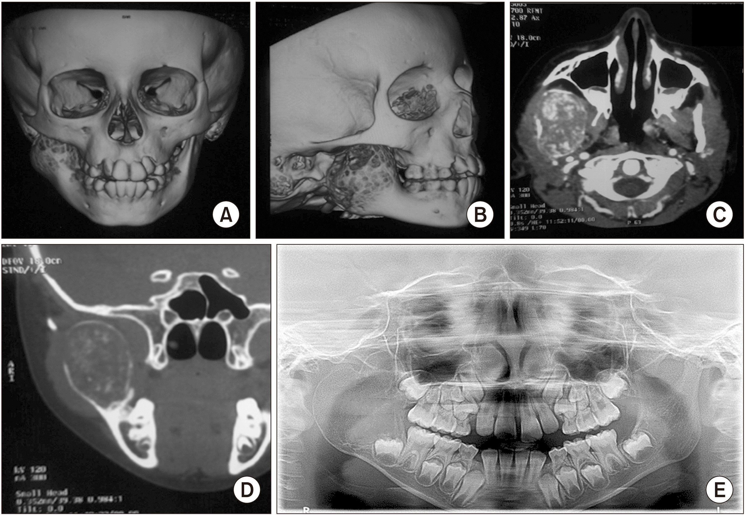
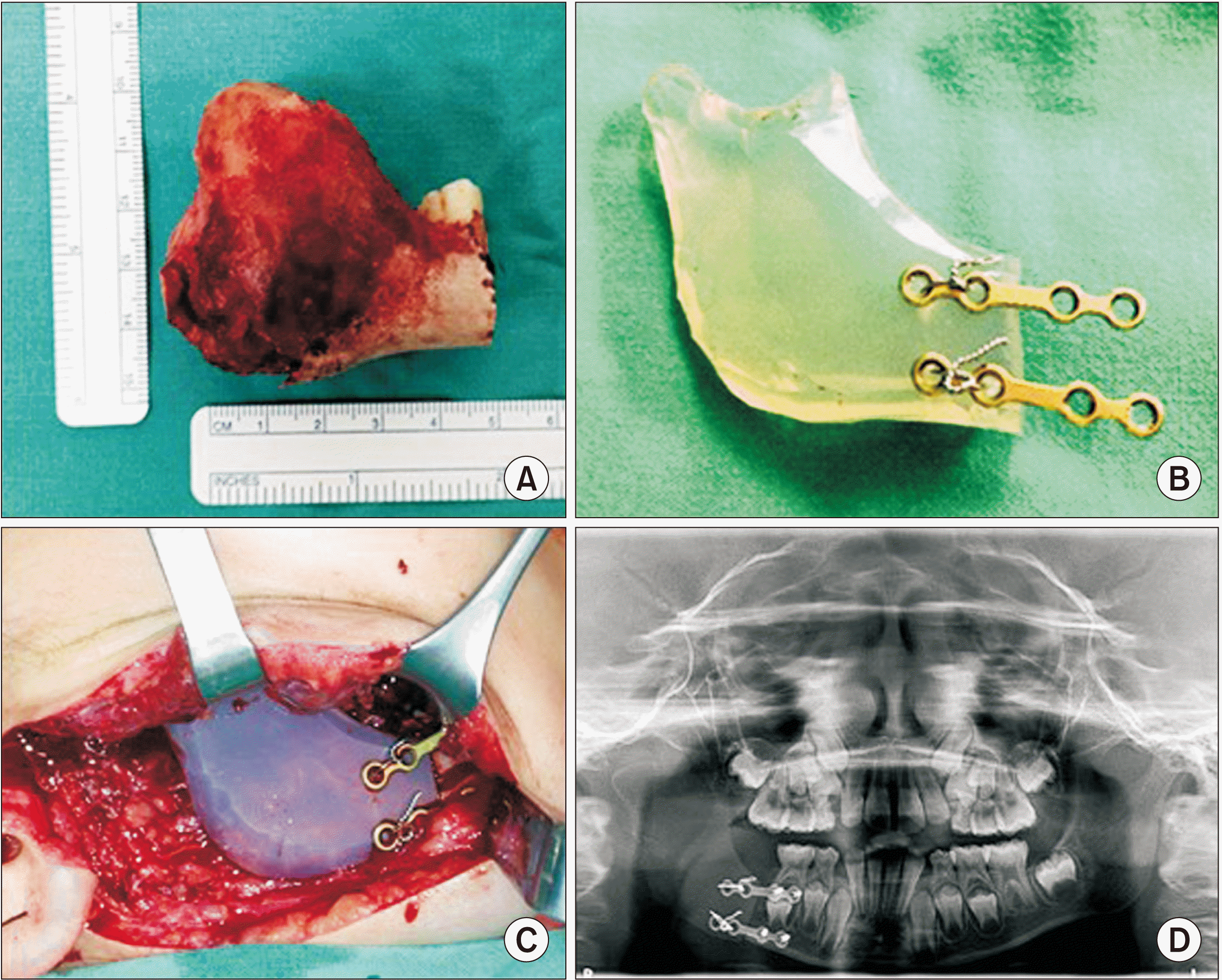
A young girl aged 8 years and 8 months was referred to the Hospital Nacional de Niños Dr. Carlos Sáenz Herrera, Caja Costarricense de Seguro Social (San José, Costa Rica) for facial asymmetry resulting from over 1-year history of osteolytic tumor in the right mandibular region. At the clinical examination, a volume increase was observed in the right side of the face, specifically in the mandibular ramus region, which on palpation corresponded to a firm, hard, non-painful mass. The oral opening was 34 mm with a slight deviation to the right. Antero-posterior radiography of the skull, orthopantomography, and computed axial tomography showed a transversely expansive osteolytic lesion ranging from the condyle of the right mandibular ramus to the first permanent right lower first molar.(Fig. 1) Histological findings after incisional biopsy indicated osteoblastoma. The treatment was the tumor removal by block resection that included condyle and right coronoid to mesial of the first lower right molar, finding, in the surgery, the perforation of the cortical of the mandibular ramus. Reconstruction was carried out immediately using a silicon spacer with the anatomical shape of the mandibular ramus, condyle and coronoid, which was fixed with osteosynthesis material in the mandibular body.(Fig. 2) During this procedure, bone marrow biopsy was performed, and no signs of neoplasia were observed on histopathology. Excisional biopsy of the mandibular tumor specimen indicated that it was composed of fibrous tissue arranged in woven bundles in some areas; at other sites the fibrovascular tissue was loose and there was a trabeculated matrix of mineralized osteoid tissue, and interlaced, irregularly-shaped bone trabeculae presenting as an osteoblastic ridge with rounded osteoblasts; furthermore, extravasation of erythrocytes and giant cells was observed in other areas, confirming the diagnosis of osteoblastoma. The resection margins were clear, indicating that removal of the tumor was complete.
The patient has not experienced recurrence more than nine years after the surgery.(Fig. 3) Dental occlusion and masticatory function with minimal facial asymmetry were maintained, and her body image and quality of life have not been significantly affected.
Osteoblastoma in the maxillofacial region accounts for 1% of primary bone tumors1-3,5-10,13-16 and 3% of all benign bone neoplasms10. There are two clinicopathological forms of osteoblastoma:
• Benign: characterized by slow growth, well-defined and vascularized sclerotic margins, with slight inflammatory response, and a size less than or equal to 4 cm2,7,8,11,12;
• Aggressive: characterized by aggressive local behavior2,7,10, a diameter larger than 4 cm7,12 and frequent recurrence (10%-21%)2,7,10, but not metastasis7; lesions grow rapidly, tend to invade adjacent tissues10, and exhibit atypical histopathological characteristics, making differentiation from low-grade osteosarcoma difficult2,7,10,11.
In the present case, osteoblastoma was diagnosed in an 8-year-old girl. The tumor’s aggressive behavior, as can be seen in Fig. 1, implies that the lesion had been present for a long time. Furthermore, the present case is diagnosed in a girl, which does not coincide with the predominant gender of this pathology cases.
The diagnosis of osteoblastoma is a challenge1,2,7,15, due to the clinical, radiological and histological similarities with osteoid osteoma1,2,5-7,10,12,13, fibrous lesions2,7,10-14, and osteosarcoma2,3,5-7,10,12-14,16, among other lesions.
According to the literature, this type of pathology can present symptomatically1-3,5-8,10,13,14, but it can also be discovered incidentally on routine examination due to facial asymmetry as a consequence of the increase in tissue volume5,8,10,11,13-15. Approximately 7.2% to 50% of osteoblastoma cases are asymptomatic10, as was our case. Osteoblastoma has variable characteristics on radiography with a variety of patterns10,15. These lesions can present in radiographs lytic or mixed images or even sclerotic masses10. The radiographic characteristics vary according to the size of the tumor and the intensity of the calcification2,6-9,14,15, and it is identified by well-defined edges2,6,8,9,16 so expansion of the cortical bones2,9,15, which is observed in Fig. 1.
Histologically, osteoblastomas exhibit well-vascularized, osteoblastic connective tissue stroma and sometimes osteoclasts along with osteoid tissue and varying degrees of calcification, as well as immature bone2,8-11,13-15.
The etiopathogenesis of this lesion remains controversial. This lesion may represent a true osteoblastic-derived neoplasm2,13,15 as well as resulting from trauma, inflammation, abnormal local tissue response, or local alterations in bone physiology2,11,13,15.
Differential diagnosis of osteblastoma includes a variety of benign to malignant tumors8,9, such as cementoblastoma5,8,9,12-16, osteoid osteoma, fibrous dysplasia, ossifying fibroma5,8,12,14-16, focal cemento-osseous dysplasia, and low-degree osteosarcoma5,8,12,14-16.
The treatment of choice is surgical excision2,6-9,13,14, either curettage2,3,5,9,12-14 or en bloc resection2-4,8-10,12-14,16, depending on tumor size, site, extent of radiographic involvement and biological behavior12. In the present case, en bloc resection and reconstruction was performed with an anatomically-shaped silicon spacer to achieve not only facial symmetry, and not affect its psychological condition and self-image, but also to normal function, taking into consideration that the patient was in the process of growth. It is worth mentioning that reconstruction is important to avoid functional problems4,8; the technique was complex in this case not only because of mandibular bone defects but also because of the involvement of the condyle, but function and quality of life were maintained without.
The possibility of recurrence2-4,7-9,11,12,14,16 and the potential for malignancy2-4,7,14,16 should be considered in treatment, including regular monitoring2,3,7. Recurrence was not observed in this case over a follow-up of more than 9 years.
In conclusion, osteoblastoma, despite being a benign tumor, can be aggressive, and its treatment will depend on early diagnosis, size and location. Early diagnosis is essential to avoid not only radical surgery, but also to minimize the risk of possible relapse and potential malignancy of a benign osteoblastoma.
Careful clinical, radiographic and histopathologic evaluation is essential to formulate a diagnosis, treatment plan and prognosis in each case.
Notes
Ethics Approval and Consent to Participate
This study was approved by the Research Bioethics Area, Centro de Desarrollo Estratégico e Información en Salud y Seguridad Social (CENDEISSS), Caja Costarricense de Seguro Social, Costa Rica (No. CENDEISSS-AB-0401-2021).
References
1. Jones AC, Prihoda TJ, Kacher JE, Odingo NA, Freedman PD. 2006; Osteoblastoma of the maxilla and mandible: a report of 24 cases, review of the literature, and discussion of its relationship to osteoid osteoma of the jaws. Oral Surg Oral Med Oral Pathol Oral Radiol Endod. 102:639–50. https://doi.org/10.1016/j.tripleo.2005.09.004. DOI: 10.1016/j.tripleo.2005.09.004. PMID: 17052641.
2. Bokhari K, Hameed MS, Ajmal M, Togoo RA. 2012; Benign osteoblastoma involving maxilla: a case report and review of the literature. Case Rep Dent. 2012:351241. https://doi.org/10.1155/2012/351241. DOI: 10.1155/2012/351241. PMID: 22593831. PMCID: PMC3347716.
3. Peters TE, Oliver DR, McDonald JS. 1995; Benign osteoblastoma of the mandible: report of a case. J Oral Maxillofac Surg. 53:1347–9. https://doi.org/10.1016/0278-2391(95)90599-5. DOI: 10.1016/0278-2391(95)90599-5. PMID: 7562203.
4. Danielidis J, Triaridis C, Demetriadis A, Psifidis A. 1980; Mandibular ramus osteoblastoma. A case report. J Maxillofac Surg. 8:251–4. https://doi.org/10.1016/s0301-0503(80)80108-1. DOI: 10.1016/S0301-0503(80)80108-1. PMID: 6932467.
5. Manjunatha BS, Sunit P, Amit M, Sanjiv S. 2011; Osteoblastoma of the jaws: report of a case and review of literature. Clin Pract. 1:e118. https://doi.org/10.4081/cp.2011.e118. DOI: 10.4081/cp.2011.e118. PMID: 24765359. PMCID: PMC3981446.
6. Miyajima D, Ishikawa A, Yagihara K, Ishida T, Yagishita H, Kasturano M, et al. 2015; A rare case of osteoblastoma of the mandible. J Oral Maxillofac Surg Med Pathol. 27:884–7. https://doi.org/10.1016/j.ajoms.2015.04.001. DOI: 10.1016/j.ajoms.2015.04.001.
7. Mardaleishvili K, Kakabadze Z, Machavariani A, Grdzelidze T, Kakabadze A, Sukhitashvili N, et al. 2014; Benign osteoblastoma of the mandible in a 12-year-old female: a case report. Oncol Lett. 8:2691–4. https://doi.org/10.3892/ol.2014.2593. DOI: 10.3892/ol.2014.2593. PMID: 25364451. PMCID: PMC4214441.
8. Castro PHS, Molinari DL, Stateri HQ, Borges AH, Volpato LER. 2016; Agressive osteoblastoma in a seven-year-old girl's mandible: treatment and six-year monitoring. Int J Surg Case Rep. 27:5–9. https://doi.org/10.1016/j.ijscr.2016.07.033. DOI: 10.1016/j.ijscr.2016.07.033. PMID: 27518431. PMCID: PMC4983634.
9. Sahu S, Padhiary S, Banerjee R, Ghosh S. 2019; Osteoblastoma of mandible: a unique entity. Contemp Clin Dent. 10:402–5. https://doi.org/10.4103/ccd.ccd_676_18. DOI: 10.4103/ccd.ccd_676_18. PMID: 32308310. PMCID: PMC7145246.
10. Pontual ML, Pontual AA, Grempel RG, Campos LR, Costa Ade L, Godoy GP. 2014; Aggressive multilocular osteoblastoma in the mandible: a rare and difficult case to diagnose. Braz Dent J. 25:451–6. https://doi.org/10.1590/0103-6440201300220. DOI: 10.1590/0103-6440201300220. PMID: 25517784.
11. Panigrahi RG, Bhuyan SK, Pati AR, Priyadarshini SR, Sagar S. 2016; Non aggressive mandibular osteoblastoma- a rare maxillofacial entity. J Clin Diagn Res. 10:ZD06–8. https://doi.org/10.7860/jcdr/2016/13709.7630. DOI: 10.7860/JCDR/2016/13709.7630. PMID: 27190965. PMCID: PMC4866263.
12. Vinuth DP, Agarwal P, Gadewar D, Dube G, Dhirawni R. 2014; A large multifocal aggressive osteoblastoma of mandible: an immunohistochemistry case study report. J Dent Res Dent Clin Dent Prospects. 8:51–5. https://doi.org/10.5681/joddd.2014.009. DOI: 10.5681/joddd.2014.009. PMID: 25024840. PMCID: PMC4091700.
13. Mahajan A, Kumar P, Desai K, Kaul RP. 2013; Osteoblastoma in the retromolar region - report of an unusual case and review of literature. J Maxillofac Oral Surg. 12:338–40. https://doi.org/10.1007/s12663-011-0263-4. DOI: 10.1007/s12663-011-0263-4. PMID: 24431864. PMCID: PMC3777043.
14. Smith RA, Hansen LS, Resnick D, Chan W. 1982; Comparison of the osteoblastoma in gnathic and extragnathic sites. Oral Surg Oral Med Oral Pathol. 54:285–98. https://doi.org/10.1016/0030-4220(82)90098-6. DOI: 10.1016/0030-4220(82)90098-6. PMID: 6957826.
15. Kim KS, Kim MJ, Seo BM, Chu SC, Lee GC. 1991; Benign osteoblastoma of the mandible: report of a case and review of the literature. J Korean Assoc Oral Maxillofac Surg. 17:54–60.
16. Ataoglu O, Oygur T, Yamalik K, Yucel E. 1994; Recurrent osteoblastoma of the mandible: a case report. J Oral Maxillofac Surg. 52:86–90. https://doi.org/10.1016/0278-2391(94)90022-1. DOI: 10.1016/0278-2391(94)90022-1. PMID: 8263651.
Fig. 1
Right mandibular ramus osteoblastoma, extending from condyle and coronoid process to permanent lower right first molar: computed axial tomography (A and B: 3D reconstruction images, C: axial image, D: coronal image) and orthopantomography (E).


 PDF
PDF Citation
 Print
Print


 XML Download