Journal List > J Korean Neurosurg Soc > v.66(2) > 1516081601

Noh, Cho, Kim, Kim, and Shin: Artificial Intelligence for Neurosurgery : Current State and Future Directions

Abstract

Artificial intelligence (AI) is a field of computer science that equips machines with human-like intelligence and enables them to learn, reason, and solve problems when presented with data in various formats. Neurosurgery is often at the forefront of innovative and disruptive technologies, which have similarly altered the course of acute and chronic diseases. In diagnostic imaging, such as X-rays, computed tomography, and magnetic resonance imaging, AI is used to analyze images. The use of robots in the field of neurosurgery is also increasing. In neurointensive care units, AI is used to analyze data and provide care to critically ill patients. Moreover, AI can be used to predict a patient’s prognosis. Several AI applications have already been introduced in the field of neurosurgery, and many more are expected in the near future. Ultimately, it is our responsibility to keep pace with this evolution to provide meaningful outcomes and personalize each patient’s care. Rather than blindly relying on AI in the future, neurosurgeons should gain a thorough understanding of it and use it to enhance their patient care.

INTRODUCTION

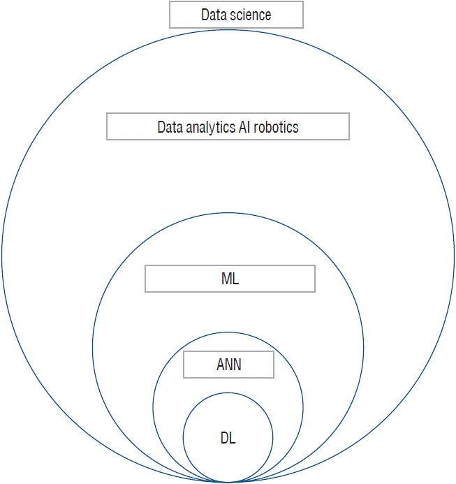
Artificial intelligence (AI) is a field of computer science that equips machines with human-like intelligence and enables them to learn, reason, and solve problems when presented with data in various formats. The term AI encompasses a wide range of words. To define the relationship between the words, there is data analytics, AI, and robotics in data science, as well as machine learning, artificial neural networks, and deep learning. AI uses a combination of machine learning algorithms, artificial neural networks, deep learning, and other data analysis techniques to achieve intelligent capabilities (Fig. 1). The healthcare sector is experiencing a paradigm shift with the recent increasing trend of mass digitization of information. Neurosurgery is often at the forefront of innovative and disruptive technologies, which have similarly altered the course of acute and chronic diseases [22]. The purpose of this study is to gain a better understanding of AI, its role in neurosurgery, and its future development directions.

HISTORY OF AI

Humans have long wanted to build thinking machines. Turing [41] suggested the possibility of developing AI and devised a test method to confirm it. The Turing Machine led John von Neumann to develop a standard computer architecture. This is considered to be the beginning of AI. Rosenblatt [31] developed an artificial neural network called the perceptron using artificial neurons that mimic human neurons. Following that, AI research received a great deal of attention and a substantial research grant from the U.S. Department of Defense. However, Minsky and Papert [24] criticized the limitations of the perceptron by publishing a mathematical proof that the perceptron can be applied to linearly separable problems such as AND or OR, but not to XOR problems. It was not until the introduction of expert systems into the industry in the 1980s that AI research, which had been stagnating for many years, received attention again. A lack of computer resources, excessively difficult management, and low cost-effectiveness, have however, led to a decline in interest in the research of AI. In the 2000s, deep learning technology based on Hinton’s Deep Belief Network began to show potential for practical use [16,25]. With this as an opportunity, deep learning technology re-emerged. This demonstration of the potential of deep learning led Google to acquire DeepMind Technology in 2014, a company that specializes in reinforcement learning. Then, AlphaGo beat Lee Sedol in 2016 and Ke Jie in 2017, and AI was once again thrust into the spotlight. Since the advent of deep learning, AI has been developing rapidly, and it is changing so rapidly that it is impossible to predict which technology will receive attention in the future.

ARTIFICIAL CONSCIOUSNESS : THE ULTIMATE GOAL OF AI

The ultimate goal of AI is to create consciousness. Thus far, humans have copied various functions of the brain, but not the consciousness. It is doubtful whether it will be possible in the future. As with any machine, AI is still a tool that produces output when input is provided. The current AI works like a slave, but does not seem to have emotions or free will. The intelligent machine can easily imitate phenomenological features of consciousness, but do not seem to be able to shape psychological features [13]. It is not possible for the machine to receive input or output by making plans on its own with subjectivity. However, human beings are aware of their existence, of the process of input and output, and of the feelings that accompany these processes. That cognitive ability is defined as consciousness. The Turing test can determine whether a machine has intelligence, but it cannot determine whether it is conscious. This has been proven with the Chinese room argument [33]. However, if AI becomes conscious, it would be very difficult to distinguish it from humans. It would also be very difficult to prove that a first-person perspective exists on the machine. The world after the realization of conscious AI will be revolutionary. Currently, the phenomenon of consciousness has been accepted as a phenomenon emerging from the complex connection of neural structures. It is advocated that consciousness can only be realized in a particular physical system because consciousness has properties that depend on physical constitution [4]. Nevertheless, proponents of artificial consciousness believe that it is possible to construct a system that can emulate this neural correlate of consciousness interoperation [12]. The mechanism of consciousness has not yet been elucidated, and the means for its research are inadequate. Computers are unable to keep up with the connectivity and complexity of the brain, but if connectivity explosively increases and complexity is overcome with the development of technologies such as quantum computing in the future, it has been carefully predicted that a consciousness phenomenon will arise in electrical systems. As neurosurgeons are privileged to have access to the human brain, they are obliged to answer these philosophical questions in a scientific manner.

THE FUNCTION OF AI IN NEUROSURGERY

Diagnosis

There are several studies in the field of neurosurgery that analyze images with AI [7,14,18,27,32,37,42] (Table 1). The use of imaging has increased dramatically over the past few decades. Xrays, computed tomography (CT), magnetic resonance imaging (MRI), etc. used in neurosurgery are essential tools for diagnosis. Manual measurements and calculations of numerous vertebral pelvic parameters in full spine X-rays require considerable time and effort. Therefore, the location of anatomical landmarks on semi-automated and automated spine X-rays and the segmentation of the spine on plain X-rays have been studied for more than 10 years [9]. Recently, a deep-learning method has been applied to automatic sagittal X-ray imaging parameter measurement, and there is a good correlation between the standard error of mild to moderate degree and manual measurements [20].
MRI is an important tool used for the detection, characterization, and monitoring of brain tumors, and typically employs a variety of sequence types [1]. Accurate tumor diagnosis is necessary to effectively determine a patient’s prognosis. The field of neuroimaging is increasingly leveraging the power of machine learning for clinically useful classification, enabling individual patient outcome prediction and semi-automated tumor description and diagnosis [6].
In the acute care setting, CT imaging is an important diagnostic tool for many urgent pathologies, such as intracranial hemorrhage. In particular for intracranial hemorrhage, rapid CT readings are needed, as half of the resulting mortality is reported to occur in the first 24 hours. However, the increase in the amount of burns places a great burden on radiologists who must maintain the accuracy and efficiency of diagnosis [23]. An AI decision support system has been developed to maintain diagnostic performance even in the face of increasing clinical volumes [43]. Recently, the U.S. Food and Drug Administration approved an AI powered by a deep learning algorithm called Viz.ai that analyzes images to detect potential large vessel occlusive strokes in CT angiography and alert specialists to initiate emergency treatment [30].

Robot in surgery

There are several studies on surgical treatment using robots in the field of neurosurgery deep brain stimulation therapy has traditionally been performed in the awake state guided by microelectrode recording, however, in recent years, asleep surgery is becoming more and more common [38]. Regardless of the technique, the goal is to position the leads as accurately as possible for the best clinical results [46]. Robot support can be used to provide high precision and precision in the embedding of electrodes while improving efficiency. Mazor Renaissance (Medtronic, Dublin, Ireland), ROSA (Zimmer Biomet, Westminster, CO, USA), Neuromate (Renishaw, West Dundee, IL, USA), and Stealth Autoguide (Medtronic) have been made and are still in use today [3]. As a result of robotic use, robot-assisted stereotaxic fixation has been able to reduce time-consuming and error-prone frame coordinate adjustments, improve consistency, reduce errors, reduce surgical time, and reduce surgical complications [28].
Robotic application supports endoscopic guided hemisphere resection. This is a well-documented surgical option for children with drug-resistant epilepsy due to hemispheric pathology. An endoscope is attached to the robotic arm ROSA (Zimmer Biomet), and disconnection of the hemisphere is performed using a hemispherical approach by a small (3×4 cm) craniotomy [44]. Other similar applications include endoscopic intranasal therapy, intraventricular tumor resection, and endoscopic third ventricle patency [8].
In summary, robots enable precise and controlled movements that are not affected by fatigue or stress. Additionally, software optimization and AI algorithms help the robot deliver consistent results across different operators through preset movements. Robots can also reduce occupational risks to operators by eliminating accumulated radiation. Finally, one of the most innovative innovations with robot assistance is the ability to perform remote interventions.

AI IN NEUROLOGICAL INTENSIVE CARE UNITS (NEURO ICU)

People working in ICU routinely interpret large, uneven patient datasets, including data types such as physiological waveforms, continuous electroencephalograms (EEG), laboratory examinations, and radiologic examinations. This has proven to be very difficult and time consuming, and ICU workers may not be able to integrate critical information into clinical decision making in a way that could affect patient outcomes. Therefore, there is an important clinical need to support clinical decision-making by using AI to automatically interpret large amounts of data collected in ICU and improve patient care.
The majority of patients in neuro ICU are bedside patients. Therefore, EEG and functional MRI, which are used to check consciousness, are necessary for ICU patients. Claassen et al. [10] used support vector machines to detect cognitive motor dissociation to rapidly read EEGs from unresponsive healthy patients.
Continuous intracranial pressure monitoring is a routinely performed method in the neuro ICU to detect increases in intracranial pressure associated with decreased cerebral perfusion [5].
Raj et al. [29] developed a dynamic multiple regression predictive model for predicting total-cause mortality 30 days after traumatic brain injury. The model used parameters collected from continuous intracranial pressure waveform measurements obtained from intraventricular catheters or intraparenchymal probes and other physiological measurements [29].
Delayed cerebral ischemia is a usual complication after subarachnoid hemorrhage in neuro ICU. Tanioka et al. [39] attempted to construct a random forest prediction model to predict vasospasm by delayed cerebral ischemia and angiography after subarachnoid hemorrhage. This study demonstrates that machine learning can improve predictive performance by integrating large numbers of multidimensional datasets [39].

PREDICTION OF PROGNOSIS

Numerous studies have demonstrated that predictions obtained from machine learning can outperform conventional predictive statistical models when applied to neurosurgery patients [11,15,17,19,21,26,34-37,40] (Table 2). Machine learning can be used not only to improve diagnosis, but also to predict patient outcomes and diagnoses.

DRUG DEVELOPMENT

Virtual development tools powered by AI allow for the development of new molecules or identification of new molecular targets within a virtual environment, as well as the selection of the most appropriate molecules for clinical trials. IBM Watson, an AI tool, was used to identify additional RNA binding proteins that were altered in amyotrophic lateral sclerosis [2]. AI can be used to identify patients who may be more responsive to specific medications and predict possible complications, thereby optimizing patient selection, speeding discovery, and reducing cost burden [45].

PREVENTION CARE

The use of AI in real-time data analysis has important clinical utility in primary and secondary prevention (i.e., disease prevention and early-stage intervention). For example, the current outbreak of COVID-19 is modeled using big data, with or without social distancing, many countries have imposed preventive measures such as tracking, quarantine, and evaluation to determine how much of an epidemic it is [47].

CONCLUSIONS

In the field of neurosurgery, AI is already being used in many areas, and there will be more in the future. Ultimately, it is our responsibility to keep pace with this evolution to provide meaningful outcomes and personalize each patient’s care. Rather than blindly relying on AI in the future, neurosurgeons should understand it thoroughly and use it to enhance their patient care.

Notes

Conflicts of interest

No potential conflict of interest relevant to this article was reported.

Informed consent

This type of study does not require informed consent.

Author contributions

Conceptualization : SHN, SHK, DAS; Data curation : SHN, SHK, DAS; Formal analysis : SHN, SHK, DAS; Funding acquisition : SHN, SHK, DAS; Methodology : SHN, SHK, DAS; Project administration : SHN, SHK, DAS; Visualization : SHN, PGC, KNK, SHK, DAS; Writing - original draft : SHN, SHK, DAS; Writing - review & editing : SHN, SHK, DAS

Data sharing

None

Preprint

None

References

1. Aerts HJ, Velazquez ER, Leijenaar RT, Parmar C, Grossmann P, Carvalho S, et al. Decoding tumour phenotype by noninvasive imaging using a quantitative radiomics approach. Nat Commun. 5:4006. 2014.
2. Bakkar N, Kovalik T, Lorenzini I, Spangler S, Lacoste A, Sponaugle K, et al. Artificial intelligence in neurodegenerative disease research: use of IBM Watson to identify additional RNA-binding proteins altered in amyotrophic lateral sclerosis. Acta Neuropathol. 135:227–247. 2018.
3. Ball T, González-Martínez J, Zemmar A, Sweid A, Chandra S, VanSickle D, et al. Robotic applications in cranial neurosurgery: current and future. Oper Neurosurg (Hagerstown). 21:371–379. 2021.
4. Bickle J. Precis of philosophy and neuroscience: a ruthlessly reductive account. Phenom Cogn Sci. 4:231–238. 2005.
5. Brain Trauma Foundation; American Association of Neurological Surgeons; Congress of Neurological Surgeons; Joint Section on Neurotrauma and Critical Care; AANS/CNS, Bratton SL, et al. Guidelines for the management of severe traumatic brain injury. II. Hyperosmolar therapy. J Neurotrauma. 24(Suppl 1):S14–S20. 2007.
6. Buchlak QD, Esmaili N, Leveque JC, Bennett C, Farrokhi F, Piccardi M. Machine learning applications to neuroimaging for glioma detection and classification: an artificial intelligence augmented systematic review. J Clin Neurosci. 89:177–198. 2021.
7. Burström G, Buerger C, Hoppenbrouwers J, Nachabe R, Lorenz C, Babic D, et al. Machine learning for automated 3-dimensional segmentation of the spine and suggested placement of pedicle screws based on intraoperative cone-beam computer tomography. J Neurosurg Spine. 31:147–154. 2019.
8. Chumnanvej S, Pillai BM, Chalongwongse S, Suthakorn J. Endonasal endoscopic transsphenoidal approach robot prototype: a cadaveric trial. Asian J Surg. 44:345–351. 2021.
9. Chwialkowski MP, Shile PE, Pfeifer D, Parkey RW, Peshock RM. Automated localization and identification of lower spinal anatomy in magnetic resonance images. Comput Biomed Res. 24:99–117. 1991.
10. Claassen J, Doyle K, Matory A, Couch C, Burger KM, Velazquez A, et al. Detection of brain activation in unresponsive patients with acute brain injury. N Engl J Med. 380:2497–2505. 2019.
11. Goyal A, Ngufor C, Kerezoudis P, McCutcheon B, Storlie C, Bydon M. Can machine learning algorithms accurately predict discharge to nonhome facility and early unplanned readmissions following spinal fusion? Analysis of a national surgical registry. J Neurosurg Spine. 31:568–578. 2019.
12. Graziano Michael SA. Consciousness and the social brain. New York: Oxford University Press;2013.
13. Haladjian HH, Montemayor C. Artificial consciousness and the consciousness-attention dissociation. Conscious Cogn. 45:210–225. 2016.
14. Hale AT, Stonko DP, Wang L, Strother MK, Chambless LB. Machine learning analyses can differentiate meningioma grade by features on magnetic resonance imaging. Neurosurg Focus. 45:E4. 2018.
15. Hernandes Rocha TA, Elahi C, Cristina da Silva N, Sakita FM, Fuller A, Mmbaga BT, et al. A traumatic brain injury prognostic model to support in-hospital triage in a low-income country: a machine learningbased approach. J Neurosurg. 132:1961–1969. 2019.
16. Hinton GE, Osindero S, Teh YW. A fast learning algorithm for deep belief nets. Neural Comput. 18:1527–1554. 2006.
17. Hopkins BS, Yamaguchi JT, Garcia R, Kesavabhotla K, Weiss H, Hsu WK, et al. Using machine learning to predict 30-day readmissions after posterior lumbar fusion: an NSQIP study involving 23,264 patients. J Neurosurg Spine. 32:399–406. 2020.
18. Huang KT, Silva MA, See AP, Wu KC, Gallerani T, Zaidi HA, et al. A computer vision approach to identifying the manufacturer and model of anterior cervical spinal hardware. J Neurosurg Spine. 31:844–850. 2019.
19. Kalagara S, Eltorai AEM, Durand WM, DePasse JM, Daniels AH. Machine learning modeling for predicting hospital readmission following lumbar laminectomy. J Neurosurg Spine. 30:344–352. 2018.
20. Korez R, Putzier M, Vrtovec T. A deep learning tool for fully automated measurements of sagittal spinopelvic balance from X-ray images: performance evaluation. Eur Spine J. 29:2295–2305. 2020.
21. Lee MH, Kim J, Kim ST, Shin HM, You HJ, Choi JW, et al. Prediction of IDH1 mutation status in glioblastoma using machine learning technique based on quantitative radiomic data. World Neurosurg. 125:e688–e696. 2019.
22. Marcus HJ, Hughes-Hallett A, Kwasnicki RM, Darzi A, Yang GZ, Nandi D. Technological innovation in neurosurgery: a quantitative study. J Neurosurg. 123:174–181. 2015.
23. McDonald RJ, Schwartz KM, Eckel LJ, Diehn FE, Hunt CH, Bartholmai BJ, et al. The effects of changes in utilization and technological advancements of cross-sectional imaging on radiologist workload. Acad Radiol. 22:1191–1198. 2015.
24. Minsky M, Papert S. Perceptrons: an introduction to computational geometry. Cambridge: MIT Press;1969.
25. Mohamed AR, Sainath TN, Dahl G, Ramabhadran B, Hinton GE, Picheny MA. Deep Belief Networks using discriminative features for phone recognition. In : 2011 IEEE International Conference on Acoustics, Speech and Signal Processing (ICASSP); 2012 Nov 1; Prague Computer Science. IEEE Signal Processing Magazine. 5060–5063. 2011.
26. Muhlestein WE, Akagi DS, Davies JM, Chambless LB. Predicting inpatient length of stay after brain tumor surgery: developing machine learning ensembles to improve predictive performance. Neurosurgery. 85:384–393. 2019.
27. Paliwal N, Jaiswal P, Tutino VM, Shallwani H, Davies JM, Siddiqui AH, et al. Outcome prediction of intracranial aneurysm treatment by flow diverters using machine learning. Neurosurg Focus. 45:E7. 2018.
28. Philipp LR, Matias CM, Thalheimer S, Mehta SH, Sharan A, Wu C. Robot-assisted stereotaxy reduces target error: a meta-analysis and metaregression of 6056 trajectories. Neurosurgery. 88:222–233. 2021.
29. Raj R, Luostarinen T, Pursiainen E, Posti JP, Takala RSK, Bendel S, et al. Machine learning-based dynamic mortality prediction after traumatic brain injury. Sci Rep. 9:17672. 2019.
30. Ratner M. FDA backs clinician-free AI imaging diagnostic tools. Nat Biotechnol. 36:673–674. 2018.
31. Rosenblatt F. The perceptron: a probabilistic model for information storage and organization in the brain. Psychol Rev. 65:386–408. 1958.
32. Scherer M, Cordes J, Younsi A, Sahin YA, Götz M, Möhlenbruch M, et al. Development and validation of an automatic segmentation algorithm for quantification of intracerebral hemorrhage. Stroke. 47:2776–2782. 2016.
33. Searle J. Chinese room argument. Scholarpedia. 4:3100. 2009.
34. Senders JT, Staples P, Mehrtash A, Cote DJ, Taphoorn MJB, Reardon DA, et al. An online calculator for the prediction of survival in glioblastoma patients using classical statistics and machine learning. Neurosurgery. 86:E184–E192. 2020.
35. Siccoli A, de Wispelaere MP, Schröder ML, Staartjes VE. Machine learning-based preoperative predictive analytics for lumbar spinal stenosis. Neurosurg Focus. 46:E5. 2019.
36. Staartjes VE, Serra C, Muscas G, Maldaner N, Akeret K, van Niftrik CHB, et al. Utility of deep neural networks in predicting gross-total resection after transsphenoidal surgery for pituitary adenoma: a pilot study. Neurosurg Focus. 45:E12. 2018.
37. Staartjes VE, Zattra CM, Akeret K, Maldaner N, Muscas G, Bas van Niftrik CH, et al. Neural network-based identification of patients at high risk for intraoperative cerebrospinal fluid leaks in endoscopic pituitary surgery. J Neurosurg. 133:329–335. 2020.
38. Starr PA, Martin AJ, Ostrem JL, Talke P, Levesque N, Larson PS. Subthalamic nucleus deep brain stimulator placement using high-field interventional magnetic resonance imaging and a skull-mounted aiming device: technique and application accuracy. J Neurosurg. 112:479–490. 2010.
39. Tanioka S, Ishida F, Nakano F, Kawakita F, Kanamaru H, Nakatsuka Y, et al. Machine learning analysis of matricellular proteins and clinical variables for early prediction of delayed cerebral ischemia after aneurysmal subarachnoid hemorrhage. Mol Neurobiol. 56:7128–7135. 2019.
40. Tunthanathip T, Sae-Heng S, Oearsakul T, Sakarunchai I, Kaewborisutsakul A, Taweesomboonyat C. Machine learning applications for the prediction of surgical site infection in neurological operations. Neurosurg Focus. 47:E7. 2019.
41. Turing AM. Computing machinery and intelligence. Mind. 59:433–460. 1950.
42. Urbizu A, Martin BA, Moncho D, Rovira A, Poca MA, Sahuquillo J, et al. Machine learning applied to neuroimaging for diagnosis of adult classic Chiari malformation: role of the basion as a key morphometric indicator. J Neurosurg. 129:779–791. 2018.
43. Voter AF, Meram E, Garrett JW, Yu JJ. Diagnostic accuracy and failure mode analysis of a deep learning algorithm for the detection of intracranial hemorrhage. J Am Coll Radiol. 18:1143–1152. 2021.
44. Wagner K, Vaz-Guimaraes F, Camstra K, Lam S. Endoscope-assisted hemispherotomy: translation of technique from cadaveric anatomical feasibility study to clinical implementation. J Neurosurg Pediatr. 23:178–186. 2018.
45. Wang L, Alexander CA. Big data in medical applications and health care. Current Research in Medicine. 6:1–8. 2015.
46. Welter ML, Schüpbach M, Czernecki V, Karachi C, Fernandez-Vidal S, Golmard JL, et al. Optimal target localization for subthalamic stimulation in patients with Parkinson disease. Neurology. 82:1352–1361. 2014.
47. Zlojutro A, Rey D, Gardner L. A decision-support framework to optimize border control for global outbreak mitigation. Sci Rep. 9:2216. 2019.

Fig. 1.
To define the relationship between the words in data science. AI : artificial intelligence, ML : machine learning, ANN : artificial neural network, DL : deep learning.
jkns-2022-0130f1.tif
Table 1.
Recent research in the field of neurosurgery by analyzing images with artificial intelligence
Study Journal Article title Algorithms used in the study
Scherer et al. [32] (2016) Stroke Development and validation of an automatic segmentation algorithm for quantification of intracerebral hemorrhage RF
Urbizu et al. [42] (2018) J Neurosurgery Machine learning applied to neuroimaging for diagnosis of adult classic Chiari malformation: role of the basion as a key morphometric indicator 7 machine learning algorithms trained : NB, DT, K-NN, LR, SVM, LDA
Paliwal et al. [27] (2018) Neurosurg Focus Outcome prediction of intracranial aneurysm treatment by flow diverters using machine learning SVM, LR, K-NN, ANN
Hale et al. [14] (2018) Neurosurg Focus Machine learning analyses can differentiate meningioma grade by features on magnetic resonance imaging K-NN, SVM, NB, ANN
Huang et al. [18] (2019) J Neurosurgery A computer vision approach to identifying the manufacturer and model of anterior cervical spinal hardware KAZE feature extractor, K-means clustering, SVM
Spine Burström et al. [7] (2019) J Neurosurgery Spine Machine learning for automated 3-dimensional segmentation of the spine and suggested placement of pedicle screws based on intraoperative cone-beam computer tomography Multiple segmentation algorithms trained (not mentioned)
Staartjes et al. [37] (2020) J Neurosurgery Neural network–based identification of patients at high risk for intraoperative cerebrospinal fluid leaks in endoscopic pituitary surgery DL

RF : random forest, NB : boosted DT, DT : decision tree, K-NN : K-nearest neighbors, LR : logistic regression, SVM : support vector machine, LDA : linear discriminant assay, ANN : artificial neural network, DL : deep learning

Table 2.
Recent research in the field of neurosurgery by predicting with artificial intelligence
Study Journal Article title Algorithms used in the study
Kalagara et al. [19] (2018) J Neurosurgery Spine Machine learning modeling for predicting hospital readmission following lumbar laminectomy DT
Staartjes et al. [36] (2018) Neurosurg Focus Utility of deep neural networks in predicting gross-total resection after transsphenoidal surgery for pituitary adenoma: a pilot study DL
Muhlestein et al. [26] (2019) Neurosurgery Predicting inpatient length of stay after brain tumor surgery: developing machine learning ensembles to improve predictive performance 29 machine learning algorithms trained
Hernandes Rocha et al. [15] (2019) J Neurosurgery A traumatic brain injur y prognostic model to suppor t in-hospital triage in a low-income countr y: a machine learning-based approach 9 machine learning algorithms trained : K-NN, Bayesian GLM, etc.
Goyal et al. [11] (2019) J Neurosurgery Spine Can machine learning algorithms accurately predict discharge to nonhome facility and early unplanned readmissions following spinal fusion? Analysis of a national surgical registry 7 machine learning algorithms trained : predictive hierarchical clustering, classification algorithm
Siccoli et al. [35] (2019) Neurosurg Focus Machine learning-based preoperative predictive analytics for lumbar spinal stenosis 7 machine learning algorithms trained : RF, XGBoost, GLMs, BDT, K-NN, GLMs, ANN
Tunthanathip et al. [40] (2019) Neurosurg Focus Machine learning applications for the prediction of surgical site infection in neurological operations DT, NB with Laplace correction, K-NN, ANN
Lee et al. [21] (2019) World Prediction of IDH1 mutation status in glioblastoma using machine learning technique based on quantitative radiomic data Classification algorithms : K-NN, SVM, DT, RF, NB, LDA, GBM
Neurosurgery
Senders et al. [34] (2020) Neurosurgery An online c alculator for the pre dic tion of sur vival in glioblastoma patients using classical statistics and machine learning 15 machine learning algorithms trained
Staartjes et al. [37] (2020) J Neurosurgery Neural network-based identification of patients at high risk for intraoperative cerebrospinal fluid leaks in endoscopic pituitary surgery DL
Hopkins et al. [17] (2020) J Neurosurgery Spine Using machine learning to predict 30-day readmissions after posterior lumbar fusion: an NSQIP study involving 23,264 patients DL

DT : decision tree, DL : deep learnin, K-NN : K-nearest neighbors, GLM : generalized linear model, RF : random forest, BDT : boosted decision tree, ANN : artificial neural network, NB : boosted DT, IDH1 : isocitrate dehydrogenase 1, SVM : support vector machine, LDA : linear discriminant assay, GBM : gradient boosting model, NSQIP : National Surgical Quality Improvement Program

TOOLS
Similar articles