

 PDF
PDF Citation
 Print
Print


Introduction
Freiberg disease, also known as a Freiberg infraction, is an uncommon disorder of the second metatarsal head of the foot1,2. It is characterized by osteonecrosis, which leads to flattening and collapse of the metatarsal head, along with degenerative changes in the metatarsophalangeal (MTP) joint1,2. It is generally accepted that the causes are multifactorial including trauma, vascular insufficiency, systemic disorder, and foot biomechanics1-3.
In the early stage, tenderness on the metatarsal head or MTP joint may be the only physical sign1. Therefore, Freiberg disease can be mimicked by a series of other disorders including stress fractures, neoplasm, and various s arthritic diseases including MTP joint synovitis3.
Conservative treatment including analgesics, activity modification, insole, and orthosis is recommended as initial management. However, due to the rarity of this disorder, there have been few reports of long-term clinical and radiologic follow-ups of Freiberg disease treated with conservative treatment. Here, we describe a long-term follow-up case of Freiberg infraction treated with conservative treatment including modification of the shoe and insole.
Go to : 
Case Report
A 24-year-old woman visited a foot clinic in the department of physical medicine and rehabilitation with right forefoot pain, which developed 4 months ago without trauma history. Three months ago, she visited a local clinic and underwent plain radiograph and ultrasonography. The plain radiograph in the local clinic showed subtle flattening and sclerotic change in the right second metatarsal head, which was regarded as normal at initial interpretation (Fig. 1A). Ultrasonography revealed synovial hypertrophy and increased joint effusion in the right second MTP joint. The patient was diagnosed with the right second MTP joint synovitis and referred to the department of orthopedic surgery in our hospital. The patient was treated conservatively with a nonsteroidal anti-inflammatory drug and short leg splint for 3 weeks. Despite 3 weeks of splinting, the symptom did not improve and she underwent magnetic resonance imaging (MRI). On MRI, there was an ill-defined low signal lesion in the right second metatarsal head on proton density-weighted image and surrounding diffuse bone marrow edema on fat-suppressed T2-weighted image, suggesting Freiberg infraction (Fig. 2A and B). Three phase bone scan showed increased uptakes in vascular, blood pool and delayed phase in the right second metatarsal head area.
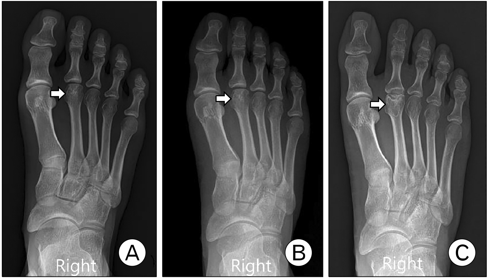 | Fig. 1Anteroposterior radiographs of right foot at initial (A), 1-month follow-up (B), and 2-year follow-up (C). Subtle joint space widening with flattening and sclerotic change in the second metatarsal head (arrow) was shown in 1-month follow-up (B). Sclerosis and collapse of the second metatarsal head (arrow) with sunken appearance and periarticular spurring were observed in 2-year follow-up (C).
|
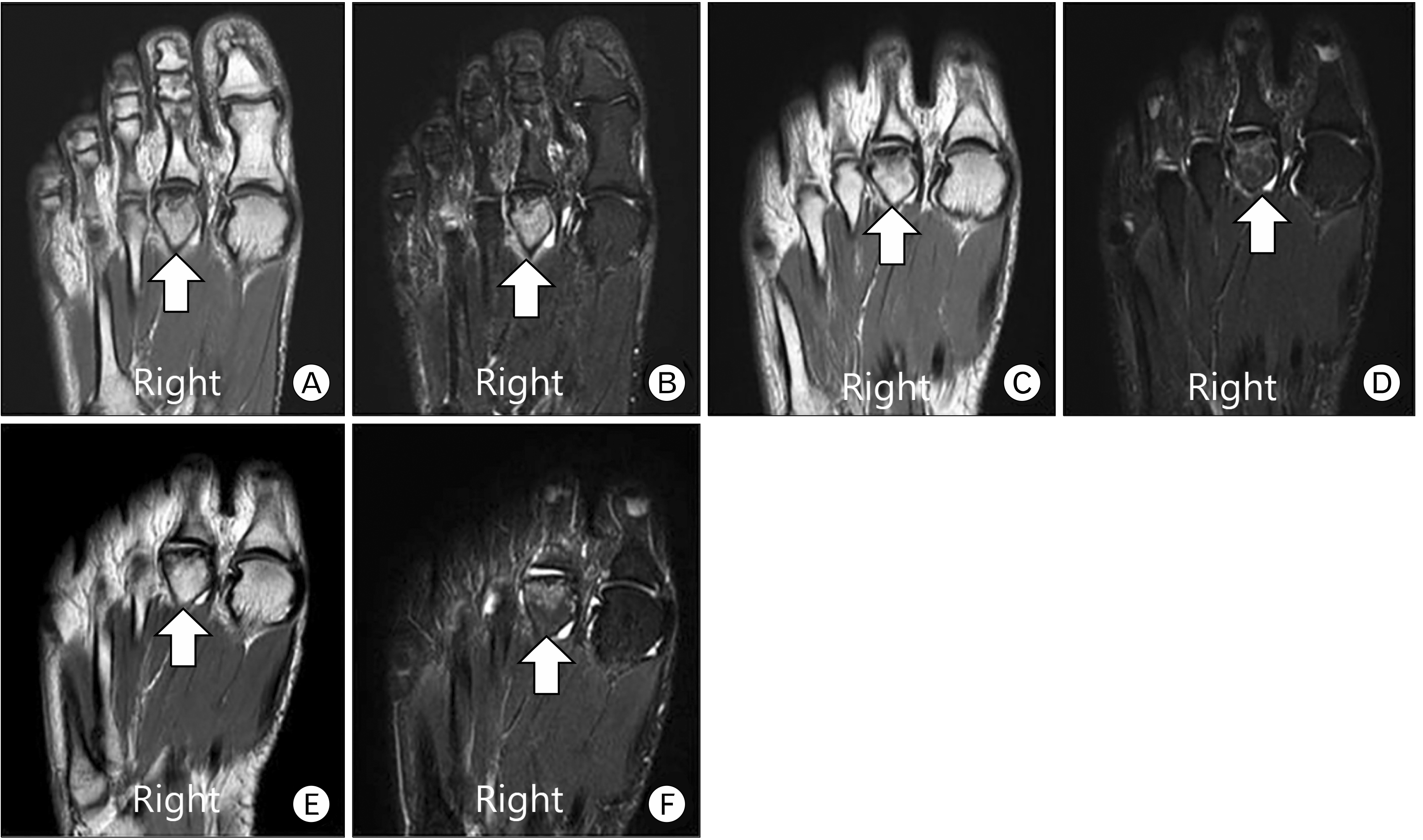 | Fig. 2Proton density-weighted image and fat-suppressed T2-weighted image at initial (A, B), 16-month follow-up (C, D), and 2-year follow-up (E, F). (A, B) Initial magnetic resonance imaging (MRI) shows an ill-defined low signal lesion in the right second metatarsal head and bone marrow edema, suggesting osteonecrosis. Sixteen-month follow-up MRI (C, D) shows a slightly decreased extent of the low signal lesion with decreased bone marrow edema, which is maintained until 2-year follow-up (E, F) (White arrow; second metatarsal head).
|
The symptom was sustained with the activity of daily living and she was referred to the department of physical medicine and rehabilitation for further comprehensive conservative management. She had no serious past medical history. She was standing and walking more than 6 hours a day wearing flat slippers in her workplace. She continued to work even after splinting in orthopedic surgery. While walking and standing, she complained of pain with the numeric rating scale (NRS) 9. On physical exam, there was local tenderness around the right second metatarsal head without redness or heating sensation. In addition, there was no associated structural or biomechanical dysfunction such as hallux valgus deformity, pronated foot, and ankle equinus. Subtle joint space widening with flattening and sclerotic change in the right second metatarsal head was shown in a 1-month follow-up X-ray (Fig. 1B).
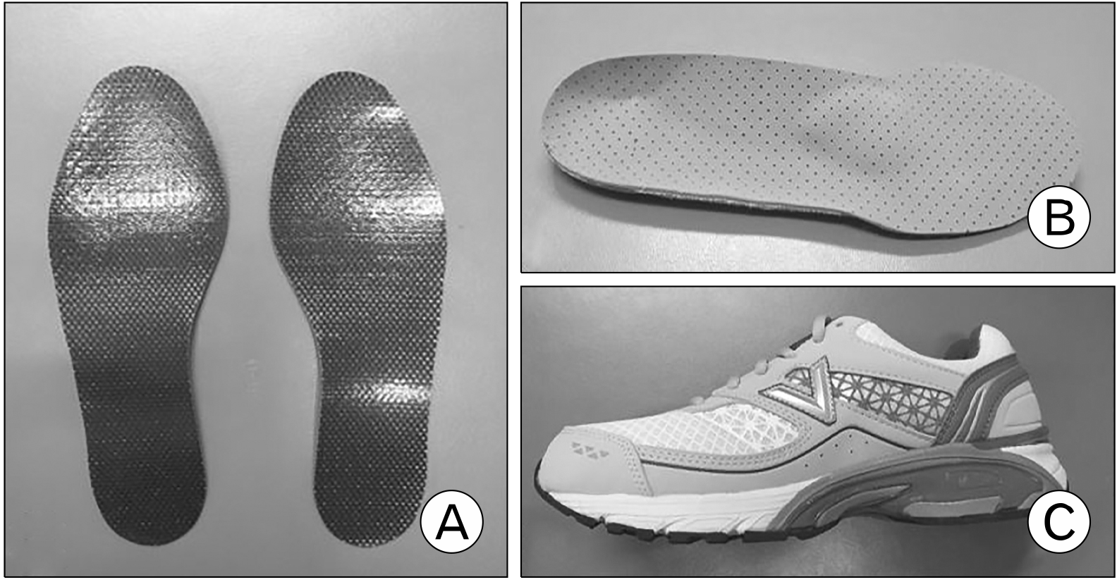
To strictly avoid the impact on forefoot, it was recommended to use crutch and suspend from work for 2 weeks. Subsequently, the pain started to improve gradually. After the acute sharp pain was relieved, a carbon fiber insert, customed semirigid insole with metatarsal dome and rocker-bottom shoes were prescribed to relieve pressure under the metatarsal heads (Fig. 3). The pain continued to improve with NRS 3 after 6 weeks. Although the pain waxed and waned according to the intensity of daily activity, it remained in well-tolerated condition until 2-year follow-up.
In a 2-year follow-up X-ray, sclerosis and collapse of the right second metatarsal head with sunken appearance and periarticular spurring were observed (Fig. 1C). In 16-month follow-up MRI, the degrees of bone marrow edema and low signal lesion were reduced compared to initial MRI without injury of articular cartilage (Fig. 2C and D). This finding was maintained at a 2-year follow-up MRI (Fig. 2E and F). In 2-year follow-up, a three-phase bone scan showed that the uptakes on the blood pool and delayed phase were decreased compared to the initial three-phase bone scan.
Go to :
Discussion
Freiberg disease is known to cause osteonecrosis, which is characterized by flattening and collapse of the metatarsal head2. Usually, nonoperative management is the first treatment focused upon the symptom relief and deformity prevention3,4. If it is treated early, this condition is self-limiting, often resolving with nonoperative management. Some patients experienced improvement in symptoms and normal range of motion2. One study reported that spontaneous healing may occur at any stage and restoration of a normal metatarsal head may be possible in the early stage5. Operative treatments are typically entertained for advanced stages or after the failure of conservative management in the early stage of the disease. There is little consensus as to the best treatment option, although recommendations can be made based on the stage2.
We reported a 2-year follow-up of Freiberg disease treated with conservative management. In our case, Freiberg disease was misconceived as the right second MTP joint synovitis after initial plain radiography and ultrasonography. Both Freiberg disease and second MTP joint synovitis begin with vague complaints of pain and tenderness of the plantar side of the second MTP joint that worsened during weight-bearing activity3,6. Because early plain radiographic findings such as joint widening cannot be detected until 3 to 6 weeks after the symptoms occur, MRI may be helpful in confirming the diagnosis in subtle or inconclusive cases1-3,6. Classically, osteonecrosis of metatarsal head on MRI shows hypointense signal on T1-weighted images and a combination of low and high signal intensities on T2-weighted images1,3.
Smillie7 proposed five staging system for Freiberg disease based on surgical findings and radiographic appearances (Table 1). Thompson and Hamilton8 described a four-type classification according to treatment recommendation (Table 2). Nonoperative management can be the first line of treatment regardless of the severity of disease at the time of presentation1-4. If the patient is not responding to nonoperative management, surgical interventions such as core decompression, joint debridement, metatarsal osteotomy, and excision arthroplasty were indicated in Smillie’s stage 3 to 5 and Thompson’s type 2 to 47,8. However, the evidence is insufficient to support the surgical intervention in Freiberg disease and there are no studies on the outcomes or prognosis of conservative treatment according to the stage and type3.
Table 1
Smillie’s staging system for Freiberg disease
![]()
Table 2
Thompson’s classification of Freiberg disease
![]()
Strategies for conservative treatment include oral anti-inflammatory medication, avoidance of high-impact activity (such as running and jumping), and modification of shoe-wear1,3,4,6. A stiff-soled shoe with rocker bottom can be helpful to offload the forefoot during toe-off4. In addition, metatarsal pad or bar can be used to relieve pressure under the metatarsal heads3. They are focused on offloading and relieving stress to alleviation of symptoms and prevention of deformity.
In our case, the sunken appearance of the metatarsal head and periarticular spurring were observed on the X-ray without loss of articular cartilage in MRI, corresponding to Smillie’s stage 3 and Thompson’s type 2. Some reports showed that open surgical debridement and osteotomy may be useful at the early stage9,10. However, our case shows that conservative management with shoe modification and insole can help to return to activity of daily life and maintain a well-tolerable condition without further progression on long-term follow-up MRI in Freiberg disease with Smillie’s stage 3 and Thompson’s type 2.
Go to :
 XML Download