Journal List > Ann Rehabil Med > v.47(1) > 1516081579

Lee, Wi, Park, Oh, Seo, and Lee: Exploratory Investigation of the Effects of Tactile Stimulation Using Air Pressure at the Auricular Vagus Nerve on Heart Rate Variability

Abstract

Objective

To explore the effects of tactile stimulation using air pressure at the auricular branch of the vagus nerve on autonomic activity in healthy individuals.

Methods

Three types of tactile stimulation were used in this study: continuous low-amplitude, continuous high-amplitude, and pulsed airflow. The tactile stimulations were provided to the cymba concha to investigate autonomic activity in 22 healthy participants. The mean heart rate (HR) and parameters of HR variability, including the standard deviation of R-R intervals (SDNN) and root mean square of successive R-R interval differences (RMSSD) were compared at baseline, stimulation, and recovery periods.

Results

Two-way repeated measures ANOVA indicated a significant main effect of time on HR (p=0.001), SDNN (p=0.003), and RMSSD (p<0.001). These parameters showed significant differences between baseline and stimulation periods and baseline and recovery periods in the post-hoc analyses. There were no significant differences in the changes induced by stimulation type and the interaction between time and stimulation type for all parameters. One-way repeated measures ANOVA showed that HR, SDNN, and RMSSD did not differ significantly among the three time periods during sham stimulation.

Conclusion

Parasympathetic activity can be enhanced by auricular tactile stimulation using air pressure, targeting the cymba concha. Further studies are warranted to investigate the optimal stimulation parameters for potential clinical significance.

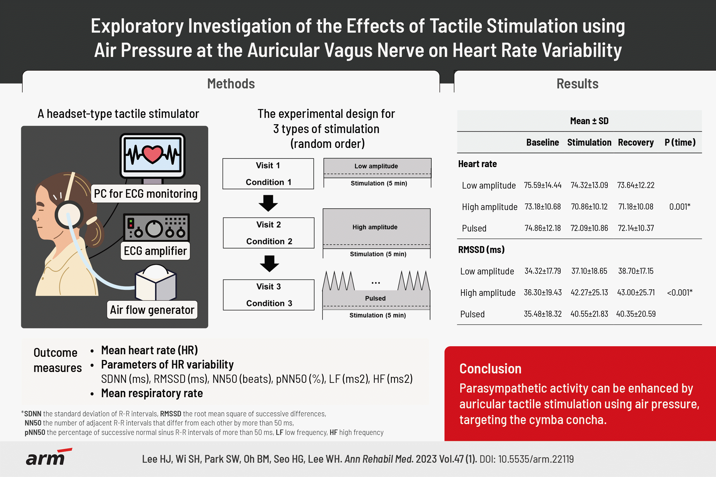
GRAPHICAL ABSTRACT

INTRODUCTION

Autonomic modulation via vagus nerve stimulation (VNS) has gained increasing interest as a potential therapeutic strategy in clinical practice. VNS is a neuromodulatory treatment that has shown clinical efficacy in various disorders, including epilepsy, depression, stroke, traumatic brain injury, Alzheimer’s disease, rheumatoid arthritis, diabetes, and cardiovascular disorders [1-3]. Prior literature has reported several benefits of VNS, including anti-adrenergic effects at central and peripheral levels, anti-apoptotic effects, an increase in nitric oxide, enhanced neuroplasticity, anti-nociception, and antiinflammatory effects [4]. It can modulate autonomic imbalance through accelerated parasympathetic activity, blunted sympathetic activity, and suppressed pro-inflammatory cytokines, making it a promising therapeutic approach [5].
Despite its potential advantages in clinical application, VNS is cautiously considered due to its requirement for invasive implantation of electrodes and stimulators. Surgical implantation is associated with increased risks of infection, bradycardia, asystole, and vocal cord paresis [6]. Stimulation-associated adverse events include voice alteration, paresthesia, cough, dyspnea, and pain [7]. As an alternate non-invasive approach, transcutaneous auricular VNS exhibits comparable effects to those of invasive VNS by stimulating the auricular vagus nerve [8,9]. The input of transcutaneous auricular VNS can be delivered through the auricular vagus nerve to the brainstem and higher brain areas via extensive projections to the second- and third-order neurons within the brain [9]. Transcutaneous auricular VNS, which can increase heart rate (HR) variability by reducing sympathetic nerve outflow, has shown promising clinical results in mitigating atrial fibrillation and ventricular arrhythmia, alleviating pain severity, reducing depression severity, and improving stroke recovery [10-14].
The auricle, a region innervated by the peripheral branches of the vagus nerve, is a plausible direct gateway for the modulation of parasympathetic activity [9]. This point is a rationale for the application of transcutaneous auricular VNS or acupuncture therapy as a non-invasive approach to enhancing parasympathetic activity in previous studies [9,12,14,15]. However, even though there has been anatomic and physiologic evidence of sensory regions innervated by the auricular branch of the vagus nerve [16,17], few previous studies have focused on the effect of tactile stimulation at the auricle as an alternative method of a non-invasive approach. The auricular branch of the vagus nerve is afferent nerve fibers, which form a cutaneous receptive field of the external ear, including the cymba concha along with antihelix, cavity of concha, tragus, crus of helix, and crura of antihelix [18]. Previous electrophysiological and functional magnetic resonance imaging studies showed that tactile stimulation can result in brain activity similar to that from transcutaneous electrical stimulation [19,20].
Thus, this study aims to investigate the effect of tactile stimulation, using air pressure, at the auricular branch of the vagus nerve on autonomic activity.

MATERIALS AND METHODS

Participants

The participants were recruited through bulletin boards at a medical school and a hospital. Participants were eligible if they had no history of cardiovascular diseases, epilepsy, severe brain injury, or mental disorders, such as depression and anxiety. Twenty-three participants were recruited for tactile stimulation, and one of them showed frequent premature ventricular contractions on an electrocardiogram (ECG) and was consequently excluded from this study. Twenty-two participants (11 female) were enrolled for auricular tactile stimulation in this study. Additional 22 participants (11 female) were enrolled for sham stimulation. There was no participant who had medical diseases or took medications that could affect the HR or HR variability. This study was approved by the Institutional Review Board of Seoul National University Hospital (no. 2104-145-1213), and all participants provided written informed consent according to the institutional guidelines.

Experimental design

Fig. 1 summarizes the experimental design of this study. Auricular tactile stimulation was performed using air pressure targeting the cymba concha of the bilateral external ears, where the auricular branch of the vagus nerve is located. The sensory threshold level was measured in all participants for auricular tactile stimulation before the experiment began to determine the tactile stimulation intensity. The sensory threshold was determined by gradually decreasing the stimulus air pressure, from the initial 0.005 psi, by 0.001 psi at intervals until the participants could no longer perceive it. The low-amplitude intensity was defined as the sensory threshold plus 0.004 psi. The high-amplitude intensity was defined as twice the low amplitude, which was determined through pilot experiments before this study commenced. The three types of auricular tactile stimulation used in this study were as follows: (1) continuous low-amplitude, (2) continuous high-amplitude, and (3) pulsed airflow. Pulsed stimulation was provided as triangular pulses, in which the air pressure increased linearly from low to high amplitude for 15 seconds, followed by a linear decrease from high to low amplitude for 15 seconds. The three types of tactile stimulation were assigned randomly to each participant. Additionally, sham stimulation was provided to a group of participants different from those who were provided with the three types of stimulation. Sham stimulation was defined as continuous high-amplitude stimulation while wearing a headset, with the air hose targeting the bilateral shoulders to avoid stimulating the cymba concha of the bilateral external ears.
After a resting period of 5 minutes, ECG data were recorded at the baseline, during stimulation, and recovery periods. Each period lasted five minutes. During the recording periods, the participants were seated with palms facing upward, knees at 90° angles, both feet flat on the floor, both hands on thighs, and eyes closed [21]. The participants were instructed not to speak or move, to stay awake, and breathe as they would regularly (comfortably and not deeply). To minimize influences of the previous stimulation, the participants attended each session three times, at least 24 hours apart from the previous session. Before the experiments began, the participants were instructed to control the confounding variables that could possibly affect HR variability, as determined by a previous study [21]. The instructions included the followings: to follow a normal sleep routine, not to exercise or consume alcohol the day before the experiment, not to consume food or caffeine at least two hours before the experiment, and to empty their urinary bladder before the experiment begins. The participants were informed to report any pain or discomfort, and the experiment was terminated if severe pain or discomfort occurred during the tactile stimulation.

Development of a tactile stimulator using air pressure

To provide the three types of tactile stimulation in terms of (1) continuous low-amplitude, (2) continuous high-amplitude, and (3) pulsed airflow, a tactile stimulator was developed to generate and control the air pressure in this study (Fig. 2). In the tactile stimulator, air pressure was generated using a BLDC blower (U71MX-024KX-4; Micronel, Tagelswangen, Switzerland), which was controlled using an Arduino UNO microcontroller board (Arduino, Monza, Italy), an open-source electronic prototyping platform, and an Arduino open-source integrated development environment. The air pressure, measured at the end of the air hose, could be modulated by 0.001 psi increments in the tactile stimulator. Air was generated from the blower and delivered via an air hose targeting the participant’s cymba concha. The inner diameter of the air hose was 11 mm, and the length from the end of the air hose to the cymba concha was approximately 3 cm. A commercial headset (HS-301; Micronics, Gimpo, Korea) was modified to allow adjustments to the end of the air hose to streamline the focus on the target area of the external ear while compensating for neck movements of the participants during the auricular tactile stimulation. The pressure and noise level generated by a tactile stimulator were measured using a digital manometer (DT-8890A; CEM, Shenzhen, China) and a sound level meter (GM1351; BENETECH, Shenzhen, China). The noise level remained below 20.0 dB, which is equivalent to the sound of rustling leaves.

Electrocardiogram data acquisition and heart rate variability analysis

ECG and HR data were recorded at 2,000 Hz using a 2-channel Biopac Student Lab version 4.1 (BIOPAC Systems, Goleta, CA, USA). Three disposable electrodes were placed under the left and right clavicles and on the lower edge of the left rib cage. After recordings, the data were exported to the Kubios HRV Premium Software version 3.5 (Kubios, Kuopio, Finland), one of the most popular software used to analyze HR variability [22]. In the timedomain measures, the standard deviation of R-R intervals (SDNN), the root mean square of successive R-R interval differences (RMSSD), the number of adjacent R-R intervals that differ from each other by more than 50ms (NN50), and the percentage of successive normal sinus R-R intervals of more than 50 ms (pNN50) were obtained. In the frequency-domain measures, the low frequency (LF) component at 0.04–0.15 Hz and high frequency (HF) component at 0.15–0.40 Hz were acquired. The respiratory rate was estimated based on ECG and R-R interval data using the respiratory rate estimation algorithm supported by Kubios HRV Premium Software [23].

Statistical analysis

Data are presented as the mean±standard deviation (SD). Two-way repeated measures ANOVA was used to examine differences in HR, the parameters of HR variability, and the respiratory rate among the three time periods (baseline, stimulation, and recovery), and among the three types of tactile stimulation (continuous lowamplitude, continuous high-amplitude, and pulsed airflow). If significant main effects were observed, posthoc tests with Bonferroni correction were conducted for pairwise comparisons. Greenhouse–Geisser correction was performed when necessary, to correct for non-sphericity. Sex was added to the two-way repeated measures ANOVA model to investigate whether differences in HR, the parameters of HR variability, and respiratory rate according to sex were observed. Age was not added to the two-way repeated measures ANOVA model as the age of the participants was in a narrow range (24–37 years). In the sham stimulation group, one-way repeated measures ANOVA was conducted to examine the differences in HR data, parameters of HR variability, and respiratory rate among the three time periods (baseline, stimulation, and recovery). The significance level was set at p<0.05. All statistical analyses were performed using IBM SPSS Statistics 19.0 (IBM Corp., Armonk, NY, USA).

RESULTS

Participants

The mean age of all participants was 29.8±4.0 years. (range: 24–37 years). Age was not different between auricular tactile stimulation and sham stimulation groups (29.2±3.9 years vs. 30.6±3.9 years, p=0.240). Only participants who thoroughly followed the instructions to control for confounding variables that could potentially affect HR variability participated in the experiments. The sensory threshold of 18 participants was measured at 0.001 psi, whereas that of the others was measured at 0.002 psi. The levels of tactile stimulation did not differ significantly between the male and female (p=0.478). One participant in auricular tactile stimulation group reported mild discomfort around the external acoustic meatus during highamplitude stimulation.

Heart rate variability analysis

Table 1 shows the mean±SD of HR, parameters of HR variability, and respiratory rate at the baseline, stimulation, and recovery periods. The within-subjects two-way repeated measures ANOVA indicated a significant main effect of time on HR (p=0.001), most parameters of HR variability including SDNN (p=0.003), RMSSD (p<0.001), NN50 (p=0.003), pNN50 (p=0.002), LF (p=0.040), and respiratory rate (p=0.018) during tactile stimulation. As shown in Table 1, most of the parameters showed significant differences between the baseline and stimulation periods, and the baseline and recovery periods in the post-hoc analyses (p<0.017 after Bonferroni correction). There was no significant main effect of stimulation type or interaction between time and stimulation type on all parameters (not included in the Table 1 due to lack of space). HR, the parameters of HR variability, and respiratory rate did not significantly differ according to sex. One-way repeated measures ANOVA showed that HR, the parameters of HR variability, and respiratory rate did not differ significantly among baseline, stimulation, and recovery periods during sham stimulation (Supplementary Table S1). The individual values of percentage changes during the baseline, stimulation, and recovery periods in the participants with continuous low-amplitude, continuous high-amplitude, pulsed, or sham stimulation are illustrated in Supplementary Figs. S1-S8.

DISCUSSION

This study aimed to explore the effects of tactile stimulation using air pressure at the auricular vagus nerve, on cardiovascular autonomic control in healthy individuals. A headset-type tactile stimulator targeting the bilateral cymba concha was developed to conduct the experiments. The results of the present study showed that auricular tactile stimulation altered cardiovascular autonomic control, causing a shift toward vagal dominance. Reduced HR and increased HR variability and respiratory rate were observed after auricular tactile stimulation, which was not evident in sham stimulation.
In this study, three types of tactile stimulation were determined to explore the effects of temporal patterns and intensity of airflow at the bilateral external ears on autonomic function. Although HR and the parameters of HR variability showed a trend of difference in continuous high and pulsed stimulation compared with continuous low stimulation, the main effect of group was not significant in this study. The results of the current study showed that auricular tactile stimulation using airflow ranging from 0.005–0.012 psi can influence cardiac parasympathetic activity. The parameters of tactile stimulation using air pressure in previous studies include the pressure, velocity, and pulse of the airflow [24-26]. In the present study, pressure (low- and high-amplitude stimulations) and pulse (continuous and pulsed stimulations) were used as the parameters of tactile stimulation, considering that the pressure of airflow might be associated with the induction of discomfort or unpleasant feelings and sensory adaptation could occur during continuous tactile stimulation. The intensity of tactile stimulation in the present study was determined by taking into consideration the variation in the sensory threshold for airflow and the potential discomfort when using the higher intensity of tactile stimulation, as observed in a pilot study, similar to transcutaneous electrical stimulation [27]. Additionally, although the target area for tactile stimulation was the cymba concha, it should be noted that the airflow was applied to the auricular and periauricular areas. It was taken into account that stimulation with airflow at the cymba concha only was not realistic, and the cutaneous distribution of the auricular branch of the vagus nerve includes the cymba concha, antihelix, cavity of concha, tragus, crus of helix, and crura of the antihelix. Interestingly, significant change in respiratory rate was observed with auditory tactile stimulation. This finding is consistent with clinical studies showing that an increased respiratory rate was observed during VNS in adults and children with epilepsy [28,29]. According to a previous preclinical study, two subtypes of vagal afferent neurons for lung-to-brain connectivity exist, one of which can cause rapid and shallow breathing after neuronal activation [30].
A limited number of studies have been performed to investigate non-electrical methods of stimulation to activate the parasympathetic activity through the auricular branch of the vagus nerve. Recently, Boehmer et al. [15] reported that acupuncture at the cymba concha can alter autonomic balance in favor of vagal tone in healthy individuals. Acupuncture at the region innervated by the auricular vagus nerve induced decreased HR and increased SDNN without changes in power spectral density parameters such as HF and LF, which is generally consistent with the results of the current study. Acupuncture at the auricular branch of the vagus nerve might increase the activity of the nucleus tractus solitarius projecting to the vagal efferent neurons of the nucleus ambiguous and dorsal motor nucleus in the medulla oblongata, consequently resulting in the activation of the parasympathetic nervous system. In addition, a pilot study outlined the anti-inflammatory effects of vibrotactile stimulation on the cymba concha in healthy individuals and patients with rheumatoid arthritis [31]. In this study, vibrotactile stimulation reduced the levels of pro-inflammatory cytokines, including tumor necrosis factor, interleukin 1 beta, and interleukin 6, in healthy individuals and decreased disease activity and pain severity in patients with rheumatoid arthritis. The anatomical basis of auricular VNS has been provided by preclinical studies using tracing techniques in rats, dogs, cats, and human cadaver studies [32-35]. The auricular tactile stimulation in the present study may activate the auricular vagus nerve, similarly to acupuncture or vibrotactile stimulation, resulting in indirect enhancement of parasympathetic activity, which should be demonstrated in further preclinical or clinical studies.
There are few studies that have explored auricular tactile stimulation to modulate parasympathetic activity via the vagus nerve, as opposed to transcutaneous electrical stimulation, which has shown promising results for clinical purposes in diverse diseases. However, there have been several studies that compared the effects of tactile and transcutaneous electrical stimulation on the peripheral and central nervous systems. The neurophysiological studies on healthy participants reported that sensory nerve action potentials from the sural and medial nerves were evoked by both tactile stimulation (using a servomotor controlled probe) and transcutaneous electrical stimulation at the peripheral regions of the limbs including dorsolateral side of the foot and the tip of the third digit [19,36]. In these studies, the peak-to-peak amplitude of the sensory nerve action potentials in tactile stimulation was approximately 5%–10% of that in supramaximal electrical stimulation. Another experimental study on neurohormonal activation in a rat model showed that tactile stimulation at the glans penis and transcutaneous electrical stimulation of the dorsal penile nerve similarly excited approximately 60% of oxytocin releasing cells in the supraoptic nucleus of the central nervous system [37]. Furthermore, an investigation of healthy individuals using functional magnetic resonance imaging also indicated that tactile stimulation with a soft brush at the index finger activated cortical activity in brain regions anatomically equivalent to those activated by transcutaneous electrical stimulation [20]. The level of electrical intensity was set above the motor threshold, and there was consequent activation of brain regions, including the primary and secondary sensorimotor cortex, insula, parietal operculum, and posterior parietal cortices. The results of the aforementioned studies suggest that tactile stimulation on the specific regions of the body may activate the cutaneous branch of the peripheral nerve and related brain regions, although the effect of tactile stimulation might be less than that of transcutaneous electrical stimulation. Further research is warranted to investigate the potential clinical significance and optimal parameters for tactile stimulation of the auricular vagus nerve to modulate parasympathetic activity for clinical applications.
This study has several limitations. First, the sample size of the present study was small. To the best of our knowledge, this is the first study to investigate the effect of auricular tactile stimulation on parasympathetic activity. It is necessary to generalize the results of this study to a larger number of participants. Second, only short-term effects of the three types of auricular tactile stimulation in terms of 5 minutes each of stimulation and recovery periods were explored in this study. Future studies are needed to investigate the long-term effects of more optimized auricular tactile stimulation, including other stimulation parameters, such as temperature or humidity of the airflow. Third, direct comparisons between the stimulation and sham conditions were not conducted because this study additionally included a sham condition, and the participants in the sham group differed from those in the experimental group. It is necessary to perform a comparison between stimulation and sham conditions with a between- and within-subject design, considering habituation to the experimental environment and inter-individual variations that can influence HR variability. Fourth, the study population only included healthy individuals. To gain clinical significance, it is mandatory to extend the study population to include older adults and patients with cardiovascular disorders.
This study reported that parasympathetic activity can be enhanced in healthy individuals by auricular tactile stimulation using air pressure, targeting the cymba concha of the bilateral external ears, where the auricular branch of the vagus nerve is distributed. Further preclinical and clinical studies are essential to investigate the exact neural mechanisms that modulate the autonomic nervous system and to determine the optimal stimulation parameters for potential clinical significance.

Notes

Byung-Mo Oh is the Editor-in-Chief of Annals of Rehabilitation Medicine. The author did not engage in any part of the review and decision-making process for this manuscript. Han Gil Seo is an Associate Editor of Annals of Rehabilitation Medicine. The author did not engage in any part of the review and decision-making process for this manuscript. Otherwise, no potential conflict of interest relevant to this article was reported.

Conceptualization: Lee WH, Oh BM, Seo HG. Methodology: Wi S, Park S. Investigation: Wi S. Formal analysis: Wi S, Lee HJ. Funding acquisition: Lee WH. Project administration: Lee WH. Visualization: Wi S. Writing – original draft: Lee WH, Lee HJ, Wi S, Park S. Writing – review and editing: Lee WH, Lee HJ, Wi S. Approval of final manuscript: all authors.

ACKNOWLEDGMENTS

This work was supported by a National Research Foundation of Korea (NRF) grant funded by the Korean government (2021R1F1A1046709) and SNUH Research Funds (04-2020-2160 and 04-2021-0330). The statistical analyses in this study were supported by the Medical Research Collaborating Center (MRCC) of the Seoul National University Hospital.

SUPPLEMENTARY MATERIALS

Supplementary materials can be found via https://doi. org/10.5535/arm.22119.

Supplementary Table S1.

The responses of mean heart rate, heart rate variability, and respiratory rate in sham stimulation
arm-22119-Supplementary-Table-S1.pdf

Supplementary Fig. S1.

The response of mean heart rate in (A) sham, (B) continuous low-amplitude, (C) continuous high-amplitude, and (D) pulsed stimulations. Bold solid lines represent average values and dotted lines represent individual values.
arm-22119-Supplementary-Fig-S1.pdf

Supplementary Fig. S2.

The response of standard deviation of R-R intervals (SDNN) in (A) sham, (B) continuous lowamplitude, (C) continuous high-amplitude, and (D) pulsed stimulations. Bold solid lines represent average values and dotted lines represent individual values.
arm-22119-Supplementary-Fig-S2.pdf

Supplementary Fig. S3.

The response of root mean square of successive R-R interval differences (RMSSD) in (A) sham, (B) continuous low-amplitude, (C) continuous high-amplitude, and (D) pulsed stimulations. Bold solid lines represent average values and dotted lines represent individual values.
arm-22119-Supplementary-Fig-S3.pdf

Supplementary Fig. S4.

The response of the number of adjacent R-R intervals that differ from each other by more than 50 ms (NN50) in (A) sham, (B) continuous low-amplitude, (C) continuous high-amplitude, and (D) pulsed stimulations. Bold solid lines represent average values and dotted lines represent individual values.
arm-22119-Supplementary-Fig-S4.pdf

Supplementary Fig. S5.

The response of percentage of successive normal sinus R-R intervals of more than 50 ms (pNN50) in (A) sham, (B) continuous low-amplitude, (C) continuous high-amplitude, and (D) pulsed stimulations. Bold solid lines represent average values and dotted lines represent individual values.
arm-22119-Supplementary-Fig-S5.pdf

Supplementary Fig. S6.

The response of low frequency (LF) in (A) sham, (B) continuous low-amplitude, (C) continuous high-amplitude, and (D) pulsed stimulations. Bold solid lines represent average values and dotted lines represent individual values.
arm-22119-Supplementary-Fig-S6.pdf

Supplementary Fig. S7.

The response of high frequency (HF) in (A) sham, (B) continuous low-amplitude, (C) continuous high-amplitude, and (D) pulsed stimulations. Bold solid lines represent average values and dotted lines represent individual values.
arm-22119-Supplementary-Fig-S7.pdf

Supplementary Fig. S8.

The response of mean respiratory rate in (A) sham, (B) continuous low-amplitude, (C) continuous high-amplitude, and (D) pulsed stimulations. Bold solid lines represent average values and dotted lines represent individual values.
arm-22119-Supplementary-Fig-S8.pdf

REFERENCES

1. Johnson RL, Wilson CG. A review of vagus nerve stimulation as a therapeutic intervention. J Inflamm Res. 2018; 11:203–13.
2. Nearing BD, Libbus I, Carlson GM, Amurthur B, KenKnight BH, Verrier RL. Chronic vagus nerve stimulation is associated with multi-year improvement in intrinsic heart rate recovery and left ventricular ejection fraction in ANTHEM-HF. Clin Auton Res. 2021; 31:453–62.
3. Garamendi-Ruiz I, Gómez-Esteban JC. Cardiovascular autonomic effects of vagus nerve stimulation. Clin Auton Res. 2019; 29:183–94.
4. Kaniusas E, Kampusch S, Tittgemeyer M, Panetsos F, Gines RF, Papa M, et al. Current directions in the auricular vagus nerve stimulation I - a physiological perspective. Front Neurosci. 2019; 13:854.
5. De Ferrari GM, Crijns HJ, Borggrefe M, Milasinovic G, Smid J, Zabel M, et al. Chronic vagus nerve stimulation: a new and promising therapeutic approach for chronic heart failure. Eur Heart J. 2011; 32:847–55.
6. Kahlow H, Olivecrona M. Complications of vagal nerve stimulation for drug-resistant epilepsy: a single center longitudinal study of 143 patients. Seizure. 2013; 22:827–33.
7. Ben-Menachem E, Revesz D, Simon BJ, Silberstein S. Surgically implanted and non-invasive vagus nerve stimulation: a review of efficacy, safety and tolerability. Eur J Neurol. 2015; 22:1260–8.
8. Beekwilder JP, Beems T. Overview of the clinical applications of vagus nerve stimulation. J Clin Neurophysiol. 2010; 27:130–8.
9. Mercante B, Ginatempo F, Manca A, Melis F, Enrico P, Deriu F. Anatomo-physiologic basis for auricular stimulation. Med Acupunct. 2018; 30:141–50.
10. Redgrave JN, Moore L, Oyekunle T, Ebrahim M, Falidas K, Snowdon N, et al. Transcutaneous auricular vagus nerve stimulation with concurrent upper limb repetitive task practice for poststroke motor recovery: a pilot study. J Stroke Cerebrovasc Dis. 2018; 27:1998–2005.
11. Busch V, Zeman F, Heckel A, Menne F, Ellrich J, Eichhammer P. The effect of transcutaneous vagus nerve stimulation on pain perception--an experimental study. Brain Stimul. 2013; 6:202–9.
12. Clancy JA, Mary DA, Witte KK, Greenwood JP, Deuchars SA, Deuchars J. Non-invasive vagus nerve stimulation in healthy humans reduces sympathetic nerve activity. Brain Stimul. 2014; 7:871–7.
13. Liu CH, Yang MH, Zhang GZ, Wang XX, Li B, Li M, et al. Neural networks and the anti-inflammatory effect of transcutaneous auricular vagus nerve stimulation in depression. J Neuroinflammation. 2020; 17:54.
14. Geng D, Liu X, Wang Y, Wang J. The effect of transcutaneous auricular vagus nerve stimulation on HRV in healthy young people. PLoS One. 2022; 17:e0263833.
15. Boehmer AA, Georgopoulos S, Nagel J, Rostock T, Bauer A, Ehrlich JR. Acupuncture at the auricular branch of the vagus nerve enhances heart rate variability in humans: an exploratory study. Heart Rhythm O2. 2020; 1:215–21.
16. Mahadi KM, Lall VK, Deuchars SA, Deuchars J. Cardiovascular autonomic effects of transcutaneous auricular nerve stimulation via the tragus in the rat involve spinal cervical sensory afferent pathways. Brain Stimul. 2019; 12:1151–8.
17. de Gurtubay IG, Bermejo P, Lopez M, Larraya I, Librero J. Evaluation of different vagus nerve stimulation anatomical targets in the ear by vagus evoked potential responses. Brain Behav. 2021; 11:e2343.
18. Peuker ET, Filler TJ. The nerve supply of the human auricle. Clin Anat. 2002; 15:35–7.
19. Krarup C, Trojaborg W. Compound sensory action potentials evoked by tactile and by electrical stimulation in normal median and sural nerves. Muscle Nerve. 1994; 17:733–40.
20. Boakye M, Huckins SC, Szeverenyi NM, Taskey BI, Hodge CJ Jr. Functional magnetic resonance imaging of somatosensory cortex activity produced by electrical stimulation of the median nerve or tactile stimulation of the index finger. J Neurosurg. 2000; 93:774–83.
21. Laborde S, Mosley E, Thayer JF. Heart rate variability and cardiac vagal tone in psychophysiological research - recommendations for experiment planning, Data analysis, and data reporting. Front Psychol. 2017; 8:213.
22. Alcantara JMA, Plaza-Florido A, Amaro-Gahete FJ, Acosta FM, Migueles JH, Molina-Garcia P, et al. Impact of using different levels of threshold-based artefact correction on the quantification of heart rate variability in three independent human cohorts. J Clin Med. 2020; 9:325.
23. Tarvainen MP, Niskanen JP, Lipponen JA, Ranta-Aho PO, Karjalainen PA. Kubios HRV--heart rate variability analysis software. Comput Methods Programs Biomed. 2014; 113:210–20.
24. Jia S, Wang L, Wang H, Lv X, Wu J, Yan T, et al. Pneumatical-mechanical tactile stimulation device for somatotopic mapping of body surface during fMRI. J Magn Reson Imaging. 2020; 52:1093–101.
25. da Cunha JC, Bordignon LA, Nohama P. Tactile communication using a CO2 flux stimulation for blind or deafblind people. Annu Int Conf IEEE Eng Med Biol Soc. 2010; 2010:5871–4.
26. Tsalamlal MY, Ouarti N, Martin JC, Ammi M. Haptic communication of dimensions of emotions using air jet based tactile stimulation. J Multimodal User Interface. 2015; 9:69–77.
27. Borges U, Laborde S, Raab M. Influence of transcutaneous vagus nerve stimulation on cardiac vagal activity: not different from sham stimulation and no effect of stimulation intensity. PLoS One. 2019; 14:e0223848.
28. Mulders DM, de Vos CC, Vosman I, van Putten MJ. The effect of vagus nerve stimulation on cardiorespiratory parameters during rest and exercise. Seizure. 2015; 33:24–8.
29. Zaaimi B, Héberlé C, Berquin P, Pruvost M, Grebe R, Wallois F. Vagus nerve stimulation induces concomitant respiratory alterations and a decrease in SaO2 in children. Epilepsia. 2005; 46:1802–9.
30. Chang RB, Strochlic DE, Williams EK, Umans BD, Liberles SD. Vagal sensory neuron subtypes that differentially control breathing. Cell. 2015; 161:622–33.
31. Addorisio ME, Imperato GH, de Vos AF, Forti S, Goldstein RS, Pavlov VA, et al. Investigational treatment of rheumatoid arthritis with a vibrotactile device applied to the external ear. Bioelectron Med. 2019; 5:4.
32. Butt MF, Albusoda A, Farmer AD, Aziz Q. The anatomical basis for transcutaneous auricular vagus nerve stimulation. J Anat. 2020; 236:588–611.
33. He W, Jing XH, Zhu B, Zhu XL, Li L, Bai WZ, et al. The auriculo-vagal afferent pathway and its role in seizure suppression in rats. BMC Neurosci. 2013; 14:85.
34. Chien CH, Shieh JY, Ling EA, Tan CK, Wen CY. The composition and central projections of the internal auricular nerves of the dog. J Anat. 1996; 189(Pt 2):349–62.
35. Nomura S, Mizuno N. Central distribution of primary afferent fibers in the Arnold’s nerve (the auricular branch of the vagus nerve): a transganglionic HRP study in the cat. Brain Res. 1984; 292:199–205.
36. Baba M, Simonetti S, Krarup C. Sensory potentials evoked by tactile stimulation of different indentation velocities at the finger and palm. Muscle Nerve. 2001; 24:1213–8.
37. Honda K, Yanagimoto M, Negoro H, Narita K, Murata T, Higuchi T. Excitation of oxytocin cells in the hypothalamic supraoptic nucleus by electrical stimulation of the dorsal penile nerve and tactile stimulation of the penis in the rat. Brain Res Bull. 1999; 48:309–13.
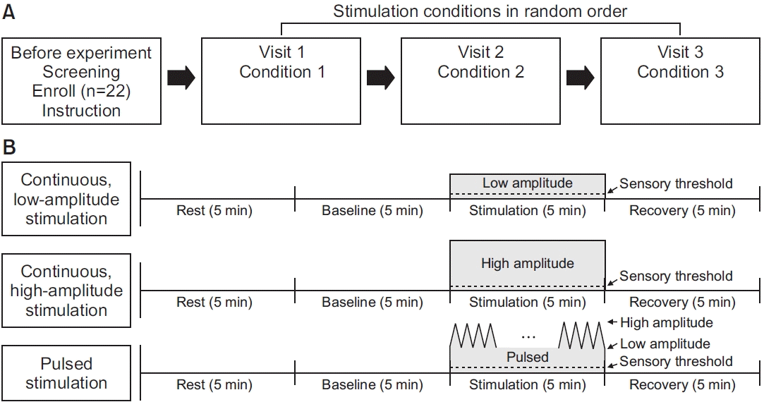
Fig. 1.
This presents schematic illustrations of the participant flow diagram (A) and the experimental design for 3 types of auricular tactile stimulation (B).
arm-22119f1.tif
Fig. 2.
The figure presents the experimental setup of a headset-type tactile stimulator using air pressure. A tactile stimulator consists of a part to generate and control air pressure and a part to provide air pressure to the external ear (A). The speaker part of the headset was replaced with the ball head and the air hose (B). The angle of the air hose could be adjusted to localize the air to the cymba concha (C).
arm-22119f2.tif
arm-22119f3.tif
Table 1.
The responses of mean heart rate, heart rate variability, and respiratory rate in (1) continuous low-amplitude, (2) continuous high-amplitude, and (3) pulsed stimulation
Parameter Mean±standard deviation
p-value (time) Post-hoc comparisons
Baseline Stimulation Recovery Baseline vs. stimulation Baseline vs. recovery Stimulation vs. recovery
Mean heart rate (bpm) 0.001* <0.001** 0.003** 0.746
Low amplitude 75.59±14.44 74.32±13.09 73.64±12.22
High amplitude 73.18±10.68 70.86±10.12 71.18±10.08
Pulsed 74.86±12.18 72.09±10.86 72.14±10.37
Heart rate variability
SDNN (ms) 0.003* 0.013** 0.004** 0.089
Low amplitude 32.53±12.27 34.80±12.41 37.37±12.98
High amplitude 36.36±15.15 40.04±17.57 41.09±16.84
Pulsed 35.21±11.76 38.26±12.48 40.11±13.74
RMSSD (ms) <0.001* 0.001** 0.001** 0.039
Low amplitude 34.32±17.79 37.10±18.65 38.70±17.15
High amplitude 36.30±19.43 42.27±25.13 43.00±25.71
Pulsed 35.48±18.32 40.55±21.83 40.35±20.59
NN50 (beats) 0.003* 0.002** 0.006** 0.313
Low amplitude 59.64±60.18 65.86±58.98 69.41±54.28
High amplitude 61.09±57.52 72.55±61.84 76.50±64.09
Pulsed 55.00±42.17 69.14±46.83 69.86±49.51
pNN50 (%) 0.002* 0.001** 0.005** 0.408
Low amplitude 18.29±20.15 20.09±19.90 20.88±18.40
High amplitude 18.84±19.51 22.63±21.26 23.89±21.81
Pulsed 16.69±16.46 21.45±18.51 21.45±18.63
LF (ms2) 0.040* 0.176 0.056 0.095
Low amplitude 384.50±434.74 460.05±435.96 693.18±707.14
High amplitude 695.73±946.03 772.50±634.28 814.91±661.82
Pulsed 486.59±527.86 641.91±513.05 823.23±809.71
HF (ms2) 0.057 0.083 0.048 0.275
Low amplitude 686.64±685.42 702.50±581.82 753.32±598.65
High amplitude 693.23±647.68 1,015.45±1,049.75 1,078.64±1,203.45
Pulsed 693.05±711.54 821.23±1136.39 840.77±951.51
Mean respiratory rate (Hz) 0.018* 0.010** 0.035 0.882
Low amplitude 0.26±0.05 0.26±0.05 0.27±0.05
High amplitude 0.26±0.04 0.27±0.04 0.26±0.04
Pulsed 0.24±0.04 0.26±0.03 0.26±0.03

SDNN, standard deviation of R-R intervals; RMSSD, root mean square of successive R-R interval differences; NN50, number of adjacent R-R intervals that differ from each other by more than 50 ms; pNN50, percentage of successive normal sinus R-R intervals of more than 50 ms; LF, low frequency; HF, high frequency.

* p<0.05;

** Bonferroni-corrected p<0.017.

TOOLS
Similar articles