Journal List > Intest Res > v.20(4) > 1516081557

Lima, Berg, and Castor: Involvement of the cannabinoid system in chronic inflammatory intestinal diseases: opportunities for new therapies

Abstract

The components of the endogenous cannabinoid system are widely expressed in the gastrointestinal tract contributing to local homeostasis. In general, cannabinoids exert inhibitory actions in the gastrointestinal tract, inducing anti-inflammatory, antiemetic, antisecretory, and antiproliferative effects. Therefore, cannabinoids are interesting pharmacological compounds for the treatment of several acute intestinal disorders, such as dysmotility, emesis, and abdominal pain. Likewise, the role of cannabinoids in the treatment of chronic intestinal diseases, such as irritable bowel syndrome and inflammatory bowel disease, is also under investigation. Patients with chronic intestinal inflammatory diseases present impaired quality of life, and mental health issues are commonly associated with long-term chronic diseases. The complex pathophysiology of these diseases contributes to difficulties in diagnosis and, therefore, in the choice of a satisfactory treatment. Thus, this article reviews the involvement of the cannabinoid system in chronic inflammatory diseases that affect the gastrointestinal tract and highlights possible therapeutic approaches related to the use of cannabinoids.

INTRODUCTION

The endocannabinoid system (ECS) is composed of the classic cannabinoid receptors 1 and 2 (CB1R and CB2R, respectively), their endogenous ligands, namely endocannabinoids, and enzymes involved in the synthesis and degradation of these molecules [1,2]. Both cannabinoid receptors (CBRs) are, in fact, part of the heptahelical G protein-coupled receptor (GPCR) family and can inhibit adenyl cyclase activity or even stimulate mitogen-activated protein kinases (MAPKs), being, therefore, classified as Gi/0 receptors [3-5]. CB1R is mainly found in the central and the peripheric nervous system (CNS and PNS, respectively) [6]. In the gastrointestinal tract, this receptor is present in the myenteric plexus and submucous plexus, endocrine cells, and enterocytes [7]. Likewise, CB2R can also be found in the CNS and PNS; however, it is also found in cardiovascular, intestinal, and reproductive systems, as well as bones and adipocytes, being largely expressed in immune cells [8].
Endocannabinoids are the endogenous ligands of the CBRs. They are fatty acid-derived molecules, synthesized by membrane phospholipids [9,10]. Calcium influx works as a synthesis trigger for these compounds in postsynaptic neurons [11], and, as they are released in the synapse, they will act as retrograde messengers, modulating the release of neurotransmitters [12,13]. Besides neurons, several other cells can also synthesize endocannabinoids, such as endothelial and mucosal cells from the intestines or even some immune cells [14-18]. Some of the most well-known and well-described endocannabinoids are N-arachidonoyl-ethanolamine (anandamide or AEA) and 2-arachidonyl-glycerol (2-AG) [19]. AEA is known to be a high-affinity CB1R agonist, with a low affinity for CB2R, being almost inactive in several models, whereas 2-AG is an agonist for both CB1R and CB2R, but with a lower affinity for the latter [20]. Both endocannabinoids will be hydrolyzed inside the cell; after reuptake, AEA is hydrolyzed by fatty acid amide hydrolase (FAAH), and 2-AG by monoacylglycerol lipase (MAGL). Both can also be oxidized by cyclooxygenase-2 (COX-2) and lipoxygenase, generating prostaglandins, ethanolamines, and several other intermediate subproducts [21,22]. Interestingly, the phytocannabinoids, too, such as cannabidiol (CBD), cannabidivarin, and cannabigerol (CBG), might activate or inhibit the CBRs or enzymes involved in the synthesis and degradation of endocannabinoids, modulating the actions of this endogenous system [23].
Activation of CBR1 and CBR2 in the CNS and actions directly in the periphery by the activation of the enteric nervous system (ENS) and resident cells control several intestinal functions, such as intestinal motility, secretion, epithelial permeability, and immunological reactions [24-26]. Both receptors may be involved in the control of gastrointestinal functions during the inflammatory process [27,28]. Thus, patients with colitis had increased levels of AEA and 2-AG that were related to controlling the local inflammation [29-31].
Therefore, several cannabinoids are being studied for the treatment of acute intestinal disorders such as dysmotility, emesis, and abdominal pain and chronic diseases such as irritable bowel syndrome and inflammatory bowel disease (IBD), which by convention is divided into 2 subtypes: Crohn’s disease (CD) and ulcerative colitis (UC) [2].
UC is characterized by inflammation in the mucosa of the large intestine from the anal orifice up to, potentially, the proximal segments. Conversely, CD is characterized by inflammation that can compromise any gastrointestinal tract region, causing ulcers, strictures, abscesses, and fistulas, but in most cases it affects the region close to the ileocecal valve [32]. The cost of IBD diagnosing, hospitalization, and treatment is usually high and treatments are not effective [33,34].
Given this context, this article aims to review the involvement of the cannabinoid system in IBD, CD, and UC and to highlight possible therapeutic approaches for these diseases with the use of cannabinoids.

METHODS

The literature review on the involvement of the cannabinoid system in IBD, with a focus on UC and CD, was carried out using the PubMed database. The descriptors used in the research were “cannabinoid chronic intestinal disease,” “cannabinoid Crohn disease,” “cannabinoid colitis,” “intestinal inflammation,” and “endocannabinoid system.” As inclusion criteria for our review, we selected publications that contained at least one of the descriptors of interest and that were in English. We used no filters for publication date and article type. Therefore, information from full texts, studies in experimental or human models, case reports, meta-analyses, and reviews were included.

ENDOCANNABINOID SYSTEM

Different subtypes of CB1R have been identified in humans and murine models. For instance, the canonical full-length CB1Ra is most commonly found in the brain and skeletal muscles, whereas CB1Rb is mainly expressed in the liver and pancreas [1,2]. Similarly, several isoforms of CB2R also display different expression sites. It was observed by Zhang et al. (2015) [3] that mouse CB2Rb is more abundant in the striatum and the spleen than CB2Ra; their expression can be induced by inflammatory events [4,5].
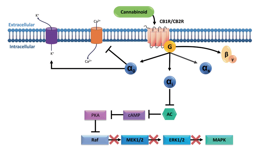
Although both CB1R (a and b) and CB2R (a and b) are members of the GPCR family, it is not unusual to find different signaling events that might be confusing at first sight. This is due to the biased agonism presented by the receptors, meaning that they can adopt multiple conformations leading to different signaling events depending on the stabilization generated by their ligands. In other words, the ligand-biased signal transduction implies that the resulting properties of the agonist or antagonist will depend on the particular signal transduction pathway that is activated (Fig. 1). Therefore, the same molecule might act as an agonist in one pathway and as an antagonist in another [6,7]. These phenomena might explain why the effects of cannabinoid molecules vary among different experimental models and different cell types.
In signaling involving the Gi/0 protein, inhibition of adenylate cyclase (AC) occurs, leading to the consequent inhibition of 3’,5’-cyclic adenosine monophosphate (cAMP) formation and inhibition of protein kinase A (PKA). The effect of PKA inhibition differs depending on cell type [8,9]. In neurons, calcium channels are blocked and potassium channels are opened, leading to hyperpolarization and inhibition of these cells [8]. In intestinal epithelial cells, it is suggested that inhibition of PKA promotes reduced phosphorylation of the cAMP response element-binding (CREB) protein, a transcription factor that regulates cell survival, and consequently a reduction in the rate of cellular apoptosis [10]. In intestinal smooth muscle cells, there is a reduction in the contractility response due to the blockage of calcium channels, which promotes a reduction in actin and myosin interaction [9,11].
Interestingly, in Zebrafish embryos, the activation of CB2R by 2-AG has been shown to inhibit leukocyte migration in response to acute injury [12]. Likewise, another study pointed out that AEA, through the activation of CB2R, promotes the suppression of the release of the inflammatory cytokines interleukin-2 (IL-2), tumor necrosis factor α (TNF-α), and interferon-γ (IFN-γ) from activated human peripheral T lymphocytes [13,14].
The phosphatidylinositol 3-kinase (PI3K) pathway is also stimulated during CB1R and CB2R activation; however, the response depends on stimulus type and microenvironment [2]. When activated, PI3K interacts with insulin receptor substrates, producing phosphatidylinositol (3,4,5)-trisphosphate (PIP3), which will regulate the location and activity of several kinases, including protein kinase B (PKB or Akt). Akt is a downstream target of PI3K and, upon phosphorylation and release into the cytoplasm, will respond to growth factors such as insulin, IL, and epidermal growth factor receptor (EGFR), modulating several cellular processes, such as migration, metabolism, and apoptosis [8,9,15].
Upon activation of the PI3K/Akt pathway, Huang et al. (2011) [15] reported an increase in p-Akt cells in the mucosal and submucosal layers of patients with UC, when compared to normal biopsy tissues. In the same study, the effects of a blocker of the PI3K/Akt pathway were analyzed, and it was found to relieve symptoms, reducing inflammation and TNF-α secretion. Similar results were found by Chen et al. (2017) [16], who described that apoptosis is dysregulated during UC progression, with proapoptotic processes in intestinal epithelial cells and antiapoptotic effect of inflammatory and immune cells.
Another interesting mechanism comprises the p38 MAPK pathway, a critical signaling pathway in bowel inflammation, modulating cellular death, growth, and differentiation [17]. In epithelial cells, MAPK is responsible for proliferative signals by EGFR [18], and p38 MAPK is also found to be increased in CD patients [19]. Furthermore, this pathway could also be involved in cytokine release [17]. However, among the MAPK family members, the extracellular signal-regulated kinase 1 and 2 (ERK1/2), p38 kinase, and Jun N-terminal kinase (JNK) are of the utmost importance in the promotion of apoptotic events, playing an important role in UC pathogenesis; therefore, the inhibition of this pathway led to a reduction in proinflammatory cytokines and cellular infiltrate [20].
In immune cells, the MAPK pathways are involved in the cells’ development, apoptosis, migration, and response against pathogenic stimuli. In microglial cells, CB2R activation through anandamide, an endocannabinoid, has been shown to induce the activation of ERK1/2 and JNK MAPK; this activation resulted in a slight increase in IL-10 levels but also in the inhibition of proinflammatory ILs [21].
The endocannabinoids are synthesized from membrane lipids in response to specific stimuli, such as neuronal activation or cellular stress, which will promote the influx of calcium into the cell, generating its depolarization [22]. There are at least 15 endogenous compounds that can target CBRs either orthosterically or allosterically. Several of them have been found to bind to cannabinoid CB1Rs in vitro at concentrations in the low or mid nanomolar range [23]. These compounds are anandamide, 2-arachidonoylglycerol, dihomo-γ-linolenylethanolamide, 2-arachidonyl glyceryl ether (noladin ether), docosatetraenoylethanolamide, N-arachidonoyl dopamine, docosahexaenoylethanolamide, and eicosapentaenoyl ethanolamide; the first 3 compounds have also been reported to display a significant affinity for CB2R [23].
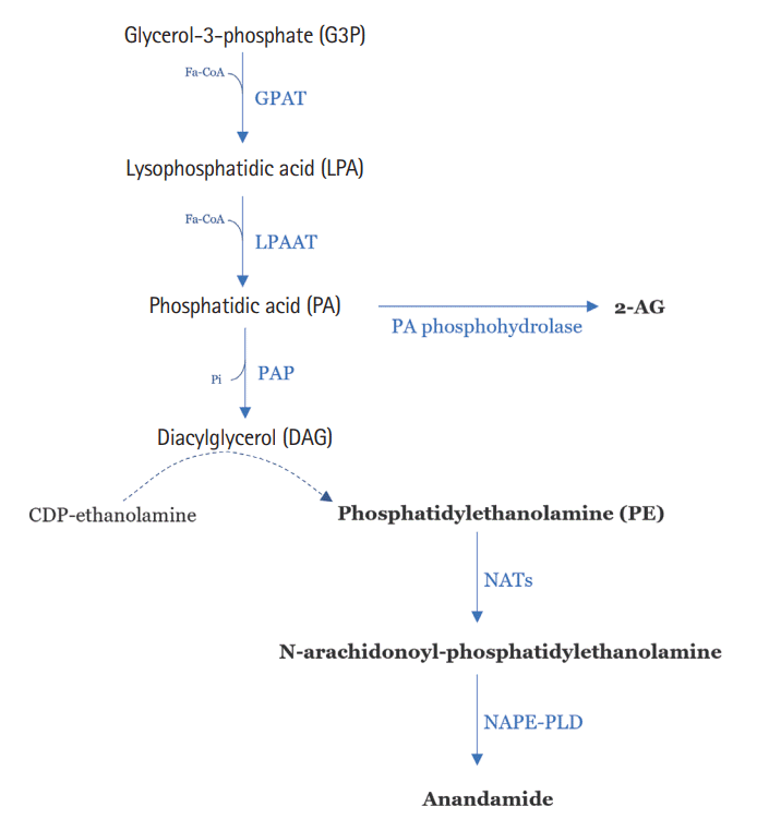
N-arachidonoyl-ethanolamine (anandamide or AEA) and 2-AG were the first ones to be discovered and are the most studied until now. The biosynthetic precursors of AEA, the N-arachidonoyl-phosphatidylethanolamines, are produced via N-acyl-transferases (NATs) by N-acyl-phosphatidylethanolamine-selective phospholipase D (NAPE-PLD) or by the sequential action of several enzymes that can also be involved in the synthesis of 2-AG, as reported in Fig. 2 [24]. Both these endocannabinoids will be hydrolyzed inside the cell; after reuptake, AEA is hydrolyzed by FAAH, and 2-AG by MAGL. Both can also be oxidized by COX-2 and lipoxygenase, generating prostaglandins, ethanolamines, and several other intermediate subproducts [25,26]. These lipidic molecules are derived from the acylation of glycerol-3-phosphate (G3P); this molecule will lead to phosphatidic acid (PA) and phosphatidylethanolamine (PE) (Fig. 2) [24,27].

ECS IN THE GASTROINTESTINAL TRACT

The actions of the ECS in the gastrointestinal tract are mainly controlled by the activation of CBRs in the CNS or the periphery, through the ENS or other tissues of the gastrointestinal tract, as shown below [28].
Both CB1R and CB2R, their endogenous agonists, and related enzymes have been detected in the gastrointestinal tract [29]. CB1R and CB2R are widely expressed in intestinal tissue and its innervations [29,30]. CB1R expression is seen in the ENS, including the submucosal plexus and myenteric plexus, and in epithelial and enteroendocrine cells. CB2Rs are expressed in the ENS, also covering the submucosal plexus and myenteric plexus, in epithelial cells in the presence of inflammation, and in immune cells [29].
In the CNS, the regulation of appetite, energy balance, and food intake can be performed by CB1Rs expressed in the lateral hypothalamus, arcuate nucleus, and paraventricular nucleus of the hypothalamus [2,31-33]. Thus, the activation of the ECS in these sites promotes increased appetite, stimulation of lipogenesis, and an increase in energy resources [34]. The control of food intake by the ECS occurs in conjunction with the action of hypothalamic hormones, such as corticotropin-releasing hormone (CRH) in the paraventricular nucleus, the cocaine-amphetamine-regulated transcribed hormone (CART) in the arcuate nucleus, and the melanin-concentrating hormone (MCH) and orexin-A in the lateral hypothalamus, which are well-known modulators of food intake [39]. Hormones produced in the periphery, such as ghrelin, leptin, insulin, and adiponectin, participate in appetite regulation along with the ECS and hypothalamic hormones [32,34]. Studies carried out in animal models have shown that the orexigenic effects are obtained from the stimulation of the ECS in the CNS and the neurons that release MCH, in addition to the increase in neuropeptide Y (NPY) and the hormones orexin-A and ghrelin [40-43].
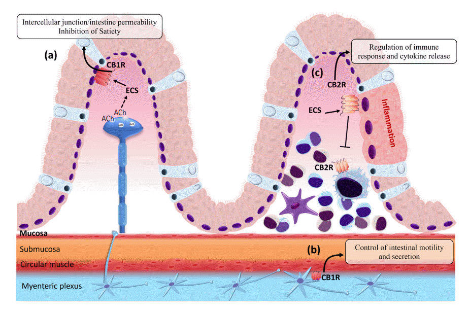
Thus, appetite control also takes place in the small intestine, where during the fasting period or in the presence of taste of dietary fats, endocannabinoid signaling is triggered by the activation of CB1Rs, aiming to inhibit satiety [44,45]. Inhibition of muscarinic receptor type 3 (M3) blocks the induced production of 2-AG in the jejunum. This can be replicated by peripheric administration of CB1R antagonists, confirming that cholinergic signaling, possibly through the vagus nerve, activates the muscarinic receptor of acetylcholine (mAChR) in the jejunum, stimulating the conversion of 1-stearoyl-2-arachidonoyl-sn-glycerol (SAG) into 2-AG by the enzyme diacylglycerol lipase (DAGL), stimulating food intake (Fig. 3, part a) [45,46]. Besides these mechanisms for controlling appetite and food intake, there are also CB1Rs expressed in the enteroendocrine cells of the small intestine, which, when activated, promote hunger during the fasting period and boost the intake of high-fat foods, inhibiting the release of cholecystokinin (CCK) that would normally bind to CCK receptors inducing satiety after a meal [44,45]. The control of intestinal motility, secretion, epithelial permeability, and immunology are also accomplished by the modulation of CB1R and CB2R expressed in the ENS and resident cells [47,48].
The activation of CB1R in the ENS is mainly related to the negative control of cholinergic neurotransmission, with consequent inhibition of the action of this neurotransmitter on muscarinic receptors and a reduction in intestinal smooth muscle contractions (Fig. 3, part b) [49-51]. Activation of CB2R by the agonist JWH-133 in rats promotes a reduction in the speed of food passage through the gastrointestinal tract, as a consequence of the decrease in muscle contractions [52]. Thus, the activation of this receptor in the ENS, as well as the activation of CB1R, promotes the inhibition of the contractility of the intestinal muscles, inhibiting the release of ACh [53].
Activation of CB1R and CB2R promotes the inhibition of proinflammatory cytokine secretion (Fig. 3, part c), suppression of macrophage and mast cell activation, modulation of TH1 and TH2 lymphocytes through induction of apoptosis, and inhibition of proliferation and consequently reduced inflammation [56-59]. Di Marzo and Izzo (2006) [60] discussed that the ECS is overstimulated in the intestines and colon during inflammatory disorders. Enhanced production of endocannabinoids is part of the gastroprotective mechanism and several other regulatory functions–including motility and secretion–that might function through CB1R [29,61]. CB2R is either absent or only weakly expressed in the human intestinal epithelium; however, in IBD it becomes evident in the luminal membranes at ulcerative margins, and immune cells in the lamina propria (LP) might also express CB2R [29,62]. Therefore, differently from CB1R, CB2R appears to be less expressed during homeostasis, but it is inducible during inflammatory disorders [21].
There is evidence that CB1R and CB2R are also related to the control of sensations such as visceral pain, emesis, and nausea, which are sensory experiences that may be linked to the functioning of the gastrointestinal tract [29]. Sanson et al. [63] demonstrated that CB1R and CB2R agonists attenuated the basal sensitivity and that this was increased by colon inflammation in rats. Moreover, the intestinal microbiota is known to contribute to several improvements in digestive health. It was demonstrated that the probiotic agent Lactobacillus acidophilus promoted an increase in the expression of CB2R in the epithelium of rats and mice and a consequent reduction in visceral sensitivity in cases of irritation and inflammation [64]. A possible mechanism for pain regulation from CB2R would be the inhibition of the release of inflammatory mediators from the epithelium that would activate the primary afferent nerves and cause visceral pain [65]. Furthermore, cannabinoids are effective antiemetics and have been used in the clinic for years; however, they are not the first-line therapeutic agents [66]. Some evidence also demonstrates the involvement of CB1R in the brainstem [67,68] and that of CB2R in the dorsal vagal complex for the regulation of emesis [69]. Additionally, in animal models, activation of the ECS in the insular cortex has been found to regulate nausea, and when endocannabinoid levels were increased by administering an FAAH or MAGL inhibitor, nausea was reduced. However, it is suggested that only MAGL inhibition is enough to generate the effect against nausea by the activation of CB1R by 2-AG, and AEA seems not to be involved in this event [70-72].
Therefore, the activation of CB1R and CB2R promotes a reduction in colonic motility; control of the secretion of neurotransmitters such as acetylcholine and hormones such as ghrelin, leptin, adiponectin, and insulin; control of the permeability of the intestinal epithelium; and regulation of visceral pain, emesis, and nausea, besides anti-inflammatory and antiproliferative effects [73,74]. Considering the various functions performed by the ECS in the gastrointestinal tract, changes in its tone can lead to several effects, which can be explored for the development of drugs for the treatment of various diseases involving disorders of the gastrointestinal tract.

ECS IMPACT IN CHRONIC IBDs

IBD is a destructive inflammatory disorder and comprises both UC and CD. Therefore, UC is an intestinal inflammatory disorder of the colon, characterized by the infiltration of immune cells and the release of inflammatory mediators that may lead to ulcers and disruptions in the mucosa [75,76]. Likewise, CD is a chronic inflammatory condition of the gastrointestinal tract, resulting from a complex interplay of factors, such as genetic susceptibility, gut microbiota, and environmental pressures, leading to dysregulations in the patient’s immune response [77]. However, UC is a confluent mucosal inflammation, being initiated at the anal verge and spreading proximally for a variable length. CD can affect any region of the gastrointestinal tract, resulting in ulcers with transmural inflammation leading to fibrosis, fistula, and strictures, usually close to the ileocecal valve. Additionally, the dispersed inflammation in CD can leave areas of normal mucosa in the midst of inflammation, called “skip areas.” [9]
IBD is a chronic disease that can reduce the life quality of patients and is correlated with extraintestinal comorbidities, such as cardiovascular diseases, osteoporosis, arthritis, and lymphomas [9,78-80]. The symptoms of IBD can be further exacerbated by stress and psychiatric diseases, such as depression and anxiety [81].
Corticosteroids and immunomodulatory drugs, which are the basis for the treatment of IBD, do not always provide satisfactory outcomes [75,82,83]. As demonstrated, the ECS is largely expressed in the gastrointestinal tract; therefore, the manipulation of this system becomes an interesting approach to be investigated.

CHANGES IN THE ECS IN UC

The expression and activation of CBR in experimental models of intestinal inflammation have been investigated by several researchers. Engel et al. (2010) [84] found that CB1R (-/-), CB2R (-/-), and CB1R/CB2R (-/-) knockout C57BL/6 mice were more susceptible to the development of UC induced by 2,4,6-trinitrobenzenesulfonic acid (TNBS). Storr et al. (2009) [85] also evaluated the participation of the ECS in an experimental model of TNBS-induced colitis in wild C57BL/6 mice and CB2R (-/-) knockout mice, and these mice were treated with the agonists JWH-133 and AM-1241 and the antagonist AM630, selective for CB2R. CB2R (-/-) knockout mice had increased inflammation and did not respond to agonist or antagonist treatments. In wild-type (WT) mice, treatment with the agonists JWH-133 and AM-1241 was found to reduce intestinal inflammation. In contrast, the use of the AM-630 antagonist in WT mice exacerbated these parameters, demonstrating an increase in inflammation. Furthermore, an increase in CB2R mRNA expression was found during inflammation in WT mice. Another study with CB2R (-/-) knockout C57BL/6 mice demonstrated that these animals have worsened colitis induced by dextran sulfate sodium (DSS) compared to WT mice [86].
Furthermore, changes in endocannabinoid metabolizing enzymes have been reported in female C57BL/6N mice, genetically modified to express less FAAH. It was shown that this enzyme deficiency generates protection against dinitrobenzene sulfonic acid (DNBS)-induced UC, as revealed by a reduction in macroscopic and microscopic damage and in myeloperoxidase (MPO) enzyme activity [61]. Storr et al. (2008) [87] demonstrated that FAAH mRNA is increased on the 3rd day after colitis induction with TNBS in mice. Recall that FAAH is the main enzyme responsible for AEA hydrolysis, so an increase in this enzyme might reduce the endocannabinoid concentration during the inflammatory process. Likewise, Shamran et al. (2017) [88] showed that FAAH inhibition ameliorates the clinical score and reduces the frequencies of activated CD4+ T cells in the colon and proximal lymphoid tissues. Furthermore, Izzo and Sharkey (2010) [89] have demonstrated that mice without or with reduced FAAH have elevated basal levels of AEA and are more resistant to TNBS-induced colitis than wild mice. Borrelli et al. (2009) [90] used male Institute of Cancer Research (ICR) mice strain and found that during DNBS-induced inflammation, the concentrations of AEA and 2-AG increased, and the mRNA expression of the FAAH enzyme in the colon decreased, revealing the physiological function of the ECS. Likewise, Tourteau et al. (2014) [91] used substances derived from 3-carboxamide-5-arylisoxazole CB2R agonists and FAAH inhibitors in the treatment of DSS-induced colitis in mice and showed significant improvement in inflammation. In another study, the use of an FAAH inhibitor, PF 3845, reduced TNBS-induced experimental UC due to increased endocannabinoid expression and activity [92]. Furthermore, a study using ARN2508, an inhibitor of FAAH, demonstrated an improvement in inflammation, a reduction in the mortality rate, and a decline in weight loss in mice with TNBS-and DSS-induced colitis. In this case, as mentioned above, the anti-inflammatory effect obtained by inhibiting FAAH is probably related to an increase in endocannabinoids such as AEA [93].
Regarding MAGL activity, in C57BL/6N mice with DSS-induced colitis, real-time polymerase chain reaction (RT-PCR) analysis demonstrated that the expression of genes encoding this enzyme was reduced during colon inflammation [94]. In addition, in mice with TNBS-induced colitis, inhibition of the MAGL enzyme by the selective inhibitor JZL184 promoted an increase in 2-AG, which was associated with a reduction in macroscopic and histological damage of the colon, as well as a decrease in the expression of inflammatory cytokines [95].
In humans, Western blot analysis and immunocytochemistry in colon tissue demonstrated an increased expression of CB2R, the enzyme DAGL, and MAGL in the colonic epithelium, mainly in patients with mild and moderate colitis. In contrast, in patients with inactive colitis, the expression of CB1R and CB2R and the DAGL enzyme was reduced, but the expression of the NAPE-PLD enzyme was increased, especially in patients treated with corticosteroids [96]. Wright et al. (2005) [62] also found an increased expression of CB2R in the human colonic epithelium during UC, demonstrating an immunomodulatory function of the ECS.
Besides the role of enzymes in the physiopathology of IBD, the endocannabinoid AEA appears to be the main modulator of colon-induced inflammation. AEA was found to be increased in colon biopsy from patients with untreated UC, as analyzed by liquid chromatography with detection by mass spectrometry [97]. Harvey et al. (2013) [98] showed that the incubation of human colon tissues with the inflammatory cytokines TNF-α and IL-1B causes damage like UC. Administration of AEA and a selective CB2R agonist, JWH-015, showed a significant improvement in inflammation, as evidenced by a reduction in mucosal crypt loss, luminal epithelial damage, and lymphocyte infiltration in the LP. These effects were partially reversed by the administration of a CB2R inverse agonist, JTE-907, which demonstrates that the anti-inflammatory effects of JWH-015 and AEA are dependent on the activation of this receptor [99].
Grill et al. (2019) [100] evaluated the regulation of the ECS in 17 patients diagnosed with UC and found an increase in AEA expression and a decrease in mRNA expression of the NAPE-PLD enzyme, which seems contradictory, considering that NAPE-PLD participates in the synthesis of this endocannabinoid. However, to explain this fact, the authors suggested that there is an allosteric feedback inhibition of NAPE-PLD by AEA or a reduced degradation of the endocannabinoid lipids. Furthermore, other enzymes besides NAPE-PLD may be responsible for the synthesis of AEA. In another study by Suárez et al. (2012) [101], human biopsy from healthy patients with untreated active colitis and from patients treated with 5-aminosalicylic acid (5-ASA), glucocorticoids, and/or immunomodulators until disease remission were analyzed using RT-PCR and immunohistochemistry to assess the expression and distribution of FAAH and NAPE-PLD in the intestine. RT-PCR analysis of the mucosa from untreated UC individuals revealed an increase in the gene expression of FAAH and no alterations in the gene expression of NAPE-PLD. Also, with immunohistochemical analysis of the colon epithelium from untreated UC patients, the authors observed a decrease in NAPE-PLD and the conservation of FAAH levels, as compared to healthy individuals. However, after treatment with 5-ASA and glucocorticoids, an increase was found in the expression of NAPE-PLD in the epithelium, indicating a role of this enzyme in UC remission [101].
Besides the above-described studies, there are several studies showing the relevancy of the ECS in the modulation of inflammation during UC by increasing the expression of receptors, by increasing the synthesis of endocannabinoids, or by inhibiting or activating the enzymes involved in the degradation or synthesis of endocannabinoids. In summary, changes in the expression of the ECS components during UC can generate anti-inflammatory effects in animal and human models, as described in Table 1.

CHANGES IN THE ECS IN CD

As in UC as discussed above, it has been suggested that in CD the ECS may act to reduce the local inflammatory response. Therefore, it has been the topic of several studies to test and evaluate changes in the expression of CBR and the synthesis and degradation of endocannabinoids during this intestinal inflammation. The single nucleotide polymorphism 1359 G/A of the CB1R gene has been shown to influence the CD phenotype. Individuals who were homozygous for the G allele showed a higher probability of developing CD before the age of 40 years, demonstrating that CB1R influences the manifestation of CD [103]. The expression of CB2R and AEA was evaluated in ileum tissue of mice and in CD4+ T cells of an experimental model of chronic ileitis. The results demonstrated an increased expression of CB2R and AEA in mice with inflammation compared to control mice. Moreover, an 11-fold increase in CB2R mRNA expression was found in regulatory T cells, demonstrating the important role of the ECS in the regulation of inflammatory response [104].
The expression of CB2R was also analyzed in histological sections of biopsy samples of the intestine of patients with CD. There was no difference in the expression of this receptor between CD patients and healthy individuals. However, the expression of CB1R was shown to be significantly increased in patients with CD [105]. Moreover, Grill et al. (2019) [100] found that the concentrations of AEA and 2-AG were markedly increased in the plasma of CD patients. Furthermore, in the intestinal mucosa, an increased gene expression of synthesizing (NAPE-PLD and DAGL) and metabolizing (FAAH and MAGL) enzymes and a decreased transcription of CB1R were identified [106].
On the other hand, Di Sabatino et al. (2011) [59] found decreased levels of AEA in the inflamed mucosa of patients with IBD compared to non-inflamed tissue, which was concomitant with lower activity of the NAPE-PLD enzyme that synthesizes this endocannabinoid and higher activity of the FAAH enzyme that degrades this ligand. Some divergences were found between these studies, and those may be related to the different tissues evaluated and to the change in the expression of the ECS according to the stage of the disease [107].
Therefore, the physiological or induced changes in the ECS during CD that can generate an anti-inflammatory effect in animal models and humans are summarized in Table 2.

CANNABINOIDS AS POSSIBLE DRUGS FOR THE TREATMENT OF CHRONIC IBDs

Given the various disadvantages and limitations of the treatments available for IBD, it is relevant to develop new pharmacological alternatives for the treatment of UC and CD. In this context, cannabinoids can be a good pharmacological strategy for the treatment of IBD because of the various functions of the ECS in maintaining intestinal homeostasis and the anti-inflammatory effects observed by the positive regulation of this system in animal and human models.
Phytocannabinoids such as CBD, Δ9-tetrahydrocannabinol (THC), CBG, cannabinol (CBN), and cannabichromene (CBC), all isolated from Cannabis sativa, popularly known as marijuana, were evaluated as possible drugs for the treatment of UC and CD. However, when using some of these substances, with the clear exception of CBD, it is necessary to pay attention to the possibility of dose-dependent psychoactive effects [119].
CBD administration was evaluated in rats and mice with intestinal inflammation induced by irritating substances, such as croton oil (CO), lipopolysaccharide (LPS), and DNBS. It was observed that CBD reduced edema, hyperemia, adhesions in the small intestine, inflammatory cell infiltration, colon weight/length ratio, and intestinal hypermotility [90,108-110]. Additionally, there was a decrease in inflammatory cytokines, such as IL-1β, and an increase in anti-inflammatory cytokines, such as IL-10 and IL-6 [90,109]. Other studies have found decreased MPO activity, reduced induced nitric oxide synthase (iNOS) expression, and reduced nitric oxide (NO) synthesis and its stable metabolites [90,110,111]. Therefore, there is evidence that the use of CBD improves intestinal inflammation in experimental models. Similarly, the administration of CBD-rich C. sativa extract in mice promoted a reduction in intestinal motility, colon weight/length ratio, and MPO activity, indicating an improvement in inflammation [113].
However, Naftali et al. (2017) [112] evaluated the administration of CBD in humans with CD and found no significant improvement in intestinal inflammation. Therefore, there is a need for more studies involving CBD in humans with CD. In contrast, the administration of CBD-rich C. sativa extract in humans with UC demonstrated greater remission of the disease, better patients’ quality of life, and a reduction in the total Mayo score, which is used to assess the UC stage by scoring the pattern and the presence of bleeding in the stools, endoscopic results, and medical evaluation [114].
THC is a cannabinoid present at high concentrations in C. sativa; it has psychoactive activity and is therefore of limited use [119]. Conversely, in some studies, the use of this phytocannabinoid presented beneficial effects on intestinal inflammation when administered alone or in conjunction with other cannabinoids.
Jamontt et al. (2010) [115] demonstrated that the administration of THC alone or in combination with CBD resulted in decreased weight gain and macroscopic damage in the intestines of rats, in addition to reducing MPO activities and improving the contractile response of the intestinal muscles. In humans with CD, the administration of THC promoted an increase in the patients’ quality of life, greater remission of the disease, and improved appetite and sleep [116]. Regarding the simultaneous use of CBD and THC, it is worth mentioning that some studies report that the negative allosteric modulation of CBD in CB1R might reduce the adverse effects of THC [128].
Another phytocannabinoid with potential application in the treatment of IBD is CBN, a product of THC oxidation, found in large quantities in C. sativa [129]. In a model of intestinal inflammation using CO, it was found that CBN inhibited the increase in intestinal motility and contributed to an anti-inflammatory effect [117]. Nonetheless, further studies are required to better understand and elucidate the applications of CBN.
Besides these compounds, another potential drug for the treatment of IBD is CBG, which, like CBD, does not display psychotropic effects [119,130,131]. Borrelli et al. (2013) [118] analyzed the effect of CBG administration in male ICR mice, in peritoneal macrophages, and in epithelial cells of the PTK6 knockout colon of these animals. In mice that received this drug, it was found that there was a reduction in the weight/length ratio of the colon, damage to the colon, intestinal permeability, MPO activity, iNOS expression, and levels of IL-1β and IFN-γ. The authors also described an increased superoxide dismutase (SOD) activity and IL-10 levels. Finally, Ki-67 cells were detected in the lower half of the mucosa, thus indicating their proliferation. In mice untreated with CBG, Ki-67 was detected mainly in inflammatory cells. In the peritoneal macrophages, the synthesis of nitrites was decreased, and the levels of iNOS were reduced. In the epithelial cells of the PTK6 knockout colon, the synthesis of reactive oxygen species (ROS) was reduced. Therefore, the authors suggested an anti-inflammatory effect of CBG in the colon.
Likewise, another phytocannabinoid, CBC, has shown a protective effect against IBD. In mice with CO-induced inflammation, it was found that the administration of CBC promoted a decrease in intestinal motility and inhibited ileum contractions induced by ACh and electrical field stimulation (EFS) [99]. In another study, in mice with DNBS-induced inflammation, the administration of CBC led to a reduction in tissue damage, the colon weight/length ratio, intestinal permeability, and MPO activity. Furthermore, CBC reduced the effect of DNBS on cell proliferation, with mitotic activity being restricted to half of the mucosa, as identified by Ki-67 immunoreactivity [106].
In Table 3, we summarize the studies that investigated the applications of phytocannabinoids as potential treatments for intestinal inflammatory disorders.
Synthetic cannabinoids, such as the non-selective CBR agonists AM-841, HU-210, and WIN55,212-2, the CB1R agonists arachidonyl-2’-chloroethylamide (ACEA) and CP55,940, and the CB2R agonists AM-1241, β-caryophyllene, compound 26, HU-308, and JHW-133, are analogs of endocannabinoids and phytocannabinoids or compounds that have been synthesized to mimic the action of natural cannabinoids [134].
Studies in mice with induced intestinal inflammation by irritating substances, such as DSS, DNBS, and TNBS, in which the mice were treated with non-selective CBR agonists (AM-841, HU-210, and WIN55 212-2), mainly showed that the administration of these drugs generated protection against intestinal inflammation, as evidenced by decreased macroscopic and microscopic damage, inflammatory infiltrate in the intestine, and MPO activity [61,120-123].
Similarly, the administration of synthetic CB1R agonists (ACEA and CP55,940) in mice with induced intestinal inflammation has shown that these substances have anti-inflammatory and antioxidant actions. For instance, treatment with ACEA led to weight gain, shortening of the colon, and reduced macroscopic damage, diarrhea, intestinal hypermotility, MPO activity, inflammatory infiltrate, and nitrite production, demonstrating the protective effect of this drug [102,106,124]. The use of CP55,940 demonstrated a reduction in intestinal motility and inflammation [117].
CB2R agonists (AM-1241, β-caryophyllene, compound 26, HU-308, JHW-133) were also evaluated in experimental models of induced inflammation. Macroscopic damage, such as ulcers and adhesions, and microscopic damage, such as thickening of the intestinal wall, shortening of the colon, inflammatory infiltrate, the expression of inflammatory mediators, and the activities of MPO and N-acetylglucosaminidase (NAG), were reduced with the administration of these drugs in mice with induced intestinal inflammation [85,86,125,126].
Furthermore, Bento et al. (2011) [125] observed that the administration of β-caryophyllene promoted a loss in the signaling pathways involving NF-κβ, CREB, ERK1/2, and IKK (as induced by MAPK and transcription factors) and an inhibition of apoptosis and proliferation in colon tissue (as demonstrated by a reduction in the levels of caspase-3 and Ki-67). Another study showed that the administration of HU-308 reduced Nod-like receptor Pyrin Domain Containing 3 (NLRP3), the ratio casp-1 p20/casp-1 p45, and the expression of proIL-1β and IL-1β mRNA (of SQSTM1 in the colon) and increased the levels of LC3-II, LC3-I, and beclin-1, which are related to increased autophagy [86].
Among the selective CB2R agonists, the synthetic drug JHW-133 is one of the most studied in the literature. The administration of this drug in mice and rats with induced intestinal inflammation resulted in a reduction in intestinal motility, weight gain, shortening of the colon, macroscopic damage, diarrhea, MPO activity, inflammatory infiltrate, bodyweight loss, and nitrite levels, leading to an improvement in the inflammatory condition of the animals [52,85,102,106,127].
Furthermore, some authors found that in CB2R-deficient mice or WT mice treated with an antagonist of this receptor, the anti-inflammatory effects promoted by AM-1241, β-caryophyllene, and JHW-133 were not observed, confirming the action of these substances through the activation of CB2R [85,125].
Table 4 shows the description of the animals, the substances used to induce intestinal inflammation, and the results obtained in each of the studies after the administration of CBR agonists (selective and non-selective).

ENZYME INHIBITORS AND CELL REUPTAKE INHIBITORS OF ENDOCANNABINOIDS, AS POTENTIAL DRUGS FOR THE TREATMENT OF CHRONIC IBDs

Several studies have shown that during intestinal inflammation, CBR is overexpressed, endocannabinoid synthesis is increased, and the activity of the metabolizing enzymes of these ligands is reduced to restore the balance in the body [84,86,92,93]. Thus, substances that inhibit the degradation of the main endocannabinoids—AEA and 2-AG by the FAAH and MAGL enzymes, respectively—and compounds capable of inhibiting the reuptake of these endocannabinoids were seen as potential drugs for the treatment of IBD.
The administration of the AEA enzyme hydrolysis inhibitor, arachidonoyl serotonin (AA-5-HT), in mice with DNBS-induced colitis promoted a reduction in macroscopic and histological scores and MPO activity [97]. The use of VDM-11, an AEA reuptake inhibitor, mainly promoted a decrease in macroscopic and microscopic damage, histological score, and MPO activity [87,97].
Moreover, the use of FAAH inhibitors (URB-597, compound 39, and PF-3845) in mice with induced intestinal inflammation demonstrated, above all, a reduction in macroscopic and microscopic damage, histological scores, bodyweight loss, MPO activity, inflammatory cytokines, and mortality rate and even normalized the architecture of intestinal tissues [87,92,132,133]. Furthermore, the administration of ARN-2508, an inhibitor of FAAH, COX-1, and COX-2, demonstrated that the bodyweight loss, mortality rate, and intestinal inflammation of the mice were reduced [93].
In another perspective, JZL-184, a selective MAGL inhibitor, when administered to mice with TNBS-induced intestinal inflammation, reduced macroscopic lesions, decreased submucosal edema, normalized mucosal structure, and reduced leukocyte infiltrates, the levels of the inflammatory cytokines MCP-1 and MIP-1, and the plasma levels of LPS [95].
Thus, several studies in experimental models have shown that the use of endocannabinoid-enzyme inhibitors and endocannabinoid reuptake inhibitors is capable of promoting improvement in the intestinal inflammatory profile, being potential drugs for the treatment of IBD (Table 5).

CONCLUSIONS

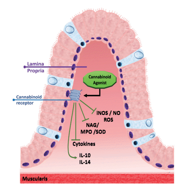
As summarized in this review, during UC and CD, the expression of CB1R and CB2R and the synthesis of endocannabinoids are increased, and the activity of cannabinoid metabolizing enzymes is decreased. Thus, potential therapeutic approaches for UC and CD have been identified with the use of CBR agonists, which include endocannabinoids, phytocannabinoids, synthetic cannabinoids, inhibitors of endocannabinoid metabolizing enzymes, and AEA reuptake inhibitors. These compounds, when evaluated in experimental models, demonstrated antiproliferative, antinociceptive, antioxidant, and anti-inflammatory actions, by regulating the levels of inflammatory cytokines, iNOS, NO, ROS, and the activities of the enzymes MPO, SOD, and NAG. In addition, they lead to an increase in the expression of anti-inflammatory cytokines, such as IL-10 and IL-4 (Fig. 4).
Studies in humans are scarce and, when performed, cover a limited sample, making it difficult to assess the efficacy of cannabinoids used in the treatment of UC and CD. Nevertheless, CBR agonists, inhibitors of the reuptake of AEA and 2-AG, and inhibitors of the enzymes that degrade endocannabinoids are promising drugs in the management of these diseases. However, comparative research on these substances is lacking, although such research is necessary to better evaluate the best therapeutic options for long-term use in the treatment of IBD.
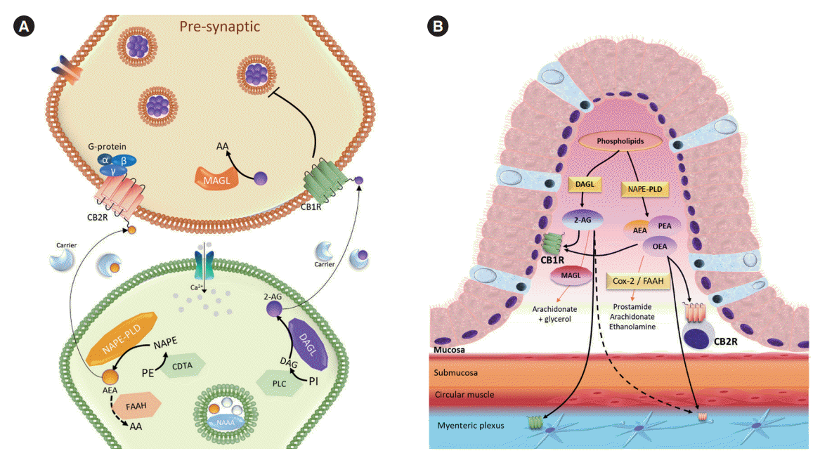
Given the importance of the cannabinoid system in gastro-intestinal tract homeostasis (Fig. 5) future clinical studies are needed to better evaluate the risks and benefits of prolonged treatment using substances that interact with this system, the possible drug interactions, and the adverse effects of using cannabinoids, to guarantee the effectiveness of treatment and patient safety.

Notes

Funding Source

The work was supported by grants from Coordenação de Aperfeiçoamento de Pessoal do Ensino Superior (CAPES): Conselho Nacional de Desenvolvimento Cientifico e Tecnológico (CNPQ), Fundação de Amparo à Pesquisa do Estado de Minas Gerais (FAPEMIG), Pró-Reitoria de Pesquisa da UFMG (PRPq) and Specialization course in Pharmacology of Department of Pharmacology, ICB-UFMG.

Conflict of Interest

No potential conflict of interest relevant to this article was reported.

Data Availability Statement

Not applicable.

Author Contribution

Conceptualization: Lima PA, Castor MGM. Data curation: Lima PA. Formal analysis: Castor MGM. Funding acquisition: Castor MGM. Investigation: Lima PA. Methodology: Lima PA, Berg BB, Castor MGM. Supervision: Castor MGM. Validation: Berg BB. Writing-original draft: Lima PA, Berg BB, Castor MGM. Writing-review & editing: Lima PA, Berg BB, Castor MGM. Approval of final manuscript: all authors.

REFERENCES

1. González-Mariscal I, Krzysik-Walker SM, Doyle ME, et al. Human CB1 receptor isoforms, present in hepatocytes and β-cells, are involved in regulating metabolism. Sci Rep. 2016; 6:33302.
crossref
2. Zou S, Kumar U. Cannabinoid receptors and the endocannabinoid system: signaling and function in the central nervous system. Int J Mol Sci. 2018; 19:833.
crossref
3. Zhang HY, Bi GH, Li X, et al. Species differences in cannabinoid receptor 2 and receptor responses to cocaine self-administration in mice and rats. Neuropsychopharmacology. 2015; 40:1037–1051.
crossref
4. Liu QR, Pan CH, Hishimoto A, et al. Species differences in cannabinoid receptor 2 (CNR2 gene): identification of novel human and rodent CB2 isoforms, differential tissue expression and regulation by cannabinoid receptor ligands. Genes Brain Behav. 2009; 8:519–530.
crossref
5. Benito C, Tolón RM, Pazos MR, Núñez E, Castillo AI, Romero J. Cannabinoid CB2 receptors in human brain inflammation. Br J Pharmacol. 2008; 153:277–285.
6. Kenakin T. Functional selectivity through protean and biased agonism: who steers the ship? Mol Pharmacol. 2007; 72:1393–1401.
crossref
7. Turu G, Hunyady L. Signal transduction of the CB1 cannabinoid receptor. J Mol Endocrinol. 2010; 44:75–85.
crossref
8. Pertwee RG. The pharmacology of cannabinoid receptors and their ligands: an overview. Int J Obes (Lond). 2006; 30 Suppl 1:S13–S18.
crossref
9. Brunton LL, Hilal-Dandan R, Knollmann BC. As Bases Farmacológicas da Terapêutica de Goodman and Gilman. 13rd ed. Porto Alegre: Artmed Editora;2018.
10. Blackwood BP, Wood DR, Yuan C, et al. A role for cAMP and protein kinase A in experimental necrotizing enterocolitis. Am J Pathol. 2017; 187:401–417.
crossref
11. Börner C, Smida M, Höllt V, Schraven B, Kraus J. Cannabinoid receptor type 1- and 2-mediated increase in cyclic AMP inhibits T cell receptor-triggered signaling. J Biol Chem. 2009; 284:35450–35460.
crossref
12. Liu YJ, Fan HB, Jin Y, et al. Cannabinoid receptor 2 suppresses leukocyte inflammatory migration by modulating the JNK/c-Jun/Alox5 pathway. J Biol Chem. 2013; 288:13551–13562.
crossref
13. Cencioni MT, Chiurchiù V, Catanzaro G, et al. Anandamide suppresses proliferation and cytokine release from primary human T-lymphocytes mainly via CB2 receptors. PLoS One. 2010; 5:e8688.
crossref
14. Maccarrone M, Bab I, Bíró T, et al. Endocannabinoid signaling at the periphery: 50 years after THC. Trends Pharmacol Sci. 2015; 36:277–296.
crossref
15. Huang XL, Xu J, Zhang XH, et al. PI3K/Akt signaling pathway is involved in the pathogenesis of ulcerative colitis. Inflamm Res. 2011; 60:727–734.
crossref
16. Chen Q, Duan X, Fan H, et al. Oxymatrine protects against DSS-induced colitis via inhibiting the PI3K/AKT signaling pathway. Int Immunopharmacol. 2017; 53:149–157.
crossref
17. Zhang D, Wang L, Yan L, et al. Vacuolar protein sorting 4B regulates apoptosis of intestinal epithelial cells via p38 MAPK in Crohn’s disease. Exp Mol Pathol. 2015; 98:55–64.
crossref
18. Miguel JC, Maxwell AA, Hsieh JJ, et al. Epidermal growth factor suppresses intestinal epithelial cell shedding through a MAPK-dependent pathway. J Cell Sci. 2017; 130:90–96.
19. Docena G, Rovedatti L, Kruidenier L, et al. Down-regulation of p38 mitogen-activated protein kinase activation and proinflammatory cytokine production by mitogen-activated protein kinase inhibitors in inflammatory bowel disease. Clin Exp Immunol. 2010; 162:108–115.
crossref
20. Gao W, Wang C, Yu L, et al. Chlorogenic acid attenuates dextran sodium sulfate-induced ulcerative colitis in mice through MAPK/ERK/JNK pathway. Biomed Res Int. 2019; 2019:6769789.
crossref
21. Basu S, Dittel BN. Unraveling the complexities of cannabinoid receptor 2 (CB2) immune regulation in health and disease. Immunol Res. 2011; 51:26–38.
crossref
22. Katona I, Freund TF. Multiple functions of endocannabinoid signaling in the brain. Annu Rev Neurosci. 2012; 35:529–558.
crossref
23. Pertwee RG. Pharmacological actions of cannabinoids. Handb Exp Pharmacol. 2005; (168):1–51.
crossref
24. Di Marzo V, Piscitelli F. The endocannabinoid system and its modulation by phytocannabinoids. Neurotherapeutics. 2015; 12:692–698.
crossref
25. Moody JS, Kozak KR, Ji C, Marnett LJ. Selective oxygenation of the endocannabinoid 2-arachidonylglycerol by leukocytetype 12-lipoxygenase. Biochemistry. 2001; 40:861–866.
crossref
26. Kozak KR, Crews BC, Morrow JD, et al. Metabolism of the endocannabinoids, 2-arachidonylglycerol and anandamide, into prostaglandin, thromboxane, and prostacyclin glycerol esters and ethanolamides. J Biol Chem. 2002; 277:44877–44885.
crossref
27. Petrie JR, Vanhercke T, Shrestha P, et al. Recruiting a new substrate for triacylglycerol synthesis in plants: the monoacylglycerol acyltransferase pathway. PLoS One. 2012; 7:e35214.
crossref
28. Sharkey KA, Wiley JW. The role of the endocannabinoid system in the brain-gut axis. Gastroenterology. 2016; 151:252–266.
crossref
29. Wright KL, Duncan M, Sharkey KA. Cannabinoid CB2 receptors in the gastrointestinal tract: a regulatory system in states of inflammation. Br J Pharmacol. 2008; 153:263–270.
crossref
30. Howlett AC, Barth F, Bonner TI, et al. International Union of Pharmacology. XXVII. Classification of cannabinoid receptors. Pharmacol Rev. 2002; 54:161–202.
crossref
31. Horvath TL. Endocannabinoids and the regulation of body fat: the smoke is clearing. J Clin Invest. 2003; 112:323–326.
crossref
32. Bellocchio L, Cervino C, Pasquali R, Pagotto U. The endocannabinoid system and energy metabolism. J Neuroendocrinol. 2008; 20:850–857.
crossref
33. Miller LK, Devi LA. The highs and lows of cannabinoid receptor expression in disease: mechanisms and their therapeutic implications. Pharmacol Rev. 2011; 63:461–470.
crossref
34. Borowska M, Czarnywojtek A, Sawicka-Gutaj N, et al. The effects of cannabinoids on the endocrine system. Endokrynol Pol. 2018; 69:705–719.
crossref
35. Saroz Y, Kho DT, Glass M, Graham ES, Grimsey NL. Cannabinoid receptor 2 (CB2) signals via G-alpha-s and Induces IL-6 and IL-10 cytokine secretion in human primary leukocytes. ACS Pharmacol Transl Sci. 2019; 2:414–428.
crossref
36. Fernández-López D, Lizasoain I, Moro MA, Martínez-Orgado J. Cannabinoids: well-suited candidates for the treatment of perinatal brain injury. Brain Sci. 2013; 3:1043–1059.
crossref
37. Sarfaraz S, Adhami VM, Syed DN, Afaq F, Mukhtar H. Cannabinoids for cancer treatment: progress and promise. Cancer Res. 2008; 68:339–342.
crossref
38. Dhopeshwarkar A, Mackie K. CB2 Cannabinoid receptors as a therapeutic target-what does the future hold? Mol Pharmacol. 2014; 86:430–437.
crossref
39. Cota D, Marsicano G, Lutz B, et al. Endogenous cannabinoid system as a modulator of food intake. Int J Obes Relat Metab Disord. 2003; 27:289–301.
crossref
40. Sakurai T, Amemiya A, Ishii M, et al. Orexins and orexin receptors: a family of hypothalamic neuropeptides and G protein-coupled receptors that regulate feeding behavior. Cell. 1998; 92:573–585.
crossref
41. Jo YH, Chen YJ, Chua SC Jr, Talmage DA, Role LW. Integration of endocannabinoid and leptin signaling in an appetiterelated neural circuit. Neuron. 2005; 48:1055–1066.
crossref
42. Gamber KM, Macarthur H, Westfall TC. Cannabinoids augment the release of neuropeptide Y in the rat hypothalamus. Neuropharmacology. 2005; 49:646–652.
crossref
43. Kola B, Farkas I, Christ-Crain M, et al. The orexigenic effect of ghrelin is mediated through central activation of the endogenous cannabinoid system. PLoS One. 2008; 3:e1797.
crossref
44. DiPatrizio NV, Astarita G, Schwartz G, Li X, Piomelli D. Endocannabinoid signal in the gut controls dietary fat intake. Proc Natl Acad Sci U S A. 2011; 108:12904–12908.
crossref
45. DiPatrizio NV, Igarashi M, Narayanaswami V, et al. Fasting stimulates 2-AG biosynthesis in the small intestine: role of cholinergic pathways. Am J Physiol Regul Integr Comp Physiol. 2015; 309:R805–13.
crossref
46. Bozelli JC Jr, Epand RM. Specificity of acyl chain composition of phosphatidylinositols. Proteomics. 2019; 19:e1900138.
crossref
47. Duncan M, Davison JS, Sharkey KA. Review article: endocannabinoids and their receptors in the enteric nervous system. Aliment Pharmacol Ther. 2005; 22:667–683.
crossref
48. Furness JB. The enteric nervous system and neurogastroenterology. Nat Rev Gastroenterol Hepatol. 2012; 9:286–294.
crossref
49. Coutts AA, Pertwee RG. Inhibition by cannabinoid receptor agonists of acetylcholine release from the guinea-pig myenteric plexus. Br J Pharmacol. 1997; 121:1557–1566.
crossref
50. Kulkarni-Narla A, Brown DR. Localization of CB1-cannabinoid receptor immunoreactivity in the porcine enteric nervous system. Cell Tissue Res. 2000; 302:73–80.
crossref
51. Coutts AA, Irving AJ, Mackie K, Pertwee RG, Anavi-Goffer S. Localisation of cannabinoid CB(1) receptor immunoreactivity in the guinea pig and rat myenteric plexus. J Comp Neurol. 2002; 448:410–422.
crossref
52. Mathison R, Ho W, Pittman QJ, Davison JS, Sharkey KA. Effects of cannabinoid receptor-2 activation on accelerated gastrointestinal transit in lipopolysaccharide-treated rats. Br J Pharmacol. 2004; 142:1247–1254.
crossref
53. Abalo R, Vera G, López-Pérez AE, Martínez-Villaluenga M, Martín-Fontelles MI. The gastrointestinal pharmacology of cannabinoids: focus on motility. Pharmacology. 2012; 90:1–10.
crossref
54. DiPatrizio NV. Endocannabinoids in the gut. Cannabis Cannabinoid Res. 2016; 1:67–77.
crossref
55. Pertwee RG. Cannabinoids and the gastrointestinal tract. Gut. 2001; 48:859–867.
crossref
56. Chang YH, Lee ST, Lin WW. Effects of cannabinoids on LPS-stimulated inflammatory mediator release from macrophages: involvement of eicosanoids. J Cell Biochem. 2001; 81:715–723.
crossref
57. Klein TW. Cannabinoid-based drugs as anti-inflammatory therapeutics. Nat Rev Immunol. 2005; 5:400–411.
crossref
58. Greineisen WE, Turner H. Immunoactive effects of cannabinoids: considerations for the therapeutic use of cannabinoid receptor agonists and antagonists. Int Immunopharmacol. 2010; 10:547–555.
crossref
59. Di Sabatino A, Battista N, Biancheri P, et al. The endogenous cannabinoid system in the gut of patients with inflammatory bowel disease. Mucosal Immunol. 2011; 4:574–583.
crossref
60. Di Marzo V, Izzo AA. Endocannabinoid overactivity and intestinal inflammation. Gut. 2006; 55:1373–1376.
crossref
61. Massa F, Marsicano G, Hermann H, et al. The endogenous cannabinoid system protects against colonic inflammation. J Clin Invest. 2004; 113:1202–1209.
crossref
62. Wright K, Rooney N, Feeney M, et al. Differential expression of cannabinoid receptors in the human colon: cannabinoids promote epithelial wound healing. Gastroenterology. 2005; 129:437–453.
crossref
63. Sanson M, Bueno L, Fioramonti J. Involvement of cannabinoid receptors in inflammatory hypersensitivity to colonic distension in rats. Neurogastroenterol Motil. 2006; 18:949–956.
crossref
64. Rousseaux C, Thuru X, Gelot A, et al. Lactobacillus acidophilus modulates intestinal pain and induces opioid and cannabinoid receptors. Nat Med. 2007; 13:35–37.
crossref
65. Ihenetu K, Molleman A, Parsons ME, Whelan CJ. Inhibition of interleukin-8 release in the human colonic epithelial cell line HT-29 by cannabinoids. Eur J Pharmacol. 2003; 458:207–215.
crossref
66. Sharkey KA, Darmani NA, Parker LA. Regulation of nausea and vomiting by cannabinoids and the endocannabinoid system. Eur J Pharmacol. 2014; 722:134–146.
crossref
67. Izzo AA, Coutts AA. Cannabinoids and the digestive tract. Handb Exp Pharmacol. 2005; (168):573–598.
crossref
68. Storr MA, Sharkey KA. The endocannabinoid system and gut-brain signalling. Curr Opin Pharmacol. 2007; 7:575–582.
crossref
69. Van Sickle MD, Duncan M, Kingsley PJ, et al. Identification and functional characterization of brainstem cannabinoid CB2 receptors. Science. 2005; 310:329–332.
crossref
70. Rock EM, Sticht MA, Limebeer CL, Parker LA. Cannabinoid regulation of acute and anticipatory nausea. Cannabis Cannabinoid Res. 2016; 1:113–121.
crossref
71. Sticht MA, Limebeer CL, Rafla BR, et al. Endocannabinoid regulation of nausea is mediated by 2-arachidonoylglycerol (2-AG) in the rat visceral insular cortex. Neuropharmacology. 2016; 102:92–102.
crossref
72. Sticht MA, Limebeer CL, Rafla BR, Parker LA. Intra-visceral insular cortex 2-arachidonoylglycerol, but not N-arachidonoylethanolamide, suppresses acute nausea-induced conditioned gaping in rats. Neuroscience. 2015; 286:338–344.
crossref
73. Izzo AA, Mascolo N, Pinto L, Capasso R, Capasso F. The role of cannabinoid receptors in intestinal motility, defaecation and diarrhoea in rats. Eur J Pharmacol. 1999; 384:37–42.
crossref
74. Uranga JA, Vera G, Abalo R. Cannabinoid pharmacology and therapy in gut disorders. Biochem Pharmacol. 2018; 157:134–147.
crossref
75. Borrelli F, Romano B, Petrosino S, et al. Palmitoylethanolamide, a naturally occurring lipid, is an orally effective intestinal anti-inflammatory agent. Br J Pharmacol. 2015; 172:142–158.
crossref
76. Hassanzadeh P, Arbabi E, Atyabi F, Dinarvand R. Application of carbon nanotubes as the carriers of the cannabinoid, 2-arachidonoylglycerol: towards a novel treatment strategy in colitis. Life Sci. 2017; 179:66–72.
crossref
77. Torres J, Mehandru S, Colombel JF, Peyrin-Biroulet L. Crohn’s disease. Lancet. 2017; 389:1741–1755.
crossref
78. Bernstein CN, Blanchard JF, Leslie W, Wajda A, Yu BN. The incidence of fracture among patients with inflammatory bowel disease: a population-based cohort study. Ann Intern Med. 2000; 133:795–799.
crossref
79. Bernstein CN, Wajda A, Blanchard JF. The clustering of other chronic inflammatory diseases in inflammatory bowel disease: a population-based study. Gastroenterology. 2005; 129:827–836.
crossref
80. Siegel CA, Marden SM, Persing SM, Larson RJ, Sands BE. Risk of lymphoma associated with combination anti-tumor necrosis factor and immunomodulator therapy for the treatment of Crohn’s disease: a meta-analysis. Clin Gastroenterol Hepatol. 2009; 7:874–881.
crossref
81. Holzer P, Hassan AM, Jain P, Reichmann F, Farzi A. Neuroimmune pharmacological approaches. Curr Opin Pharmacol. 2015; 25:13–22.
crossref
82. Burger D, Travis S. Conventional medical management of inflammatory bowel disease. Gastroenterology. 2011; 140:1827–1837.
crossref
83. Ioannidis O, Varnalidis I, Paraskevas G, Botsios D. Nutritional modulation of the inflammatory bowel response. Digestion. 2011; 84:89–101.
crossref
84. Engel MA, Kellermann CA, Burnat G, Hahn EG, Rau T, Konturek PC. Mice lacking cannabinoid CB1-, CB2-receptors or both receptors show increased susceptibility to trinitrobenzene sulfonic acid (TNBS)-induced colitis. J Physiol Pharmacol. 2010; 61:89–97.
85. Storr MA, Keenan CM, Zhang H, Patel KD, Makriyannis A, Sharkey KA. Activation of the cannabinoid 2 receptor (CB2) protects against experimental colitis. Inflamm Bowel Dis. 2009; 15:1678–1685.
crossref
86. Ke P, Shao BZ, Xu ZQ, et al. Activation of cannabinoid receptor 2 ameliorates DSS-induced colitis through inhibiting NLRP3 inflammasome in macrophages. PLoS One. 2016; 11:e0155076.
crossref
87. Storr MA, Keenan CM, Emmerdinger D, et al. Targeting endocannabinoid degradation protects against experimental colitis in mice: involvement of CB1 and CB2 receptors. J Mol Med (Berl). 2008; 86:925–936.
crossref
88. Shamran H, Singh NP, Zumbrun EE, et al. Fatty acid amide hydrolase (FAAH) blockade ameliorates experimental colitis by altering microRNA expression and suppressing inflammation. Brain Behav Immun. 2017; 59:10–20.
crossref
89. Izzo AA, Sharkey KA. Cannabinoids and the gut: new developments and emerging concepts. Pharmacol Ther. 2010; 126:21–38.
crossref
90. Borrelli F, Aviello G, Romano B, et al. Cannabidiol, a safe and non-psychotropic ingredient of the marijuana plant Cannabis sativa, is protective in a murine model of colitis. J Mol Med (Berl). 2009; 87:1111–1121.
crossref
91. Tourteau A, Leleu-Chavain N, Body-Malapel M, et al. Switching cannabinoid response from CB(2) agonists to FAAH inhibitors. Bioorg Med Chem Lett. 2014; 24:1322–1326.
crossref
92. Sałaga M, Mokrowiecka A, Zakrzewski PK, et al. Experimental colitis in mice is attenuated by changes in the levels of endocannabinoid metabolites induced by selective inhibition of fatty acid amide hydrolase (FAAH). J Crohns Colitis. 2014; 8:998–1009.
crossref
93. Sasso O, Migliore M, Habrant D, et al. Multitarget fatty acid amide hydrolase/cyclooxygenase blockade suppresses intestinal inflammation and protects against nonsteroidal anti-inflammatory drug-dependent gastrointestinal damage. FASEB J. 2015; 29:2616–2627.
crossref
94. Grill M, Hasenoehrl C, Kienzl M, Kargl J, Schicho R. Cellular localization and regulation of receptors and enzymes of the endocannabinoid system in intestinal and systemic inflammation. Histochem Cell Biol. 2019; 151:5–20.
crossref
95. Alhouayek M, Lambert DM, Delzenne NM, Cani PD, Muccioli GG. Increasing endogenous 2-arachidonoylglycerol levels counteracts colitis and related systemic inflammation. FASEB J. 2011; 25:2711–2721.
crossref
96. Marquéz L, Suárez J, Iglesias M, Bermudez-Silva FJ, Rodríguez de Fonseca F, Andreu M. Ulcerative colitis induces changes on the expression of the endocannabinoid system in the human colonic tissue. PLoS One. 2009; 4:e6893.
crossref
97. D’Argenio G, Valenti M, Scaglione G, Cosenza V, Sorrentini I, Di Marzo V. Up-regulation of anandamide levels as an endogenous mechanism and a pharmacological strategy to limit colon inflammation. FASEB J. 2006; 20:568–570.
crossref
98. Harvey BS, Nicotra LL, Vu M, Smid SD. Cannabinoid CB2 receptor activation attenuates cytokine-evoked mucosal damage in a human colonic explant model without changing epithelial permeability. Cytokine. 2013; 63:209–217.
crossref
99. Izzo AA, Capasso R, Aviello G, et al. Inhibitory effect of cannabichromene, a major non-psychotropic cannabinoid extracted from Cannabis sativa, on inflammation-induced hypermotility in mice. Br J Pharmacol. 2012; 166:1444–1460.
crossref
100. Grill M, Högenauer C, Blesl A, et al. Members of the endocannabinoid system are distinctly regulated in inflammatory bowel disease and colorectal cancer. Sci Rep. 2019; 9:2358.
crossref
101. Suárez J, Romero-Zerbo Y, Márquez L, et al. Ulcerative colitis impairs the acylethanolamide-based anti-inflammatory system reversal by 5-aminosalicylic acid and glucocorticoids. PLoS One. 2012; 7:e37729.
crossref
102. Kimball ES, Schneider CR, Wallace NH, Hornby PJ. Agonists of cannabinoid receptor 1 and 2 inhibit experimental colitis induced by oil of mustard and by dextran sulfate sodium. Am J Physiol Gastrointest Liver Physiol. 2006; 291:G364–G371.
crossref
103. Storr M, Emmerdinger D, Diegelmann J, et al. The cannabinoid 1 receptor (CNR1) 1359 G/A polymorphism modulates susceptibility to ulcerative colitis and the phenotype in Crohn’s disease. PLoS One. 2010; 5:e9453.
crossref
104. Leinwand KL, Jones AA, Huang RH, et al. Cannabinoid receptor-2 ameliorates inflammation in murine model of Crohn’s disease. J Crohns Colitis. 2017; 11:1369–1380.
crossref
105. Stintzing S, Wissniowski TT, Lohwasser C, Alinger B, Neureiter D, Ocker M. Role of cannabinoid receptors and RAGE in inflammatory bowel disease. Histol Histopathol. 2011; 26:735–745.
106. Romano B, Borrelli F, Fasolino I, et al. The cannabinoid TRPA1 agonist cannabichromene inhibits nitric oxide production in macrophages and ameliorates murine colitis. Br J Pharmacol. 2013; 169:213–229.
crossref
107. Alhouayek M, Muccioli GG. The endocannabinoid system in inflammatory bowel diseases: from pathophysiology to therapeutic opportunity. Trends Mol Med. 2012; 18:615–625.
crossref
108. Capasso R, Borrelli F, Aviello G, et al. Cannabidiol, extracted from Cannabis sativa, selectively inhibits inflammatory hypermotility in mice. Br J Pharmacol. 2008; 154:1001–1008.
crossref
109. Lin XH, Yuece B, Li YY, et al. A novel CB receptor GPR55 and its ligands are involved in regulation of gut movement in rodents. Neurogastroenterol Motil. 2011; 23:862–e342.
crossref
110. De Filippis D, Esposito G, Cirillo C, et al. Cannabidiol reduces intestinal inflammation through the control of neuroimmune axis. PLoS One. 2011; 6:e28159.
crossref
111. Schicho R, Storr M. Topical and systemic cannabidiol improves trinitrobenzene sulfonic acid colitis in mice. Pharmacology. 2012; 89:149–155.
crossref
112. Naftali T, Mechulam R, Marii A, et al. Low-dose cannabidiol is safe but not effective in the treatment for Crohn’s disease, a randomized controlled trial. Dig Dis Sci. 2017; 62:1615–1620.
crossref
113. Pagano E, Capasso R, Piscitelli F, et al. An orally active Cannabis extract with high content in cannabidiol attenuates chemically-induced intestinal inflammation and hypermotility in the mouse. Front Pharmacol. 2016; 7:341.
crossref
114. Irving PM, Iqbal T, Nwokolo C, et al. A randomized, double-blind, placebo-controlled, parallel-group, pilot study of cannabidiol-rich botanical extract in the symptomatic treatment of ulcerative colitis. Inflamm Bowel Dis. 2018; 24:714–724.
crossref
115. Jamontt JM, Molleman A, Pertwee RG, Parsons ME. The effects of delta-tetrahydrocannabinol and cannabidiol alone and in combination on damage, inflammation and in vitro motility disturbances in rat colitis. Br J Pharmacol. 2010; 160:712–723.
crossref
116. Naftali T, Bar-Lev Schleider L, Dotan I, Lansky EP, Sklerovsky Benjaminov F, Konikoff FM. Cannabis induces a clinical response in patients with Crohn’s disease: a prospective placebo-controlled study. Clin Gastroenterol Hepatol. 2013; 11:1276–1280.
crossref
117. Izzo AA, Fezza F, Capasso R, et al. Cannabinoid CB1-receptor mediated regulation of gastrointestinal motility in mice in a model of intestinal inflammation. Br J Pharmacol. 2001; 134:563–570.
crossref
118. Borrelli F, Fasolino I, Romano B, et al. Beneficial effect of the non-psychotropic plant cannabinoid cannabigerol on experimental inflammatory bowel disease. Biochem Pharmacol. 2013; 85:1306–1316.
crossref
119. Casajuana Köguel C, López-Pelayo H, Balcells-Olivero MM, Colom J, Gual A. Psychoactive constituents of cannabis and their clinical implications: a systematic review. Adicciones. 2018; 30:140–151.
120. Fichna J, Bawa M, Thakur GA, et al. Cannabinoids alleviate experimentally induced intestinal inflammation by acting at central and peripheral receptors. PLoS One. 2014; 9:e109115.
crossref
121. Lin S, Li Y, Shen L, et al. The anti-inflammatory effect and intestinal barrier protection of HU210 differentially depend on tlr4 signaling in dextran sulfate sodium-induced murine colitis. Dig Dis Sci. 2017; 62:372–386.
crossref
122. Li K, Fichna J, Schicho R, et al. A role for O-1602 and G protein-coupled receptor GPR55 in the control of colonic motility in mice. Neuropharmacology. 2013; 71:255–263.
crossref
123. Feng YJ, Li YY, Lin XH, Li K, Cao MH. Anti-inflammatory effect of cannabinoid agonist WIN55, 212 on mouse experimental colitis is related to inhibition of p38MAPK. World J Gastroenterol. 2016; 22:9515–9524.
crossref
124. Kimball ES, Wallace NH, Schneider CR, D’Andrea MR, Hornby PJ. Small intestinal cannabinoid receptor changes following a single colonic insult with oil of mustard in mice. Front Pharmacol. 2010; 1:132.
crossref
125. Bento AF, Marcon R, Dutra RC, et al. β-Caryophyllene inhibits dextran sulfate sodium-induced colitis in mice through CB2 receptor activation and PPARγ pathway. Am J Pathol. 2011; 178:1153–1166.
crossref
126. El Bakali J, Muccioli GG, Body-Malapel M, et al. Conformational restriction leading to a selective CB2 cannabinoid receptor agonist orally active against colitis. ACS Med Chem Lett. 2014; 6:198–203.
crossref
127. Singh UP, Singh NP, Singh B, Price RL, Nagarkatti M, Nagarkatti PS. Cannabinoid receptor-2 (CB2) agonist ameliorates colitis in IL-10(-/-) mice by attenuating the activation of T cells and promoting their apoptosis. Toxicol Appl Pharmacol. 2012; 258:256–267.
crossref
128. Laprairie RB, Bagher AM, Kelly ME, Denovan-Wright EM. Cannabidiol is a negative allosteric modulator of the cannabinoid CB1 receptor. Br J Pharmacol. 2015; 172:4790–4805.
crossref
129. Rhee MH, Vogel Z, Barg J, et al. Cannabinol derivatives: binding to cannabinoid receptors and inhibition of adenylylcyclase. J Med Chem. 1997; 40:3228–3233.
crossref
130. Gugliandolo A, Pollastro F, Grassi G, Bramanti P, Mazzon E. In vitro model of neuroinflammation: efficacy of cannabigerol, a non-psychoactive cannabinoid. Int J Mol Sci. 2018; 19:1992.
crossref
131. Amin MR, Ali DW. Pharmacology of medical cannabis. Adv Exp Med Biol. 2019; 1162:151–165.
crossref
132. Andrzejak V, Muccioli GG, Body-Malapel M, et al. New FAAH inhibitors based on 3-carboxamido-5-aryl-isoxazole scaffold that protect against experimental colitis. Bioorg Med Chem. 2011; 19:3777–3786.
crossref
133. Fichna J, Sałaga M, Stuart J, et al. Selective inhibition of FAAH produces antidiarrheal and antinociceptive effect mediated by endocannabinoids and cannabinoid-like fatty acid amides. Neurogastroenterol Motil. 2014; 26:470–481.
crossref
134. Pacher P, Bátkai S, Kunos G. The endocannabinoid system as an emerging target of pharmacotherapy. Pharmacol Rev. 2006; 58:389–462.
crossref
135. Casu MA, Porcella A, Ruiu S, et al. Differential distribution of functional cannabinoid CB1 receptors in the mouse gastroenteric tract. Eur J Pharmacol. 2003; 459:97–105.
crossref
136. Duncan M, Mouihate A, Mackie K, et al. Cannabinoid CB2 receptors in the enteric nervous system modulate gastrointestinal contractility in lipopolysaccharide-treated rats. Am J Physiol Gastrointest Liver Physiol. 2008; 295:G78–G87.
137. Lee Y, Jo J, Chung HY, Pothoulakis C, Im E. Endocannabinoids in the gastrointestinal tract. Am J Physiol Gastrointest Liver Physiol. 2016; 311:G655–G666.
crossref

Fig. 1.
The canonical signaling pathway of cannabinoid receptors (CBRs). Here we display a largely simplified example of the main signaling pathways initiated by the binding of a cannabinoid agonist to a CBR, either CB1R or CB2R. The canonical binding pathway involves the coupling of Gi/0 proteins. The α subunits will display different actions, as α0 will lead to inactivation of Ca2+ voltage related channels and K+ channels. Activation; αi will lead to inhibition of adenylate cyclase (AC) and 3’,5’-cyclic adenosine monophosphate (cAMP) formation, this will inhibit the MEK/ERK/MAPK cascade, resulting in the cellular survival/death decision. In some instances, CBRs might couple to Gs proteins and, therefore, αs will stimulate cAMP [6,23,35-38]. PKA, protein kinase A; MEK, mitogen-activated protein kinase kinase; ERK, extracellular signal-regulated kinase; MAPK, mitogen-activated protein kinase.
ir-2021-00160f1.tif
Fig. 2.
Synthesis of 2-arachidonyl glycerol (2-AG) and anandamide (AEA). Glycerol-3-phosphate (G3P) by acyl-CoA:glycerol-3-phosphate acyltransferase (GPAT) action produces lysophosphatidic acid (LPA), and its acylation by acyl-CoA:lysophosphatidic acid acyltransferase (LPAAT) creates phosphatidic acid (PA). This molecule can be converted into 2-AG by PA phosphohydrolase or into diacylglycerol (DAG) by phosphatidic acid phosphatase (PAP). DAG will then interact with cytidine 5’-diphosphate (CDP)-ethanolamine, producing phosphatidylethanolamine (PE) that will be converted into N-arachidonoyl-phosphatidylethanolamine (NAPE) by NAT. Finally, NAPE-PLD will convert NAPE into AEA.
ir-2021-00160f2.tif
Fig. 3.
Role of cannabinoid receptor in the gut. (a) Control of endocannabinoid signaling in the intestines by cholinergic innervation of efferent vagal fibers, releasing and acetylcholine (ACh), and activating muscarinic receptors that, in time, will stimulate the conversion of 1-stearoyl-2-arachidonoyl-sn-glycerol (SAG) in 2-arachidonyl glycerol (2-AG), by the enzyme diacylglycerol lipase (DAGL). 2-AG will bind to cannabinoid receptor 1 (CBR1), promoting the inhibition of satiety [54]. (b) CB1R receptors in the nervous fibers located in the myenteric plexus will regulate intestinal motility [53,55]. (c) Cannabinoid receptor 2 (CBR2) is expressed in the submucous-plexus and immune cells. Its expression is further induced in epithelial cells during inflammatory events [29]. ECS, endocannabinoid system.
ir-2021-00160f3.tif
Fig. 4.
Cannabinoid agonists effects in the regulations of intestinal inflammatory diseases. Upon activation, both cannabinoid receptors 1 and 2 (CB1R and CB2R) will modulate several pathways and endogenous mechanisms leading to the reductions of proinflammatory cytokines, interleukins (ILs), oxidative stress molecules and inflammatory infiltrate. iNOS, induced nitric oxide synthase; NO, nitric oxide; ROS, reactive oxygen species; NAG, N-acetylglucosaminidase; MPO, myeloperoxidase; SOD, superoxide dismutase.
ir-2021-00160f4.tif
Fig. 5.
Schematic representation of endocannabinoid metabolic pathways. (A) Synthesis of the endocannabinoid system (ECs) and enzymes enrolled in endocannabinoid hydrolysis in neurons; (B) overall representation of synthesis and degradation of endocannabinoids in the gastrointestinal tract (GI) tract. The cannabinoid receptor 1 (CB1R) is expressed in mostly all the GI tract, especially in the colon, where it can be found in epithelial cells, smooth muscle, and submucosal–myenteric plexus [135]. Although cannabinoid receptor 2 (CB2R) expression is found in immune cells, such as lymphocytes and antigen-presenting cells (APCs), it can also be found in the enteric nervous system [62,136]. The endocannabinoid precursors are produced by the remodeling of phospholipids and will be produced on demand. N-acyl-phosphatidylethanolamine-selective phospholipase D (NAPE-PLD) is responsible for the synthesis of ECs such as anandamide (AEA), oeoylethanolamide (OEA), and palmitoylethanolamide (PEA); and diacylglycerol lipase (DAGL) will lead to 2-arachidonyl glycerol (2-AG). The catabolism of these first ECs will be mediated by fatty acid amide hydrolase (FAAH), expressed through the GI tract, but it can also occur by other enzymes such as cyclooxygenase 2 (COX-2). 2-AG is mainly catabolized by monoacylglycerol lipase (MAGL), and this enzyme is expressed from the epithelium to the muscle layers and enteric neurons [24,137]. AA, arachidonic acid; NAAA, N-acylethanolamine acid amide hydrolase; PLC, phospholipase C; DAG, diacylglycerol; PI, phosphatidylinositol; CDTA, calcium-dependent transacylase; PE, phosphatidylethanolamine.
ir-2021-00160f5.tif
Table 1.
Physiological or Induced Changes of the ECS in Ulcerative Colitis
Components of the ECS Expression Tissue analyzed First author (year)
Receptor
 CB1R Increased CHO-GLP-1R cells transduced with lentiviral of the CB1R isoforms Kimball (2006) [102], Engel (2010) [84]
 CB2R Increased C57BL/6j mice brain Storr (2009) [85], Marquéz (2009) [96], Engel (2010) [84], Harvey (2013) [98], Ke (2016) [86]
Human brain tissues
C57BL/10J and the BTBR T+tF/J peripheral tissues and brain
Endocannabinoids binders
 AEA Increased Colon sample from DNBS-treated mice D'Argenio (2006) [97], Harvey (2013) [98], Sasso (2015) [93], Grill (2019) [100]
CD1 mice colon mucosa
ICR mice colon
 2-AG Increased ICR mice colon Borrelli (2009) [90]
Synthesizing enzymes
 NAPE-PLD Increased Human colonic endoscopic biopsies Marquéz (2009) [96], Suárez (2012) [101]
Decreased Human colonic endoscopic biopsies Grill (2019) [100], Suárez (2012) [101]
 DAGL Increased Human colonic tissue Marquéz (2009) [96]
Metabolizing enzymes
 FAAH Decreased ICR mice colon Storr (2008) [87], Borrelli (2009) [90], Sałaga (2014) [92], Tourteau (2014) [91], Sasso (2015) [93]
C57B1/6 colon samples
Rat brain homogenates
Increased Human colonic tissue Suárez (2012) [101]
 MAGL Decreased C57BL/6 mice colon or ileum Alhouayek (2011) [95], Grill (2019) [94]
Increased Human colonic tissue Marquéz (2009) [96]

ECS, endocannabinoid system; CB1R, cannabinoid receptor 1; CB2R, cannabinoid receptor 2; AEA, anandamide; DNBS, dinitrobenzene sulphonic acid; ICR, Institute of Cancer Research; 2-AG, 2-arachidonyl glycerol; NAPE-PLD, N-acyl phosphatidylethanolamine-specific phospholipase D; DAGL, diacylglycerol lipase; FAAH, fatty acid amide hydrolase; MAGL, monoacylglycerol lipase.

Table 2.
Physiological or Induced Changes of the ECS in CD
Components of the endocannabinoid system Expression Tissue analyzed First author (year)
Receptor
 CB1R Increased Human colonic tissue Stintzing (2011) [105]
 CB2R Increased TNFΔARE/+ mice ileum Leinwand (2017) [104]
Binders
 AEA Increased TNFΔARE/+ mice ileum Leinwand (2017) [104], Grill (2019) [100]
Human intestinal mucosal biopsy from patients with UC
Decreased Intestinal mucosa biopsy from patients with CD Di Sabatino (2011) [59]
 2-AG Increased Human intestinal mucosa biopsy from patients with UC Grill (2019) [100]
Synthesizing enzymes
 NAPE-PLD Increased Human intestinal mucosa biopsy from patients with UC Grill (2019) [100]
Decreased Intestinal mucosa biopsy from patients with CD Di Sabatino (2011) [59]
 DAGL Increased Human intestinal mucosa biopsy from patients with UC Grill (2019) [100]
Metabolizing enzymes
 FAAH Increased Human intestinal mucosal biopsy from patients with UC or CD Di Sabatino (2011) [59], Grill (2019) [100]
 MAGL Increased Human intestinal mucosal biopsy from patients with UC Grill (2019) [100]

ECS, endocannabinoid system; CD, Crohn's disease; CB1R, cannabinoid receptor 1; CB2R, cannabinoid receptor 2; TNFΔARE, tumor necrosis factor modified AUUUA-pentanucleotide rich elements; AEA, anandamide; UC, ulcerative colitis; 2-AG, 2-arachidonyl glycerol; NAPE-PLD, N-acyl phosphatidylethanolamine-specific phospholipase D; DAGL, diacylglycerol lipase; FAAH, fatty acid amide hydrolase; MAGL, monoacylglycerol lipase.

Table 3.
Phytocannabinoids Used in the Treatment of Intestinal Inflammation
Animal Inflammation induction Results after phytocannabinoid administration First author (year)
CBD–CBR agonist and FAAH inhibitor
Male ICR mice CO - Hypermotility induced by CO was reduced. Capasso (2008) [108]
- The administration of rimonabant (CB1R antagonist) neutralized The action of CBD, however, The administration of SR144528 (CB2R antagonist) did not neutralize The effect of CBD. Indicating that CBD acts predominantly on CB1R receptors.
- The effects of the joint administration of CBD, loperamide, and AA-5-HT were not additives to improve inflammation.
- Inhibition of ACh-induced contractions in the ileum from mice treated with CO.
Matured Sprague–Dawley rats and C57/BL mice LPS - Improvement of edema and infiltration of inflammatory cells. Lin (2011) [109]
- Prevention of increased levels of TNF-α and IL-6 in rats and mice. In rats, the action on TNF-α was greater and in mice, the action was greater on IL-6.
- Normalization of ileum and colon contractions.
Male ICR mice DNBS - Weight gain in mice. Borrelli (2009) [90]
- Reduced colon weight /colon length ratio.
- Macroscopic damage, including edema, mild hyperemia, and adhesions of the small intestine were decreased.
- iNOS expression was reduced.
- Decrease in the synthesis of nitrite and NO stable metabolites.
- IL-1β levels reduced.
- IL-10 levels increased.
- AEA and 2-AG levels normalized.
- It was not cytotoxic to CACO-2 cells.
- Antioxidant effect observed in CACO-2 cells.
Human rectal cells LPS+IFN-γ - S100B protein expression was reduced. De Filippis (2011) [110]
- iNOS expression, NO levels, and stable metabolites were reduced.
Male Swiss OF1 mice LPS - S100B protein expression was decreased.
- Decrease in mast cells in the intestinal tissue.
- MAC3 in the intestine was reduced.
- TNF-α was reduced.
- Reduced immunoreactivity for caspase-3.
Males CD1 mice TNBS - Improved colitis score. Schicho (2012) [111]
- Reduced MPO activity.
- Less destruction of the epithelial lining.
- Reduction in the thickness of the colon.
- Decreased immunocyte infiltration.
Humans with CD - - Absence of significant changes. Naftali (2017) [112]
Cannabis sativa extract rich in CBD–CBR agonist and FAAH inhibitor
Male ICR mice CO - Reduction of intestinal motility. Pagano (2016) [113]
DNBS - Reduced colon weight/colon length ratio.
- Reduced MPO activity.
- Intestinal motility was reduced.
Humans with mild to moderate UC - - Greater remission of the disease. Irving (2018) [114]
- Better quality of life.
- Reduction in the Mayo total score.
CBD and THC–CBR agonist and FAAH inhibitor
Male Charles River Wistar rats TNBS - THC+CBD resulted in a reduction in weight gain. Jamontt (2010) [115]
- THC+CBD promoted a reduction in macroscopic damage.
- THC+CBD reduced the MPO activity.
- THC+CBD increased the range of low-frequency spontaneous contractions in the colon.
- The substances improved the contraction response to carbachol.
- Increased contractile and relaxing responses in the colon by EFS, with the administration of THC and its combination with CBD.
THC–CBR agonist
Humans with CD - - Greater remission of the disease. Naftali (2013) [116]
- Better quality of life.
- Improves patients' appetite and sleep.
CBN–CBR agonist
Male ICR mice CO - Intestinal motility was reduced. Izzo (2001) [117]
- Improvement of inflammation.
CBG–indirect activation of CBR
Male ICR mice DNBS - Reduced colon weight/colon length ratio. Borrelli (2013) [118]
- Signs of colon damage (glands in regeneration, reduced edema, only superficial erosion) were reduced.
- Ki-67 immunopositive cells in the lower half of the mucosa.
- Intestinal permeability was reduced.
- Reduced MPO activity.
- Increased SOD activity.
- iNOS expression reduced.
- IL-1β and IFN-γ levels were decreased.
- Increased levels of IL-10.
Peritoneal macrophages LPS - Decreased nitrite synthesis.
- iNOS levels reduced.
Ptk6 null colonic epithelial cells Fenton reagent (H2O2+Fe2+) - ROS synthesis reduced.
CBC–weak inhibitor from MAGL
Male ICR mice CO - Intestinal motility was reduced. Izzo (2012) [99]
- Reduced ACh-induced contractions and EFS-induced contractions too.
Peritoneal macrophages LPS - Reduced nitrite levels. Romano (2013) [106]
- Did not affect iNOS and COX-2 expression.
- IFN-γ and IL-10 were reduced.
- Did not decrease IL-1β levels.
Male ICR mice DNBS - Reduced colon weight/colon length ratio.
- Decrease in intestinal permeability.
- Reduced MPO activity.
- Improvement in tissue damage.
- Ki-67 immunoreactivity revealed the mitotic activity is restricted to one-half of the mucosa.

CBD, cannabidiol; CBR, cannabinoid receptor; FAAH, fatty acid amide hydrolase; ICR, Institute of Cancer Research; CO, croton oil; CB1R, cannabinoid receptor 1; CB2R, cannabinoid receptor 2; AA-5-HT, N-arachidonoyl-serotonin; ACh, acetylcholine; LPS, lipopolysaccharide; TNF-α, tumor necrosis factor α; IL, interleukin; DNBS, dinitrobenzene sulphonic acid; iNOS, inducible nitric oxide synthase; NO, nitric oxide; AEA, anandamide; 2-AG, 2-arachidonyl glycerol; CACO-2 cells, human colon adenocarcinoma cells; IFN-γ, interferon γ; MAC3, macrophage differentiation antigen; TNBS, 2,4,6-trinitrobenzene sulfonic acid; MPO, myeloperoxidase; CD, Crohn's disease; UC, ulcerative colitis; THC, tetrahydrocannabinol; EFS, electrical field stimulation; SOD, superoxide dismutase; ROS, reactive oxygen species; MAGL, monoacylglycerol lipase; COX-2, cyclooxygenase-2.

Table 4.
Synthetic Cannabinoids Used in the Treatment of Intestinal Inflammation
Synthetic cannabinoid Animal Inflammation induction Results after administration of synthetic cannabinoids First author (year)
CB1R and CB2R agonists
AM-841 Males CD1 WT mice and knockout CB1R -/-, CB2R -/-, CB1/2R -/- DSS - Decrease of macroscopic and microscopic colonic damage. Fichna (2014) [120]
- Decreased immune cell infiltrate.
- Reduced MPO activity.
- Absence of an anti-inflammatory effect of AM-841 in knockout mice.
Males CD1 mice TNBS - Decrease of macroscopic and microscopic damage.
- Reduced MPO activity.
- No changes were seen in ulcer score or body weight.
HU-210 Female C57BL/6N WT mice, CB1R-deficient and FAAH-deficient DNBS - Mice with CB1R-deficient had the most pronounced inflammation. Massa (2004) [61]
- Decrease of macroscopic and microscopic damage.
- Reduced MPO activity.
C57BL/10J WT and knockout (Tlr-4 -/-) mice DSS - In both mice, the HU-210 promoted a reduction in the disease activity index and the histopathological score. Lin (2017) [121]
- Decreased loss of goblet cells in the colon and ileum of WT mice.
- In Tlr-4 -/- mice, the loss of goblet cells was less and occurred only in the colon. The HU-210 reversed the loss in this case as well.
- Intestinal microbiota decrease in both strains of mice.
- Decreased CD3+ T cells in the Peyer’s patches in both strains.
- The HU-210 promoted a reduction in the ratio of CD4+/CD8+ T cells in the WT mice. A lower ratio of CD4+/CD8+ T cells was founded in Tlr-4 -/- mice.
- Increased expression in zonula occludens-1.
- Bacterial translocation was inhibited and plasma LPS levels were reduced.
- Levels of inflammatory cytokines (IL-17, IL-6, IL-1β, TNF-α) were decreased in WT mice.
- Reduced MPO activity in WT mice.
- Positive regulation of p38a and pp38 in the colon was inhibited in the WT mice.
WIN55,212-2 Males CD1 mice - - Delayed gastric emptying. Li (2013) [122]
- Decreased intestinal motility.
- Contractile responses generated by EFS were reduced in the ileum and colon segments.
C57BL/6 mice (half males and half females) DSS - Colon weight reduced. Feng (2016) [123]
- Plasma levels of TNF-α and IL-6 reduced.
- Reduced MPO activity in the colon.
- Inhibited expression of p-38 and p-p38.
- Increased claudin-1 expression.
CB1R agonists
ACEA Male CD1 mice OM - Colon weight gain, colon shortening, inflammation score, macroscopic damage, and diarrhea were reduced. Kimball (2006) [102]
- Almost complete resolution of epithelial damage, almost no inflammatory infiltrate, elimination of submucosal edema, and smooth muscle normalization.
- Reduced MPO activity.
Female BALB/c mice DSS - Larger doses were needed to attenuate the inflammation when compared to the inflammation induced by OM.
- Colon shortening and inflammatory damage were reduced.
- Decreased smooth muscle thickening, normalized epithelium, and reduced inflammatory infiltrate.
Male CD1 mice OM - Decreased intestinal hypermotility. Kimball (2010) [124]
- Reduced inflammation.
Male ICR mice peritoneal macrophages LPS - Nitrite production was reduced. Romano (2013) [106]
- Absence of cytotoxic effects.
CP55,940 Male ICR mice CO - Reduced intestinal motility. Izzo (2001) [117]
- Reduced inflammation.
CB2R agonists
AM-1241 C57BL/6N and knockout (CB2R -/-) mice TNBS - Macroscopic damage score (ulcers and colonic adhesions), colon shortening, and MPO activity were reduced. Storr (2009) [85]
- Mice with suppression in the expression of CB2 receptors do not react to treatment.
β-Caryophyllene Male CD1 mice DSS, oxazolone - Reduction of the disease activity score. Bento (2011) [125]
- Reduction of the macroscopic damages (ulcers, hyperemia, colon shortening) and the microscopic damages (bowel wall thickening).
- MPO and NAG activity was reduced.
- mRNA and levels colonic proteins expression of inflammatory mediators TNF-α, IL-1, IFN-γ, CXCL1/KC, and keratinocytes were reduced.
- Increased levels of IL-4.
- Less activation of signaling pathways involving NF-κB, CREB, ERK1/2, and IKK, induced by MAPK and transcription factors.
- Inhibition of apoptosis and cell proliferation in colonic tissue, which was assessed by reducing the levels of caspase-3 and Ki-67, respectively.
- The administration of the CB2R antagonist inhibited the beneficial effects of β-caryophyllene.
Compound 26 Mice TNBS - Mortality rate and body weight reduced. El Bakali (2014) [126]
- Decreased bowel thickening and ulceration areas.
- IL-1β mRNA expression reduced.
HU-308 Male C57BL/6 mice DSS - Decreased weight loss, bloody diarrhea, colon shortening, and inflammatory infiltrate. Ke (2016) [86]
- NLRP3 proteins, casp-1 p20/casp-1 p45 ratio, mRNA proIL-1β, and IL-1β were reduced.
- Colon SQSTM1 reduced.
- The increased proportion of LC3-II, LC3-I, and beclin-1.
JHW-133 Male C57BL/6 mice peritoneal macrophages LPS/DSS - NLRP3 proteins, casp-1 p20/casp-1 p45 ratio, mRNA proIL-1β, and IL-1β were reduced.
- Proportion of LC3-II, LC3-I, beclin-1, and SQSTM1increased.
- Increased level of p-AMPK.
- Decreased level of p-mTOR and p-P70S6K.
Male Sprague–Dawley rats LPS - Reduced Intestinal motility. Mathison (2004) [52]
Males CD1 mice OM - Colon weight gain, colon shortening, inflammation score, macroscopic damage, and diarrhea were reduced. Kimball (2006) [102]
- Demonstrated nearly complete resolution of epithelial damage, a near absence of an inflammatory infiltrate, elimination of submucosal edema, and normal smooth muscle appearance.
- Reduced MPO activity.
Female DSS - It required higher doses and more frequent dosing than in OM-induced colitis to decrease inflammation. Kimball (2006) [102]
BALB/c mice - Macroscopic damage was reduced.
- Smooth muscle thickening decreased.
- Epithelium normalized.
- Inflammatory infiltrate reduced.
C57BL/6N mice TNBS - Macroscopic damage score (ulcers and colonic adhesions) and colon shortening were reduced. Storr (2009) [85]
- Reduced MPO activity.
- Mice with CB2R suppression do not react to treatment.
Female IL-10 -/- mice Spontaneous chronic colitis - Bodyweight loss was reduced. Singh (2012) [127]
- Decreased in leukocyte infiltrates, mainly lymphocytes, in the colon's LP.
- Partially restored glandular and goblet cell architecture.
- Increased the expression of CD80, and CD86 by B220 cells.
- Down-regulated the expression of 4-1BB, a T cell activation marker.
- Decreased CD4+ T cells in the MLN and LP.
- Increased CD4+ T cells in the spleen.
- No change in CD8+ T cells, except in the MLN.
- Declined CD4+CD69+T cells in the spleen, MLN and LP.
- Reduced NK 1.1 cells in the LP.
- Decreased mast cells in the spleen and LP.
- Induced apoptosis of activated T cells through the action of JHW-133 in CB2R.
- Reduced LY6G+expression in the MLN, LP, and spleen.
C57BL/6 WT mice DSS - Bodyweight loss was reduced.
- Decreased lymphocyte infiltration.
- Inflammation scores and histological damage were reduced.
- Decreased CD11b+ expression in the MLN and LP.
- Reduced IFN-γ in the spleen and LP.
- Protective effect against colitis through activation CB2 receptors.
Male ICR mice peritoneal macrophages LPS - Reduced nitrite levels. Romano (2013) [106]

CB1R, cannabinoid receptor 1; CB2R, cannabinoid receptor 2; WT, wild type; DSS, dextran sulfate sodium; MPO, myeloperoxidase; TNBS, 2,4,6-trinitrobenzene sulfonic acid; FAAH, fatty acid amide hydrolase; DNBS, dinitrobenzene sulphonic acid; LPS, lipopolysaccharide; IL, interleukin; TNF-α, tumor necrosis factor α; EFS, electrical field stimulation; ACEA, arachidonyl-2’-chloroethylamide; OM, oil of mustard; ICR, Institute of Cancer Research; CO, croton oil; NAG, N-acetylglucosaminidase; CREB, cAMP response element-binding; ERK1/2, extracellular signal-regulated kinase 1 and 2; IKK, Inhibitor of nuclear factor kappa-B kinase; MAPK, mitogen-activated protein kinase; NLRP2, Nod-like receptor Pyrin Domain Containing 2; SQSTM1, sequestosome 1; LC3, microtubule-associated protein light chain 3; p-AMPK, phospho-adenosine monophosphate-activated protein kinase; p-mTOR, phospho-mammalian target of rapamycin; LP, lamina propria; MLN, mesenteric lymph node.

Table 5.
Enzyme Inhibitors and Endocannabinoid Reuptake Inhibitors Used in the Treatment of Intestinal Changes or Inflammation
Enzyme or reuptake inhibitor Animal Inflammation induction Results after administration of enzyme or reuptake inhibitor Location of the enzymes or inhibitors First author (year)
AEA enzyme hydrolysis inhibitor
AA-5-HT Male C57/BJ mice DNBS - Macroscopic and histological scores were reduced. Mice colon mucosa and submucosa by liquid chromatography-mass spectrometry. D'Argenio (2006) [97]
- Reduced MPO activity.
- No changes in the levels of AEA and 2-AG.
AEA reuptake inhibitor
VDM-11 Male C57/BJ mice DBNS - Complete reversal of colon inflammation. Mice colon mucosa and submucosa, detected by liquid chromatography-mass spectrometry. D'Argenio (2006) [97]
- Macroscopic and histological scores were reduced.
- Reduced MPO activity.
- Increased AEA levels in the colon.
Male C57Bl/6N mice TNBS - Macroscopic and microscopic damage was reduced in the colon (ulcers and adhesions). Mice colon, detected by real-time PCR. Storr (2008) [87]
- Decreased colon shortening.
- Reduced MPO activity.
- Reduced mortality rate.
FAAH inhibitor
URB-597 Male C57Bl/6N mice TNBS - Macroscopic and microscopic damage was reduced in the colon (ulcers and adhesions). Mice colon, detected by real-time PCR. Storr (2008) [87]
- Decreased colon shortening.
- Reduced MPO activity.
- Reduced mortality rate.
Compound 39 C57Bl/6 mice TNBS - Bodyweight loss was reduced. In vitro assay with human recombinant FAAH to determine IC50. Andrzejak (2011) [132]
- Macroscopic scores were reduced.
- Histological scores were decreased.
- TNF-α and IL1-β were decreased in the colon.
PF-3845 Male C57Bl/6 mice TNBS - Macroscopic damage score was reduced. LC/MS/MS spectromety in mouse colon. Sałaga (2014) [92]
- Microscopic damage, such as loss of mucosal architecture, thickening of smooth muscle, abscesses in the crypts, and extensive cellular infiltration were decreased.
Male albino mice, FAAH -/-, C57Bl/6 background OM - Smooth muscle contractility was reduced in the colon. LC/MS/MS in samples of the brainstem, ileum, and colon. Fichna (2014) [133]
- Antidiarrheal action mediated through the CB1R.
- Anti-nociceptive action.
- No changes in the locomotor activity.
FAAH, COX-1, and COX-2 inhibitor
ARN-2508 Male adult CD1 mice TNBS/DSS - Reduced inflammation. PGF2a levels were measured in rat brain homogenates using a competitive enzyme immunoassay. Sasso (2015) [93]
- Decreased mortality rate.
- Bodyweight loss was decreased.
MAGL selective inhibitor
JZL-184 Male C57Bl/6 mice TNBS - Macroscopic lesions are prevented in the colon. In vitro assay in radiolabeled 2-oleoylglycerol was used as substrate for MAGL activity in mice colon samples. Alhouayek (2011) [95]
- Edema reduced in the submucosa.
- Mucosa structure normalized.
- Leukocyte infiltration reduced.
- mRNA levels of IL-12, IL-6, IL-1, TNF-α, IP-10 were reduced.
- mRNA levels of MCP-1 and MIP-1 were decreased.
- Plasma LPS levels were reduced.

AEA, anandamide; AA-5-HA, N-arachidonoyl-serotonin; DNBS, dinitrobenzene sulphonic acid; MPO, myeloperoxidase; 2-AG, 2-arachidonyl glycerol; TNBS, 2,4,6-trinitrobenzene sulfonic acid; PCR, polymerase chain reaction; FAAH, fatty acid amide hydrolase; TNF-α, tumor necrosis factor α; IL, interleukin; IC50, half-maximal inhibitory concentration; LC/MS/MS, high performance liquid chromatography tandem mass spectrometry; OM, oil of mustard; CB1R, cannabinoid receptor 1; COX, cyclooxygenase; DSS, dextran sulfate sodium; PGF, prostaglandin F2a; IP-10, IFN-gamma-inducible protein-10; MCP-1, monocyte chemoattractant protein-1; MIP-1, macrophage inflammatory protein-1; LPS, lipopolysaccharide; MAGL, monoacylglycerol lipase.

TOOLS
Similar articles