INTRODUCTION
Infliximab (IFX) is a chimeric antibody preparation against tumor necrosis factor α, and, although it demonstrates a strong therapeutic effect in CD, loss of response (LOR) occurs in about 30% to 50% of patients during IFX maintenance therapy after remission induction.
12 The presence of antibodies to IFX (ATI), which correlate strongly to infusion reactions, is believed to be a factor inducing LOR.
3 However, there are few detailed comparisons of whether trough level of IFX (TLI) or ATI is useful in determining LOR.
45 Moreover, the goal of CD treatment has recently been shifting away from achieving clinical remission through IFX treatment and toward mucosal healing (MH), though the TLI required to achieve this goal has yet to be established.
Accordingly, in the present study, we conducted a prospective trial to determine whether TLI or ATI is more effective in judging LOR. We also conducted a prospective trial of whether TLI is associated with achieving MH.
METHODS
1. Patients and Study Design
The present study was a single-site, prospective study that was conducted in 215 CD patients who received IFX maintenance therapy (IFX infusions [5 or 10 mg/kg] every 6 to 8 weeks) at Fukuoka University Chikushi Hospital, Department of Gastroenterology, between November 2012 and November 2014. The protocol was approved by the Institutional Review Board for Clinical Research of Fukuoka University Chikushi Hospital (November 2012, R12-036).
Subjects were patients 18 years and older in whom initial treatment induced remission, were undergoing maintenance therapy, and had been receiving IFX treatment for more than 14 weeks and no longer than 5 years. The IFX dose (IFX, 5 or 10 mg/kg) and concomitant immunomodulatory use were not criteria for exclusion.
In addition, the TLI and ATI measurements used in this study and the assessment of endoscopic mucosal activity were performed blind, without knowledge of the results of either.
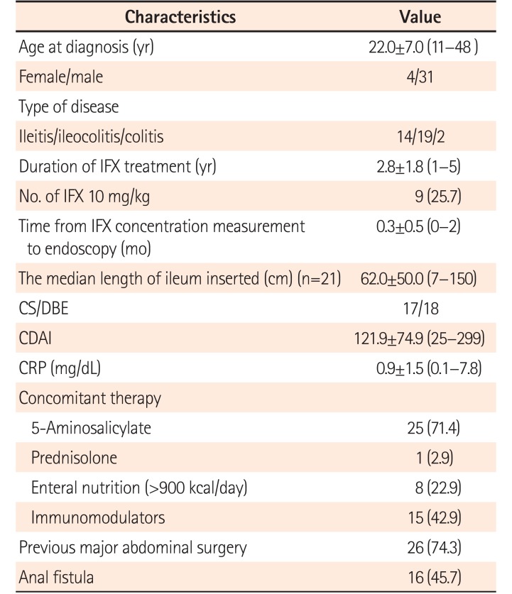
A total of 108 patients were enrolled in Study 1, in which the objective was to investigate the relationships of TLI and ATI with the clinical demographics. In Study 2, 35 patients were enrolled to investigate the relationships of TLI with endoscopic MH. The inclusion criteria for each of these studies are shown in
Fig. 1. Study 1 included 108 patients and Study 2 included 35 patients who met the following criteria: (1) efficacy of initial infusion of IFX was response; (2) provided informed consent to blood sampling to measure IFX blood concentrations and to endoscopy; (3) their course could be followed up sufficiently; (4) their CDAI could be measured; and (5) were able to undergo colonoscopy (CS) or double-balloon enteroscopy (DBE) within 2 months before or after the date of IFX blood concentration measurement. Exclusion criteria were: (1) continuous administration of IFX for ≤14 weeks or >5 years (49 patients); (2) a stoma (19 patients); or (3) not obtaining consent (4 patients).
Fig. 1
Overview of study protocol, subject selection and inclusion criteria in Study. The inclusion criteria in Study 1 and Study 2 were: (1) efficacy of initial infusion of infliximab (IFX) was response, were undergoing maintenance therapy; (2) provided informed consent to blood sampling to measure IFX blood concentrations and to endoscopy; (3) their course could be followed up sufficiently; (4) their CDAI could be measured; and (5) were able to undergo colonoscopy or double-balloon enteroscopy within 2 months before or after the date of IFX blood concentration measurement. Exclusion criteria were: (1) continuous administration of IFX for ≤14 weeks or ≥5 years; (2) a stoma; or (3) not obtaining consent. A total of 72 patients were excluded. In the study design in Study 1, the first assay (assay A and B) was performed with patients divided into loss of response (LOR) group and remission group. Assay A and clinical symptoms in the antibodies to IFX (ATI)-positive patents in the remission group were checked after 1 year. In Study 2, endoscopic examination and assay A were performed after enrollment.
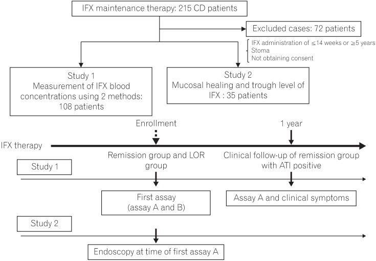

In the design of Study 1, the first assay (assays A and B) was performed with patients divided clinically using CDAI into a remission group and a LOR group after patient enrollment. Then, after about 1 year, we conducted a follow-up evaluation of ATI-positive patients in the remission group whose course had been closely followed. In Study 2, endoscopic examination and the first assay (assay A) were performed within 2 months of each other.
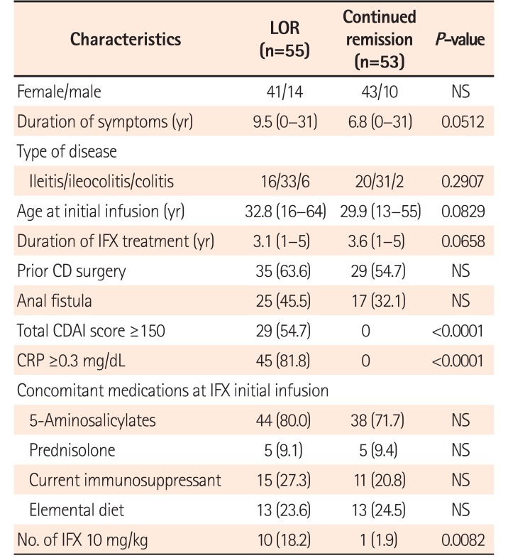
Eleven of the 108 patients (10.2%) in Study 1 were receiving an IFX dose of 10 mg/kg. Nine of the 35 patients (25.7%) in Study 2 were receiving an IFX dose of 10 mg/kg.
2. Measurements of IFX Concentrations
Serum taken immediately before IFX infusion was used for TLI measurements. TLI measurements were conducted at Tanabe R&D Service Co., Ltd. (Saitama, Japan; assay A), and Shiga University of Medical Science (assay B). Measurements were performed blind, without disclosing patient background or clinical results.
Serum TLI measurements with assay A were conducted with an ELISA using a monoclonal antibody against IFX obtained from Jansen Biotech Inc. (Horsham, PA, USA). The detection limit was 0.1 µg/mL.
6
Serum TLI measurements with assay B were conducted using an ELISA system using an avidin ELISA plate (blocking-less type; Sumitomo Bakelite Co., Ltd., Tokyo, Japan).
7
3. ATI Measurement
With assay A, measurements were performed using an ELISA method based on a double-antigen format. If IFX is present in the blood it will compete with the labeled-IFX, making accurate measurement of ATI impossible. As a result, to obtain a positive or negative result for ATI, the determination can only be made under conditions in which IFX is not present in the blood.
On the other hand, with assay B, ATI measurements were conducted using an original method developed by Shiga University of Medical Science called modified Direct-ELISA.
8 In modified direct-ELISA, the IFX-ATI immune complexes were initially dissociated, and the binding capacities of ATIs were recovered. ATIs were then immobilized onto ELISA plates and detected with horseradish peroxidase-labeled IFX.
4. Measurement of Clinical Laboratory Data
Biochemical markers such as CRP were measured by the Laboratory Test Department of Fukuoka University Chikushi Hospital. Blood samples taken immediately before IFX infusion were also used for these measurements.
5. Assessment of Clinical Activity
The clinical activity index for IFX was assessed according to the CDAI.
9 A CDAI ≤150 indicates a clinically inactive state, while ≥150 indicates the active phase. In this study, the CDAI was measured at the time IFX trough levels were measured, following infusion of IFX.
In Study 1, because the objective was to evaluate the clinical usefulness for diagnosing LOR, patients were classified as LOR or remission strictly based on the CRP level and CDAI score at the time IFX blood concentrations were measured. Remission was defined as CDAI <150 points and CRP <0.3 mg/dL. LOR was defined as CDAI ≥150 points and/or CRP ≥0.3 mg/dL.
6. Endoscopic Examination
The DBE models used were the Fujinon EN-580T, EN-450P5, and EN-450T5 (Fujinon Inc., Saitama, Japan); the CS models used were the Olympus PCF-240AI, PCF-PQ260I, PCF-Q260AI, and PCF-290I (Olympus, Tokyo, Japan). A transanal approach was used in all patients. DBE was performed in 18 patients, and CS was performed in 17 patients. The mean distance of small intestinal observation after passing through Bauhin's valve was 62 cm (range, 7–150 cm). Lesions were assessed at the site where activity was the strongest that could be confirmed endoscopically.
7. Measurement of Endoscopic Activity
The Fukuoka index was used to evaluate endoscopic mucosal activity. There are essentially 3 components to this index: stenosis, polyposis, and ulcer.
10 In this study, ulcer scores were used to assess ileal and colorectal mucosa. Without using the polyposis score, Beppu et al.
11 reported no link between the stenosis score and MH assessment. For ileal and colorectal lesions, the sites where activity was the highest were assessed for the activity index. No lesion (0 point) or ulcer scarring (1 point) was defined as “mucosal healing (MH),” and an ulcer score of 2 to 4 points was defined as “non-mucosal healing (nMH).” For small intestinal lesions, the sites evaluated were the small intestinal mucosa in patients with ileitis CD and ileocolitis CD. For colonic lesions, the colonic mucosa in colitis CD and the colonic mucosa in ileocolitis CD were the sites evaluated.
8. Statistical Analyses
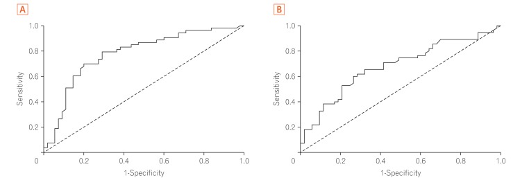
Fisher exact test or the Mann-Whitney U-test was used in 2-group comparisons, and to analyze the diagnostic ability of TLI and ATI, cutoff values were established for each using the minimum distance criteria from the area under the receiver operating characteristic curve (AUROC). Significance was defined as a P-value ≤0.05. Statistical analysis was performed using SPSS version 21.0 (IBM Corp., Armonk, NY, USA).
DISCUSSION
This prospective study examined whether TLI or ATI was useful for the evaluation of LOR. It was found that TLI was statistically more useful than ATI in judging LOR. Furthermore, in the relationship between MH and TLI, TLI was shown to be useful in MH of the large intestine. This is the first report to separately assess MH in the large and small intestines.
In the clinical background for this study, the mean period from the start of IFX until assay or endoscopy was performed was about 3 years. In a report by Beppu et al.,
11 the IFX treatment course was relatively long, at about 4 years. In other recent reports IFX was also administered for long durations of 40.0 to 68.7 months.
1213 The treatment period is therefore consistent between these reports and that in Studies 1 and 2 described herein.
In Study 1, TLI or ATI was useful for discrimination of LOR was determined using AUROC analysis.
There are various methods for the measurement of TLI and ATI. However, there have been few reports that use 2 or more measuring methods in a clinical trial. The present study compared the results of 2 typical measurement methods (Tanabe R & D, assay A; Shiga University of Medical Science, assay B) using the same serum. This was done because there is a significant problem in ATI measurement with assay A. It is known that there are inconclusive cases in which ATI cannot be measured, although it can be measured in 54% to 70% of cases; there are cases, however, that do not satisfy the requirements of assay A for measuring ATI.
3614 Therefore, development of a method to measure ATI that does not depend on the serum IFX density was urgently needed. Imaeda et al.
8 may have solved this problem by raising the sensitivity of the ATI value using a new measurement method (Direct ELISA; DA ELISA and IC-based ELISA). The present study examined the clinical value of ATI measurement using this method and the DA ELISA method.
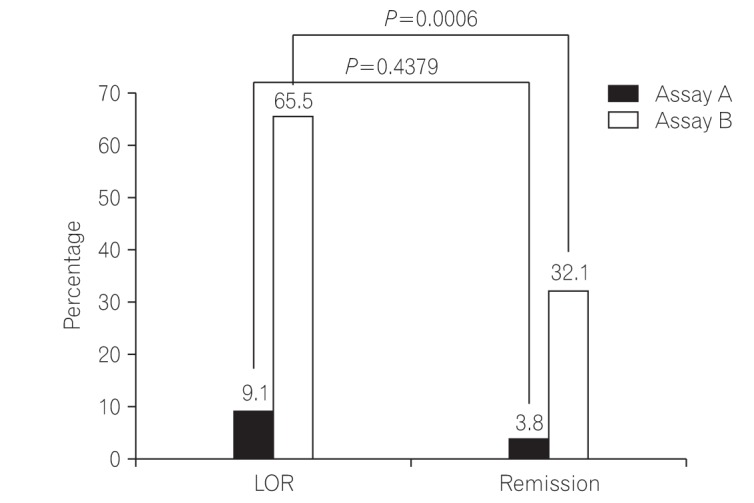
The results showed that assay A had clearly lower sensitivity, specificity, and negative predictive value for ATI than assay B. In the LOR group, the ATI positive rate was higher in assay B than in assay A. We therefore judged that clinical activity could not be satisfactorily evaluated with the ATI results from assay A.
In addition, the ATI level in assay B was significantly higher in the LOR group, while TLI levels were significantly lower in the LOR group than in the remission group. These results show that the presence of ATI is related to a drop in TLI when the ATI level does not depend on serum IFX, as is the case with assay B. However, there have been few reports that compared TLI and ATI evaluations of clinical LOR with high discrimination ability. AUROC analysis is useful for evaluating discrimination ability. There have been only 3 reports, including the present study that compared TLI with ATI by AUROC analysis (
Table 3).
45 In both of the other reports, the AUROC of TLI was larger than the AUROC of ATI. Those results are not inconsistent with this study. This is certain to depend on the measurement technique of ATI and has the possibility of greatly contributing to the clarification of LOR. However, the present results suggest that measurement of TLI alone is sufficient for the evaluation of clinical LOR. ATI appears to have a supplementary role; it may be appropriate to measure ATI when the TLI value is low and clinical LOR is suspected or when an IR may have occurred. A low ATI did not affect the long-term prognosis when the investigation of ATI positivity included many cases of ATI positivity in the remission group in the present study and in the remission group at 1year.
Table 3
Comparison of TLI and ATI Studies Using ROC Curve Analysis

|
Author |
Year |
No. |
LOR vs. remission |
TLI |
AUC |
Se/Sp (%) |
ATI level |
AUC |
Se/Sp (%) |
|
Steenholdt et al.4
|
2011 |
85 |
26 vs. 59 |
0.50 |
0.930 |
86.0/85.0 |
10.00 U/mL |
0.890 |
81.0/90.0 |
|
Vande Casteele et al.5
|
2015 |
483 |
NA |
2.79 |
0.681 |
52.5/77.6 |
3.15 U/mL |
0.632 |
38.0/87.4 |
|
Present study |
2015 |
108 |
55 vs. 53 |
2.60 |
0.778 |
70.9/79.2 |
4.90 μg/mL |
0.679 |
65.5/67.9 |

In Study 2, the relationship between endoscopic MH and TLI was examined based on the results of Study 1. The TLI necessary for small intestinal MH and colonic MH was examined in this study based on the supposition that it was different. In a recent report MH was defined using a different endoscopic score than that used in the present study. However, most reports define MH as disappearance of the ulcer lesion. The definition of MH in the present study used the ulcer score of the Fukuoka index. The reason why this definition was used in the present study was that, using the Fukuoka index, Beppu et al.
11 enumerated the points for calculating the scores at which the small intestinal lesions and the colon change to a morbid state were separately appreciable. In addition, it was reported that small intestinal MH and colonic MH were related to clinical remission. The results of Study 2 appeared to show that colonic MH and TLI were causally related, while there was no significant relationship between small intestinal MH and TLI.
The reasons why no significant differences were seen in TLI and MH in small intestinal lesions are thought to be the following: (1) endoscopic observation is easy in large intestinal lesions and detailed lesions can be identified. As a result, findings that agree with clinical symptoms can be obtained. With small intestinal lesions, however, it is not easy to observe the entire small intestine and the lesion areas are small. It is possible that clinical symptoms and small intestinal lesions do not agree because of the tendency to identify very small lesions. (2) The effectiveness of IFX for small intestinal lesions may be lower than that in the large intestine. Imaeda et al.
12 reported that TLI of ≥4.0 µg/mL was needed in MH. Additionally, Ungar et al.
15 reported that 80% to 90% of patients achieve MH with a TLI of 6 to 10 µg/mL and that the MH achievement rate becomes higher as the TLI value increases. Although those authors did not classify and score small intestinal lesions and large intestinal lesions, considering those reports our findings suggest that higher TLI is needed in order to achieve small intestinal MH. The above reasons may therefore explain why no significant differences were seen between small intestinal MH and TLI. At the same time, no significant relationship was seen between the MH group and the nMH group in either the small intestine or the large intestine with ATI, although this was a comparison using assay A. In the large intestine there was also a tendency to achieve MH when the CRP value was low, but no relationship was seen between other background factors and achieving MH. Imaeda et al.
12 reported that ATI and MH had only a weak relationship; in the present study, MH and ATI also had a weak relationship.
Whether combined therapy with an immunomodulator is related to TLI was evaluated. TLI was not higher in LOR and remission groups even with combined use of an immunomodulator.
This study has several limitations. Although Study 1 was a prospective study, there was no follow up from the first IFX administration. Furthermore, with limitation to the cross-sectional period only patients receiving long-term IFX were enrolled. Recent reports have shown that ATI can exist as stable ATI or transient ATI, and that transient ATI sometimes appears coincidentally during the time a patient is receiving IFX and does not affect LOR. Stable ATI, however, is reported to affect LOR. Therefore, multiple ATI measurements are recommended as it cannot be determined whether ATI is stable or transient with a single measurement. There are also reports that in determining LOR, a more accurate prediction is possible with a combination of CRP, TLI, and stable ATI.
161718 As ATI was measured only once in this study, it could not be determined whether it was stable or transient. Moreover, ATI-positive patients in the remission group were taken to be patients who would not experience LOR in at least one year and in whom TLI would not significantly decrease; however, the possibility cannot be ruled out that many cases of transient ATI were also included. Nevertheless, from reports that transient ATI does not affect LOR and that multiple ATI measurements are recommended, we recommend measurement of TLI for clinical purposes. TLI measurement means that LOR can be determined with a single measurement, rather than having to perform multiple measurements to determine whether ATI is stable or transient.
The limitations of Study 2 are thought to be that the number of patients who could participate in the study was small and that the entire small intestine could not be observed.
In conclusion, the present study showed that TLI was more useful for diagnosis and evaluation of LOR in CD during IFX maintenance therapy than ATI; ATI appears to have a supporting role in LOR evaluation. In addition, remission could be evaluated only by TLI. As for colonic MH, a relationship with TLI was observed; for remission, TLI needed to be greater than 2.6 µg/mL.



 PDF
PDF ePub
ePub Citation
 Print
Print


 XML Download