Abstract
Background
A proper depth of percutaneous central venous catheter (PCVC) is very important to reduce procedural time and prevent various complications in very low birth weight (VLBW) infants who require minimal handling or have a sensitive skin. The objective of this study was to suggest a formula for faster and proper insertion of PCVC in VLBWIs to prevent unintended consequences of patients’ conditions.
Methods
Prospective data of VLBW infants admitted from June 2015 to January 2018 who had PCVC inserted via the great saphenous vein within seven days after birth were analyzed. Correlations of length of inserted PCVC with body weight, body length, and postmenstrual age at the date of PCVC insertion were determined with a linear regression analysis. Using results of this analysis, a formula to determine the optimal insertion length of PCVC was derived. Coefficient of determination was used to assess how well outcomes were replicated by the formula.
Results
The formula to predict the proper insertion length of PCVC via the great saphenous vein at popliteal crease level was obtained as follows: Optimal Length (cm) = 3.8 × Body Weight (kg) + 11.1. With everyday movements such as flexion and extension of the lower extremities, the mean difference in catheter tip position was 7.0 ± 3.9 mm, which was not significant enough to escalate the risk of catheter tip displacement. The rate of catheter-related complications was as low as 4.9% in this study.
Graphical Abstract
Very low birth weight (VLBW) infants often show delayed establishment of enteral feeding, hence requiring prolonged intravenous access. Percutaneous central venous catheter (PCVC) or peripherally inserted central catheter (PICC) is essential for medications, parenteral nutrition, and fluid administration in VLBW infants weighing less than 1,500 g.12 PCVC is preferable to peripheral venous catheter for VLBW infants in a neonatal intensive care unit (NICU), especially for those who need prolonged intravenous therapy as they can tolerate concentrated fluid without requiring frequent insertion.345 PCVC is a safe and effective technique for prolonged intravenous therapy in NICU.67 Patients in NICU are premature and vulnerable who need considerable efforts for minimal handling and strict limitation of intervention including insertion of PCVC.8 PCVC insertion should also be done in a short period of time with few to no failed trials. The process should avoid skin irritation.9 However, in the actual field, multiple steps are taken during PCVC insertion. Before skin puncture, the indirect length of the blood vessel is measured with a sterile tape measure. After the catheter is inserted, the depth of the catheter is checked via X-ray imaging. If the depth is not appropriate, the catheter is adjusted and checked with X-ray imaging again. These steps are repeated as necessary until the catheter has a proper position. However, such steps can prolong the procedure time and cause catheter related complications including line-associated bloodstream infections, phlebitis, occlusion, external catheter leakage, and effusion.1011 Previous studies have found that movement of extremities is related to the migration of the catheter tip.12 Furthermore, incorrect tip placement has been associated with a high frequency of mechanical complications.1314 Therefore, it is very important to perform a precise and brief PCVC insertion. It is also important to minimally irritate the weak skin barrier because the outcome of VLBW infants depends on proper and minimal handling with fewer complications. Thus, in this study, we recorded the length of PCVC measured in VLBW infants and analyzed different demographic and clinical factors to suggest a guideline for fast and proper insertion of PCVC via the great saphenous vein. We also examined possible complications of improper catheter position with day-to-day limb movements.
This prospective cohort study included patients who were admitted to the NICU of Jeonbuk National University Hospital from June 2015 to January 2018 with birth weights less than 1,500 g who had PCVC insertion done within seven days after birth. Patients whose PCVCs were not inserted in the great saphenous vein and those who were born with a congenital cardiac anomaly were excluded. Each patient’s gestational age, birth weight, gender, weight, and length on the date of PCVC insertion, post menstrual age (PMA) on the date of PCVC insertion, duration of PCVC, PCVC tip culture results, and complications were recorded. All PCVCs were inserted using a 1 Fr (28G) single-lumen polyurethane catheter (Premicath®; Vygon, Aachen, Germany) via the great saphenous vein. The catheter was inserted near the popliteal crease level. The procedure was carried out in an aseptic environment by skilled nurses and doctors. The catheter was initially inserted via a cannula into the vein. X-ray was taken to confirm the location of the tip. Once a proper position was confirmed, the catheter was fixed to the skin following the manufacturer’s instruction.
Previous studies have shown that an ideal catheter tip location is 1 cm outside the heart in premature infants.15 The majority of catheters placed at T8–T9 vertebra will lie at the junction of the right atrium and the inferior vena cava.16 Therefore, in this study, optimal PCVC catheter length was defined as the length inserted through the great saphenous vein at the popliteal crease level to the inferior vena cava at the level of T9 vertebra body. To predict or calculate the optimal catheter length with a formula, we first measured the total indwelling length of the catheter after inserting it with infant in a supine, straight-legged position. The length between the catheter tip and the center of T9 vertebral body was measured through an X-ray image using a picture archiving and communication system (PACS, Marosis M-view® 5.4; Marotech, Seoul, Korea). The length from the skin puncture site to the popliteal crease level was measured with a tape measure for value adjustments. To evaluate the difference in catheter tip position with leg movements, radiographs were taken again with the hip and knee joints in 90-degree flexion for the infant in a supine position with knees touching the floor. The difference was measured with the PACS. All X-rays were taken at a distance of 1.2 meters with a focus on the umbilicus.
Characteristics of study participants and associated variables are described as mean ± standard deviation (SD) for continuous variables. Linear regression analysis was used to evaluate the optimal length of the catheter. Coefficient of determination (R2) was calculated to determine how well observed outcomes could be replicated by the regression equation. A P value of less than 0.05 was considered statistically significant. All statistical analyses were carried out with Statistical Package for the Social Sciences (SPSS) version 23 for Mac OS (IBM Corp., Armonk, NY, USA).
This study was conducted in accordance with the principles of the Declaration of Helsinki. It was approved by the Institutional Review Board (IRB) of Jeonbuk National University Hospital (IRB file No. 2014-05-001-004). Written informed consent was obtained from all infants’ parents.
Among 91 VLBW infants admitted to the NICU of Jeonbuk National University Hospital, 30 infants were excluded. Twenty-one infants were excluded because PCVC was inserted in the upper extremities. Two were excluded due to congenital cardiac anomaly. Seven were excluded due to the lack of consent for participation in this study. Finally, a total of 61 patients were enrolled. The mean gestational age of these infants was 29.3 ± 2.6 weeks. Their average birth weight and birth length were 1,155.6 ± 248.9 g and 37.2 ± 2.5 cm, respectively. Catheter insertion was performed on postnatal day 3.0 ± 1.0. On the day of PCVC insertion, mean body weight, length, and PMA of infants were 1,106.9 ± 248.0 g, 37.0 ± 2.3 cm, and 29.6 ± 2.6 weeks, respectively. The mean duration of PCVC placement was 16.1 ± 8.7 days (Table 1).
In simple regression analysis, optimal length was calculated with three different formulas based on body weight, body length, or PMA on the date of PCVC insertion (Fig. 1) as shown below:
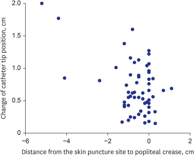
The depth of the catheter tip tended to increase with flexion of the lower extremities. Mean difference in catheter tip position was 7.0 ± 3.9 mm. No other variables were related to the length of catheter tip movement. However, there were two outliers, with catheter tip movements being the greatest in two subjects whose skin puncture sites were the furthest (5.2 and 4.4 cm) from the popliteal crease (Fig. 2).
There were three (4.9%) cases in this study in which the PCVC had to be removed due to complications. One (1.6%) was due to phlebitis. One (1.6%) was due to mechanical obstruction. The other one (1.6%) showed clinical signs of sepsis. Culture of the removed catheter tip and blood yielded growth of methicillin resistant Staphylococcus aureus. Therefore, central line-associated bloodstream infection (CLABSI) was diagnosed. Ampicillin and gentamicin were used empirically. They were replaced by vancomycin after confirming the growth of methicillin resistant S. aureus.
A formula using body weight on the date of PCVC insertion as a variable was obtained to predict the proper depth of PCVC in VLBW infants by measuring the length of PCVC insertion through the great saphenous vein: Optimal Length = 3.8 × Body Weight (kg) + 11.1 (R2 = 0.78, P < 0.001). Body weight was used as a variable in the formula because it had the highest R2 among the three variables (body weight, length, and PMA on the date of PCVC insertion).
Chen et al.17 have reported a formula for optimal length of catheter placement in lower extremities based on weight and length, with weight (from foot, R2 = 0.84; from femoral vein, R2 = 0.77) being more relevant than the length (from popliteal vein, R2 = 0.70). However, both R2 values were higher than 0.7, showing high correlations with optimal length of PCVC. Kim et al.18 have also developed a formula for distance from the skin puncture site to the superior vena cava-right atrium junction based on age, height, and length, with height (R2 = 0.77) being the most relevant, followed by weight (R2 = 0.58) and age (R2 = 0.40). Unlike these previous findings, weight was far more relevant than length or PMA in our study. Although body weight might have fluctuated on a daily basis, PCVC was inserted at the time of physiologic weight loss and body weight was measured in grams using a scale for accuracy. This might be more accurate than the length of an infant because the length was measured with a tape measure while the patient was in supine position with both lower extremities held in extension manually.
It is known that a central venous catheter is typically inserted into a vein in the upper arm, which is relatively more sanitary than areas such as the neck or groin.19 However, Ohki et al.20 have considered that saphenous PCVC can be safely used in critically ill neonates due to no fatal complications regarding hygiene. In pediatric patients, the upper or lower extremities or the scalp (in neonates or young infants) can be used as a catheter insertion site.21 However, it is more beneficial to use the lower extremity because when the venous catheter is inserted in the upper extremity, the tip is more likely to be displaced into a non-central blood vessel. In addition, there were no significant differences in complications between upper (27%) and lower (21%) extremity placed PCVC.22 Furthermore, PCVCs placed in the lower extremity were less likely to be removed for catheter-related complications compared with those placed in the upper extremity (39.9% in PCVCs placed in the upper extremity vs. 26.4% in those placed in the lower extremity, relative risk, 1.51; 95% confidence interval, 1.12–2.03; P = 0.007).23 Upper extremity placed PCVC has higher risks of malposition and non-elective removal for complications than lower extremity placed PCVC.13 In addition, upper extremity placed PCVC was associated with a higher risk of pleural or pericardial effusions together with malposition, dislodgement, and non-elective removal compared to lower extremity placed PCVC.132324 In the present study using lower extremity placed PCVC, pleural effusion, pericardial effusion, and superior vena cava syndrome were not found. However, these complications were found in those with upper extremity placed PCVC. Another benefit of using the lower extremities for PCVC is that both the left and right show no significant difference in length, unlike the upper extremities, making it more advantageous for applying the formula.
It is thought that movement of catheter tip in different positions such as knee flexion and extension of the lower extremities can displace the catheter and cause unwanted events. In this study, the mean difference in catheter tip position between flexion and extension of the lower extremity was 7.0 ± 3.9 mm. The length of displacement of the catheter was just about a vertebra’s width, considering that the mean length between the middles of the 8th and 9th thoracic vertebrae of subjects in this study was 6.0 ± 0.6 mm. Considering the average range of catheter tip movement, targeting the 9th thoracic vertebra would give the optimal parameter of 8th to 10th thoracic vertebrae and minimize the risk of catheter tip misplacement. Other factors that affected the range of catheter tip movement were skin puncture site and its distance from the popliteal crease (Fig. 2). In a similar study by Ohki et al.,20 the catheter was inserted at the medial malleolus and a wider range of catheter tip movement was recorded than that in our study in which the skin was penetrated near the popliteal crease. This might be the reason why there were no fatal side effects associated with catheter tip displacement during movement of lower extremities in the present study.
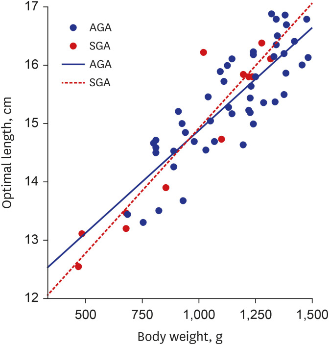
When groups were further subdivided, values of optimal length of PCVC were compared between small for gestational age (SGA) and appropriate for gestational age (AGA) infants, no significant difference in slope or R2 value was observed (AGA: R2 = 0.70, SGA: R2 = 0.89) (Fig. 3). Such high R2 values in both groups indicate that the weight is suitable variable for predicting the appropriate depth.
The present study has some limitations. First, most catheters inserted were positioned slightly distal to the popliteal crease. Only a few cases were inserted proximally. Such a skewed population was a limiting factor when analyzing whether the insertion site position was a variable in catheter tip displacement during joint movement. Second, PCVC is made of polyurethane, which is a material that stretches relatively well. Polyurethane catheters might have stretched out and seemed longer. However, no specific corrections could be made.25 This could make the measurements less accurate. Third, once the catheter was placed, it was used for more than two weeks. During this time, the infant not only gained weight, but also grew in length. The rate of growth was different for each infant, making it impossible to adjust for this when analyzing results. Lastly, the change in length over time was not followed up. It could not be reflected in the formula. Therefore, further study with a larger number of subjects needs to be done to prove this relativity.
In summary, using body weight as a factor, the formula developed in this study was feasible for predicting the optimal PCVC insertion length with fewer complications. We believe that this formula can benefit premature infants who require strict minimal handling or have a very weak and sensitive skin by facilitating more accurate PCVC insertion in a short period of time.
Notes
References
1. Gavelli V, Wackernagel D. Peripherally inserted central catheters in extremely preterm infants: Placement success rates and complications. Acta Paediatr. 2022; 111(3):554–556. PMID: 34757656.
2. Kim H, Kim SH, Byun HS, Choi YY. Clinical use and complications of percutaneous central venous catheterization in very low birth weight infants. Korean J Pediatr. 2005; 48(9):953–959.
3. Huang HC, Su LT, Liu YC, Chang HY, Ou-Yang MC, Chung MY, et al. The role of ultrasonography for detecting tip location of percutaneous central venous catheters in neonates-a single-center, prospective cohort study. Pediatr Neonatol. 2021; 62(3):265–270. PMID: 33637475.
4. Lee ES, Lee YH, Shin SM. Clinical experiences on PCVC (pereutaneous central venous cathetization) in newborn infants. J Korean Soc Neonatol. 1995; 2(1):34–41.
5. Lee J, Kim M, Cho CY, Choi YY. Effects of percutaneous central venous catheterization in very low birth weight infants. J Korean Soc Neonatol. 2000; 7(2):81–88.
6. Soong WJ, Hwang B. Percutaneous central venous catheterization: five year experiment in a neonatal intensive care unit. Zhonghua Min Guo Xiao Er Ke Yi Xue Hui Za Zhi. 1993; 34(5):356–366. PMID: 8237354.
7. Thiagarajan RR, Ramamoorthy C, Gettmann T, Bratton SL. Survey of the use of peripherally inserted central venous catheters in children. Pediatrics. 1997; 99(2):E4.
8. Sung SI, Ahn SY, Seo HJ, Yoo HS, Han YM, Lee MS, et al. Insensible water loss during the first week of life of extremely low birth weight infants less than 25 gestational weeks under high humidification. Neonatal Med. 2013; 20(1):51–57.
9. Kristoff K, Wang R, Munson D, Dysart K, Stracuzzi L, Wade K, Birnbaum S. A quality improvement initiative to provide timely central vascular access in a neonatal intensive care unit. Adv Neonatal Care. 2022; 22(3):203–209. PMID: 34407057.
10. Alhatem A, Estrella Y, Jones A, Algarrahi K, Fofah O, Heller DS. Percutaneous route of life: chylothorax or total parenteral nutrition-related bilateral pleural effusion in a neonate? Fetal Pediatr Pathol. 2021; 40(5):505–510. PMID: 32000556.
11. Colacchio K, Deng Y, Northrup V, Bizzarro MJ. Complications associated with central and non-central venous catheters in a neonatal intensive care unit. J Perinatol. 2012; 32(12):941–946. PMID: 22343397.
12. Nadroo AM, Glass RB, Lin J, Green RS, Holzman IR. Changes in upper extremity position cause migration of peripherally inserted central catheters in neonates. Pediatrics. 2002; 110(1 Pt 1):131–136. PMID: 12093958.
13. Chen H, Zhang X, Wang H, Hu X. Complications of upper extremity versus lower extremity placed peripherally inserted central catheters in neonatal intensive care units: A meta-analysis. Intensive Crit Care Nurs. 2020; 56:102753. PMID: 31445794.
14. Dhillon SS, Connolly B, Shearkhani O, Brown M, Hamilton R. Arrhythmias in children with peripherally inserted central catheters (PICCs). Pediatr Cardiol. 2020; 41(2):407–413. PMID: 31853581.
15. Perin G. PICC placement in the neonate. N Engl J Med. 2014; 370(22):2153–2154.
16. Finn D, Kinoshita H, Livingstone V, Dempsey EM. Optimal line and tube placement in very preterm neonates: an audit of practice. Children (Basel). 2017; 4(11):99. PMID: 29149032.
17. Chen IL, Ou-Yang MC, Chen FS, Chung MY, Chen CC, Liu YC, et al. The equations of the inserted length of percutaneous central venous catheters on neonates in NICU. Pediatr Neonatol. 2019; 60(3):305–310. PMID: 30217481.
18. Kim JH, Kim CS, Bahk JH, Cha KJ, Park YS, Jeon YT, et al. The optimal depth of central venous catheter for infants less than 5 kg. Anesth Analg. 2005; 101(5):1301–1303. PMID: 16243984.
19. Mussa B. Advantages, disadvantages, and indications of PICCs in inpatients and outpatients. Sandrucci S, Mussa B, editors. Peripherally Inserted Central Venous Catheters. Milano, Italia: Springer;2014. p. 43–51.
20. Ohki Y, Nako Y, Morikawa A, Maruyama K, Koizumi T. Percutaneous central venous catheterization via the great saphenous vein in neonates. Acta Paediatr Jpn. 1997; 39(3):312–316. PMID: 9241891.
21. O’Grady NP, Alexander M, Burns LA, Dellinger EP, Garland J, Heard SO, et al. Summary of recommendations: guidelines for the prevention of intravascular catheter-related infections. Clin Infect Dis. 2011; 52(9):1087–1099. PMID: 21467014.
22. Wrightson DD. Peripherally inserted central catheter complications in neonates with upper versus lower extremity insertion sites. Adv Neonatal Care. 2013; 13(3):198–204. PMID: 23722492.
23. Elmekkawi A, Maulidi H, Mak W, Aziz A, Lee KS. Outcomes of upper extremity versus lower extremity placed peripherally inserted central catheters in a medical-surgical neonatal intensive care unit1. J Neonatal Perinatal Med. 2019; 12(1):57–63. PMID: 30149479.
24. Kwak KJ, Park JH, Choi HJ, Kim CS, Lee SL. Early-onset pericardial effusion after peripherally inserted central venous catheterization in a preterm infant. Korean J Perinatol. 2015; 26(4):355.
25. Ateş Y, Yücesoy CA, Unlü MA, Saygin B, Akkaş N. The mechanical properties of intact and traumatized epidural catheters. Anesth Analg. 2000; 90(2):393–399. PMID: 10648328.