This article has been
cited by other articles in ScienceCentral.
Abstract
This case report describes skeletal anchorage-supported maxillary protraction performed with the Alternate Rapid Maxillary Expansion and Constriction (Alt-RAMEC) protocol over a treatment duration of 14 months in a 16-year-old female patient who was in the late growth-development period. Miniplates were applied to the patient's aperture piriformis area to apply force from the protraction appliance. After 9 weeks of following the Alt-RAMEC protocol, miniplates were used to transfer a unilateral 500-g protraction force to a Petit-type face mask. A significant improvement was observed in the soft tissue profile in measurements made both cephalometrically and in three dimensional photographs. Subsequently, the second phase of fixed orthodontic treatment was started and the treatment was completed with the retention phase. Following treatment completion, occlusion, smile esthetics, and soft tissue profile improved significantly in response to orthopedic and orthodontic treatment.
Keywords: Face mask, Class III treatment, Miniplate, Alt-RAMEC
INTRODUCTION
Skeletal Class III malocclusions are one of the most difficult cases to solve. These malocclusions may occur due to maxillary retrognathia, mandibular prognathia, or both. Maxillary retrognathia is the main cause of the problem in approximately 40% of Class III individuals.
1
Treatment of Class III malocclusions is relatively easy when the problem is limited to the alveolar bone, but in cases involving the basal bone due to retrognathia of the maxilla or prognathia of the mandible, the malocclusion does not respond easily to treatment and is likely to recur following completion. The patient's age and growth period are the determining factors in the treatment of this craniofacial deformity. Orthopedic treatment in the early period aims to reduce the therapeutic needs in the future.
2 Protraction appliances are used in cases involving maxillary retrognathia to support the growth of an inadequate maxilla by the application of extraoral force. This force facilitates in the growth during the developmental period. For this purpose, face masks are most commonly used.
3
Treatment methods for this malocclusion in adults can involve a combination of orthodontics and surgery or only orthodontic camouflage. Orthognathic surgery is usually the ideal treatment for adults with Class III malocclusion; however, many patients do not accept the surgical option because of financial constraints or the invasiveness of the procedure. Alternatives to orthognathic surgery include camouflage treatments with distalization of the mandibular dental arch using temporary skeletal anchorage units and Class III intermaxillary elastics.
4-6
This case report presents the use of the Alternate Rapid Maxillary Expansion and Constriction (Alt-RAMEC) protocol and face-mask treatment with skeletal anchorage units to correct the current status of a late adolescent patient with anterior crossbite and skeletal Class III malocclusion.
DIAGNOSIS AND ETIOLOGY
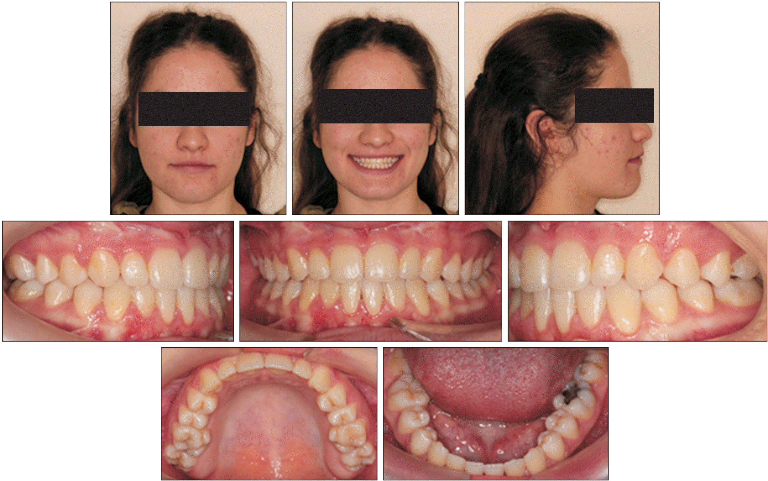
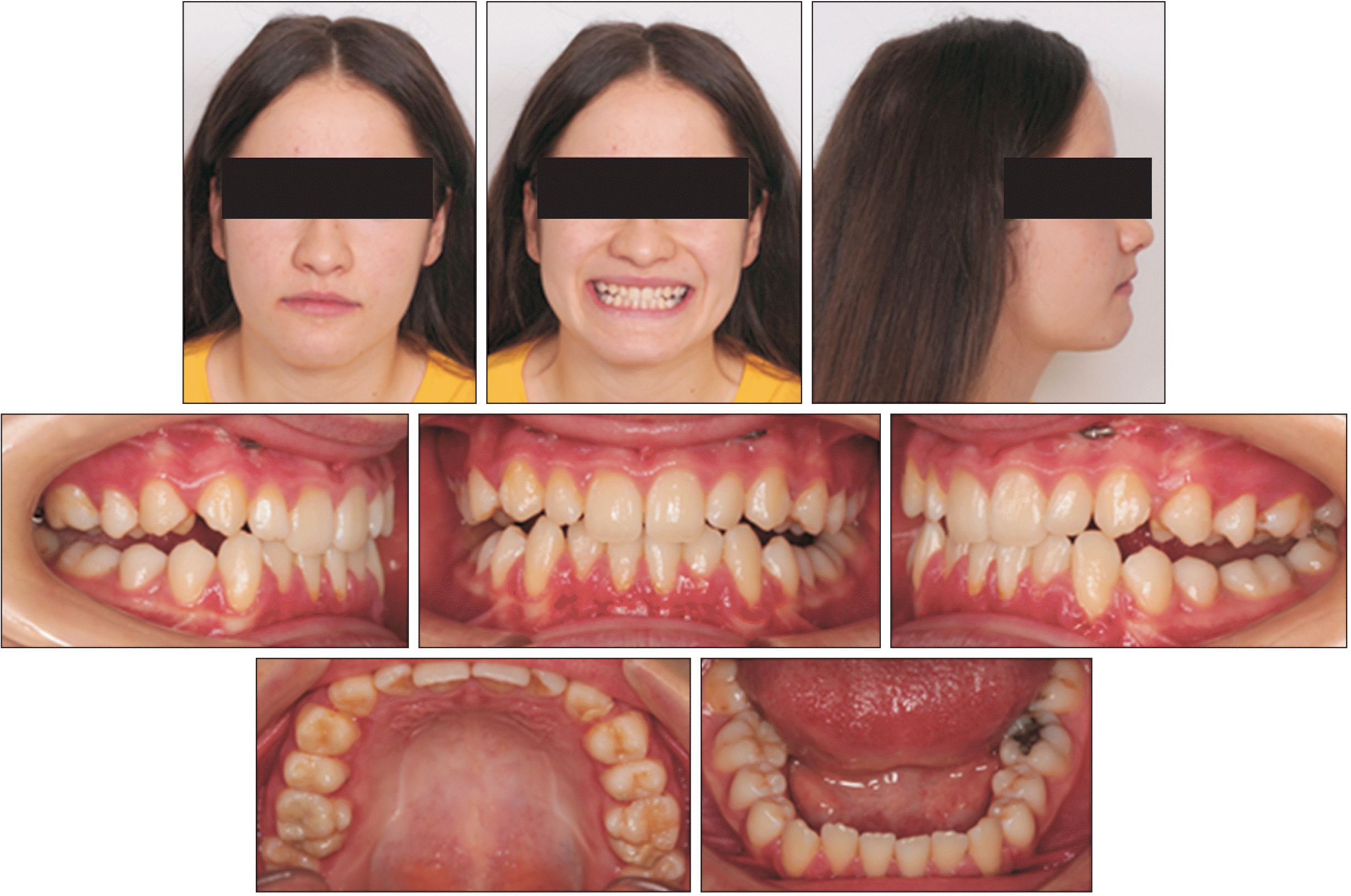
The patient was a 16-year and 7-month-old girl who presented to the Suleyman Demirel University, Department of Orthodontics with the chief complaint of a protruding mandible. Clinically, she had a concave facial profile and a retrusive maxilla (
Figure 1). Intraorally, she had an anterior crossbite with no crowding in the lower dental arch and minimal crowding in the upper dental arch. Panoramic radiographs showed no concerns.
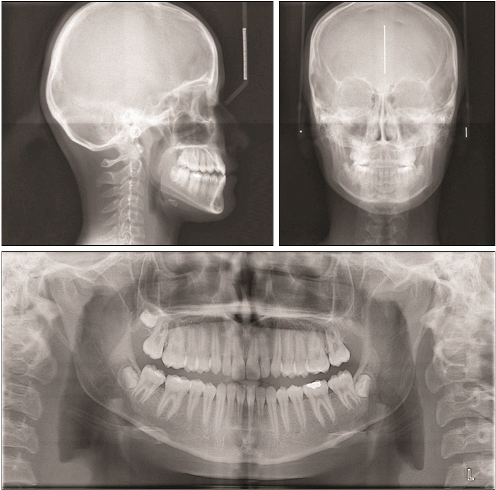
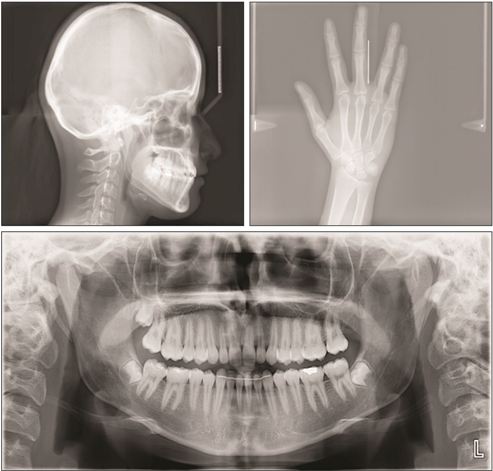
The cephalometric analysis showed a skeletal Class III malocclusion with maxillary deficiency (A point-nasion-B point [ANB], –0.9°) and a normal mandibular plane angle (gonion-menton [Go-Me] to Frankfort horizontal plane [FMA], 25.44°). The maxillary incisors were normal (upper incisor [U1] to sella-nasion [SN], 102.85°), and the mandibular incisors were retroclined (lower incisor [L1] to Go-Me [IMPA], 83.37°), compensating for the skeletal malocclusion (
Figure 2,
Table 1). The patient had a family history of mandibular prognathism. A written informed consent was obtained from the patient for the publication of this case report.
TREATMENT OBJECTIVES
The treatment objectives in this case were as follows: (1) to improve the profile and facial appearance, which was the primary complaint of the patient, (2) to provide Class I functional ideal occlusal relationship, and (3) to correct anterior crossbite and Class III malocclusion.
TREATMENT ALTERNATIVES
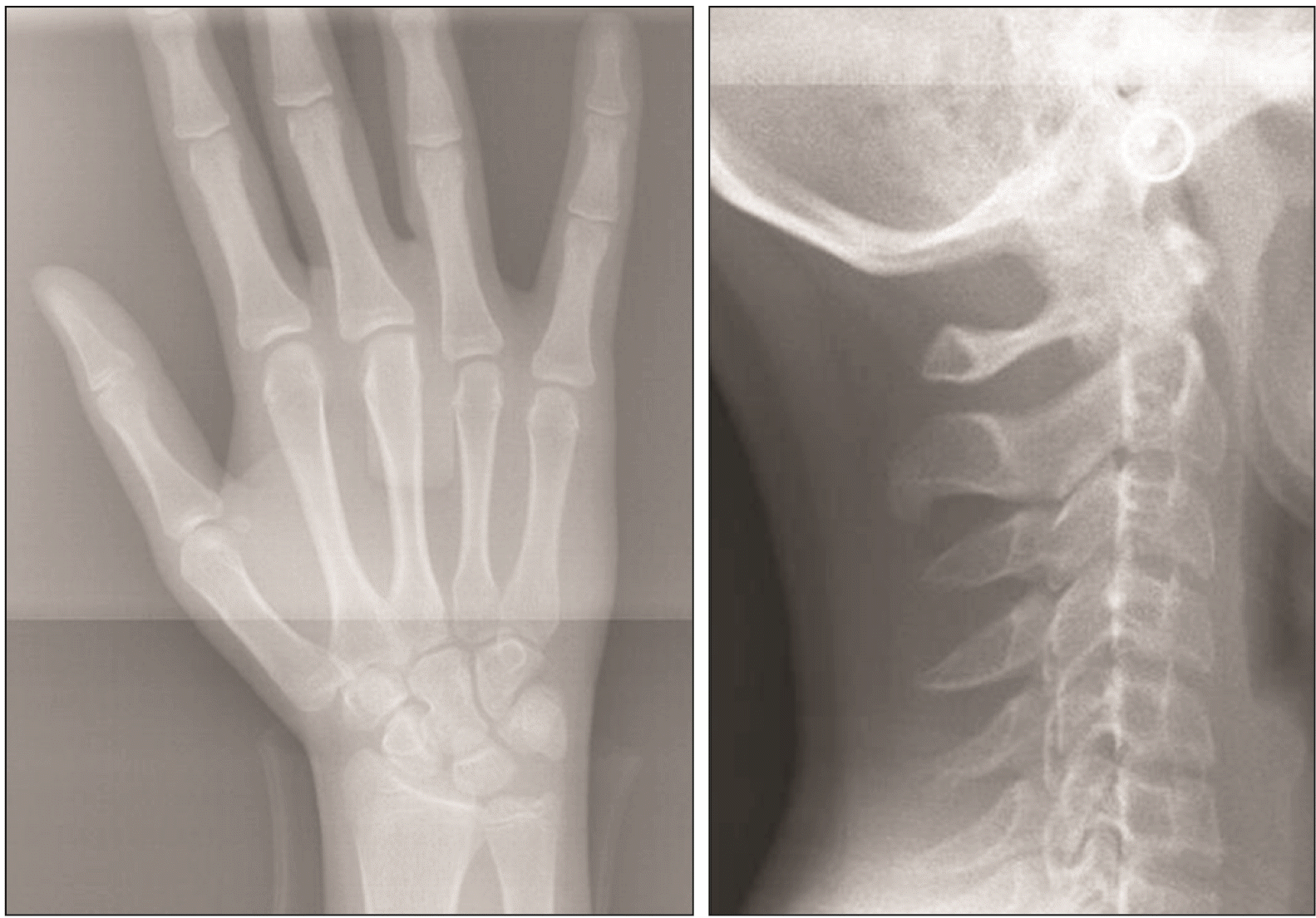
Radiographs of the hand-wrist and cervical vertebrae showed that the patient was in the post-pubertal period (
Figure 3). Since the chief complaint of our patient was the profile appearance, which would not change much with camouflage treatment, orthognathic surgery was suggested as a definitive solution. Orthognathic surgery including maxillary advancement and mandibular set back planning of fixed orthodontic treatment were discussed with the patient. This approach would allow correction of the skeletal concern and create an ideal occlusion with good facial and dental esthetics. However, the patient refused the surgical approach. Other treatment options included: (1) distalization of the mandibular teeth with skeletal anchorage units (2), mandibular interproximal reduction (3), Class III elastics (4), and/or maxillary protraction with a face mask. The patient evaluated the advantages and disadvantages of each of these treatment options.
Since the primary complaint of the patient was related to the profile and facial appearance, a treatment plan that could affect the skeletal soft tissue as well as achieve dental correction was emphasized. We planned to start with the Alt-RAMEC protocol to increase both the skeletal changes and to ensure the effectiveness of the face mask by providing mobilization in the sutures, and subsequently apply a face mask with skeletal anchorage units for improving the skeletal changes. We decided to apply fixed orthodontic treatment after maxillary protraction.
TREATMENT PROGRESS
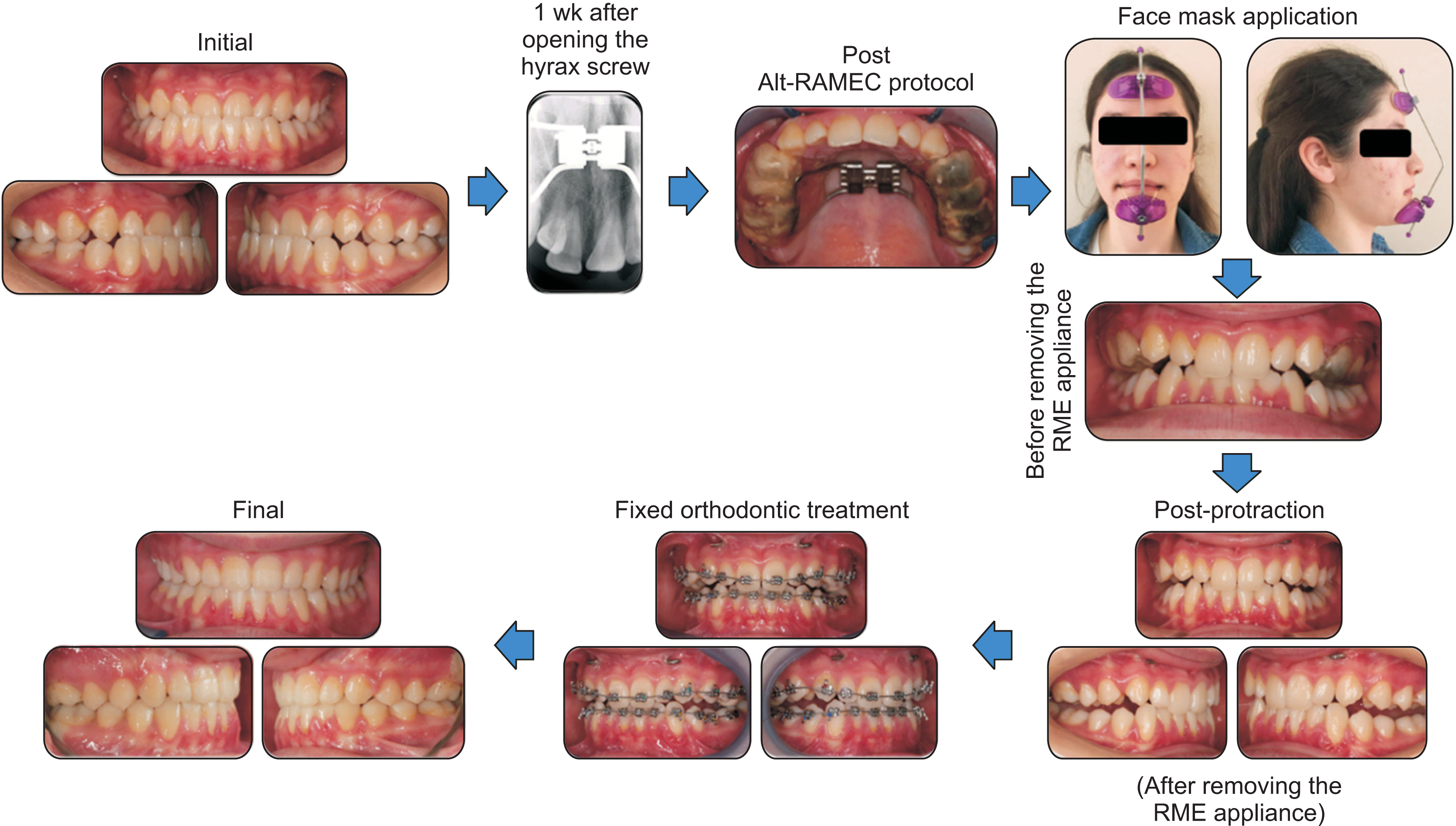
The treatment was started with the Alt-RAMEC protocol before protraction. The acrylic coverage rapid maxillary expansion (RME) appliance was cemented, and the Alt-RAMEC protocol continued for 9 weeks. The parents of the patient were instructed to open the screw (Leone A0620-19, Florence, Italy) twice per day for 1 week and to close it twice per day for the following week (0.25 mm per turn). This protocol for alternate opening and closing was repeated for eight consecutive weeks.
During the protocol, miniplates were placed surgically. Under local anesthesia, two I-shaped miniplates were placed in the apertura piriformis region of the maxilla with six screws. The tip of the I-shaped plate was bent to form a hook. After 10 days, healing was observed and sutures were removed.
In the 9th week, the screw was opened for 1 week, and a Petit-type face mask was used with a force of approximately 500 g applied bilaterally with an anteroinferior force vector of approximately 30° to the occlusal plane from the hooks of the I-shaped miniplates. The patient was evaluated at monthly intervals, and she was instructed to wear the appliances for at least 20 hours per day until at least a 2-mm positive overjet was achieved. Protraction treatment was completed in 6 months (
Figures 4 and
5).
Subsequently, the acrylic coverage RME appliance was removed and the fixed treatment phase was started (
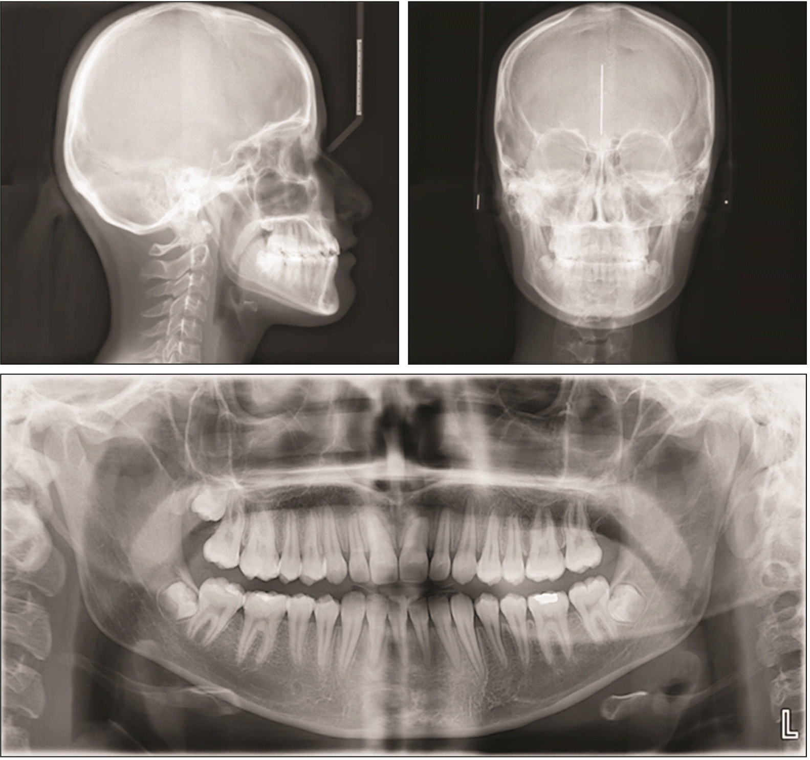
Figure 6). Miniplates were used for retention and during fixed treatment at night. After the protraction phase, the fixed orthodontic phase was started. After the application of MBT 0.22 slot metal brackets (Mini Ovation, Dentsply GAC International, Bohemia, NY, USA), leveling was performed with 0.016, 0.016 × 0.016-inch (in), 0.016 × 0.022-in, 0.017 × 0.025-in, and 0.019 × 0.025-in Nickel-Titanium archwires arch wires and 0.019 × 0.025-in steel arch wires, respectively. After ensuring that the molar and canine teeth were in Class I occlusion at the finishing stage, digitation elastics were applied to the mandibular canines and first premolars from the maxillary canines. Lastly, an essix plate was placed for stabilization of both arches. The total treatment period was approximately 14 months (
Figures 7 and
8).
RESULTS
Post-treatment data revealed that the treatment goals were achieved. Since the patient did not want to undergo orthognathic surgery or tooth extraction, the initial complaint was largely addressed to and the malocclusion was resolved, although the results were not ideal. Smile esthetics and anterior crossbite improved and tooth midlines were aligned with the facial midline. Acceptable overbite and overjet were obtained with Class I canine and molar relationships. No significant root or bone resorption was observed in the post-treatment panoramic image, and good root parallelism was visible.
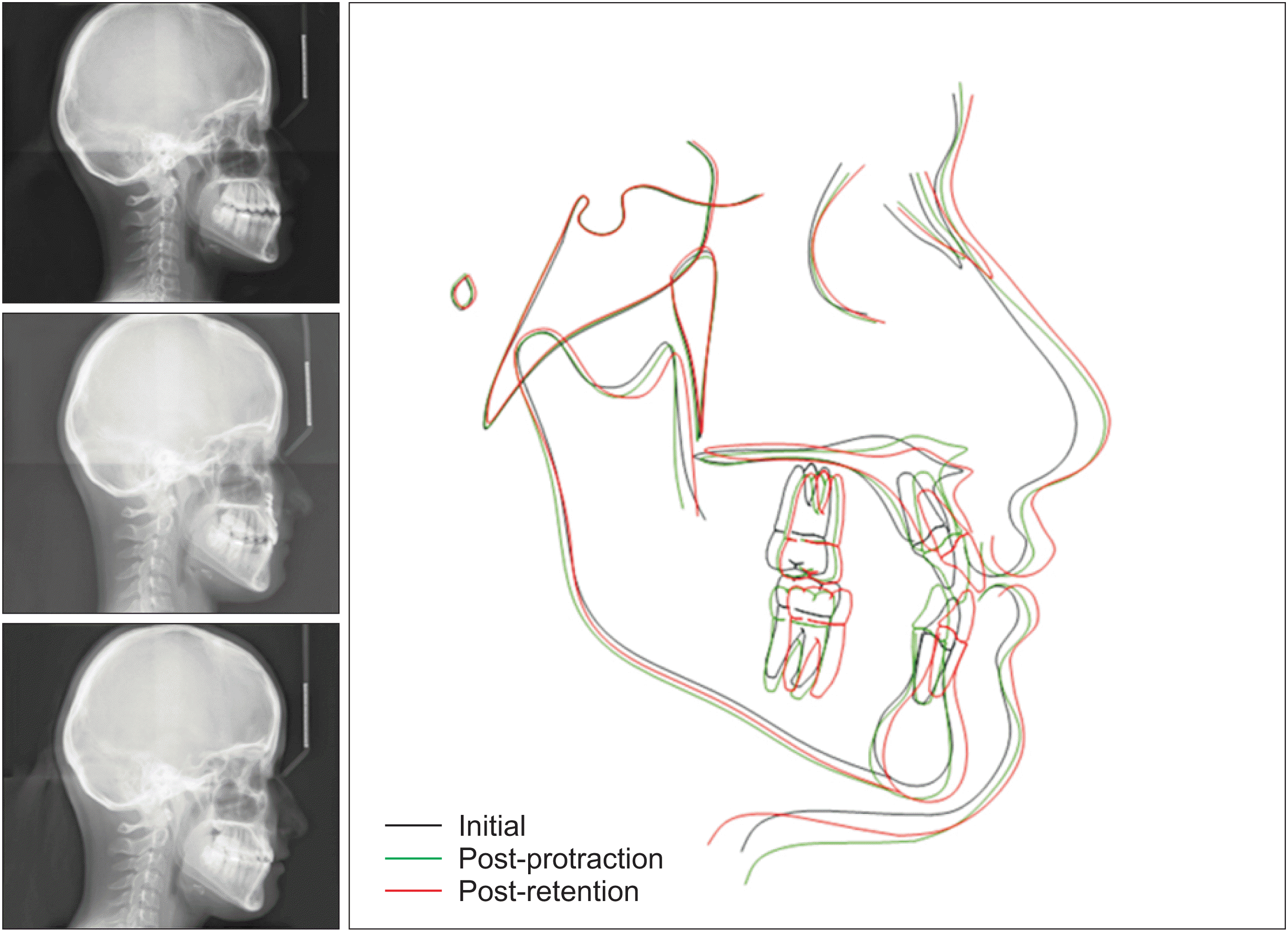
In the lateral cephalometric analysis, skeletal changes were observed with forward movement of the base of the maxilla (sella-nasion-A point [SNA]: 79.87° to 84.58°) and slight forward movement of the mandible (sella-nasion-B point [SNB]: 80.77° to 81.43°). A minimal increase in vertical dimensions (sella-nasion to gonion-gnathion [SN/GoGn]: 30.85° to 31.03°) was also observed. This can be explained by the posterior rotation of the mandible due to the effect of the face mask. Dentoalveolar changes included minimal protrusion with the face mask in the upper incisors and retrusion after fixed treatment and after retention. Although slight retrusion was observed with the face mask in the lower incisors, the crowding was corrected with protrusion at the end of the treatment. An increase in overjet (from –0.83 mm to 4.29 mm) and overbite (0.26 mm to 1.71 mm) was observed (
Table 1).
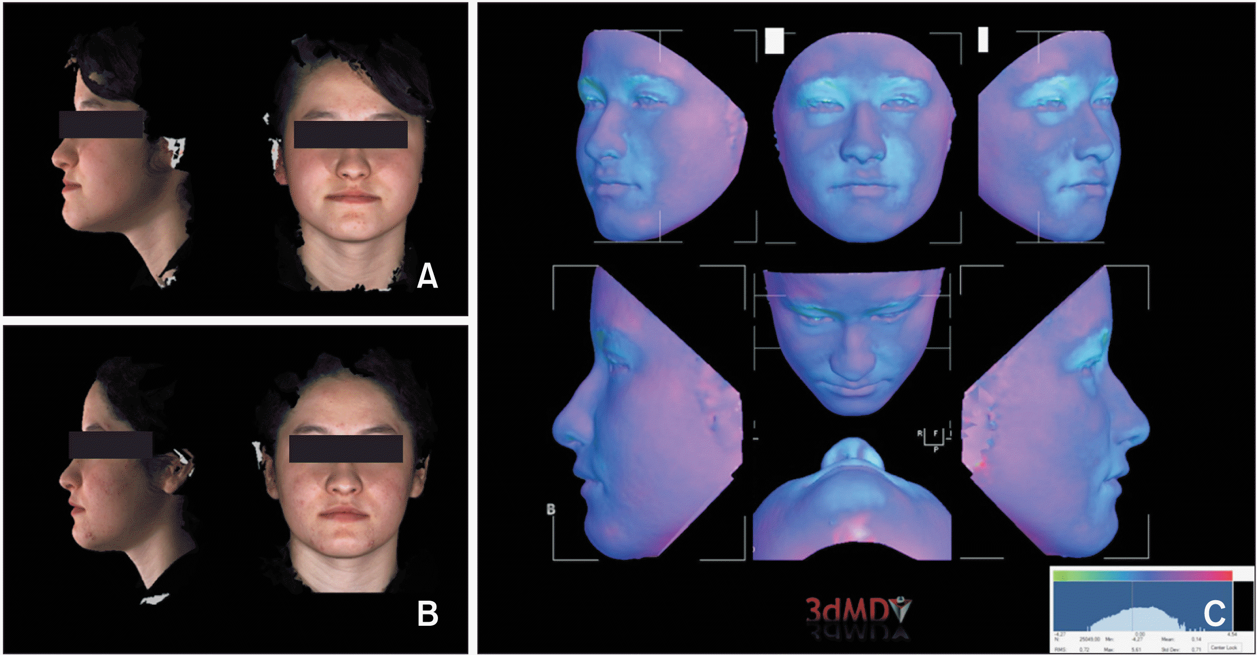
In the three dimensional photographs captured before and after protraction of our patient, both measurements and volumetric registration were obtained with the 3dMD Vultus program (3dMD VultusR software version 2.3.0.2; 3dMD, Atlanta, GA, USA). In 3dMD measurements, both volumetric (midface volume: pre, 28,113 mm
3; post, 28,665 mm
3) and linear increments were observed in the middle part of the face. These changes were followed by changes in the measurements in the upper lip and nasal region (
Table 2). Although the measurements of the lower part of the face and the mandible showed a reduction, there was no clinically significant change (
Figure 9).
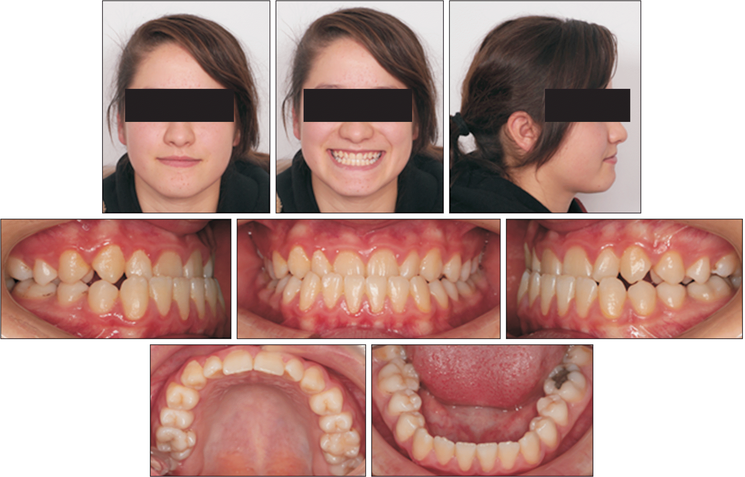
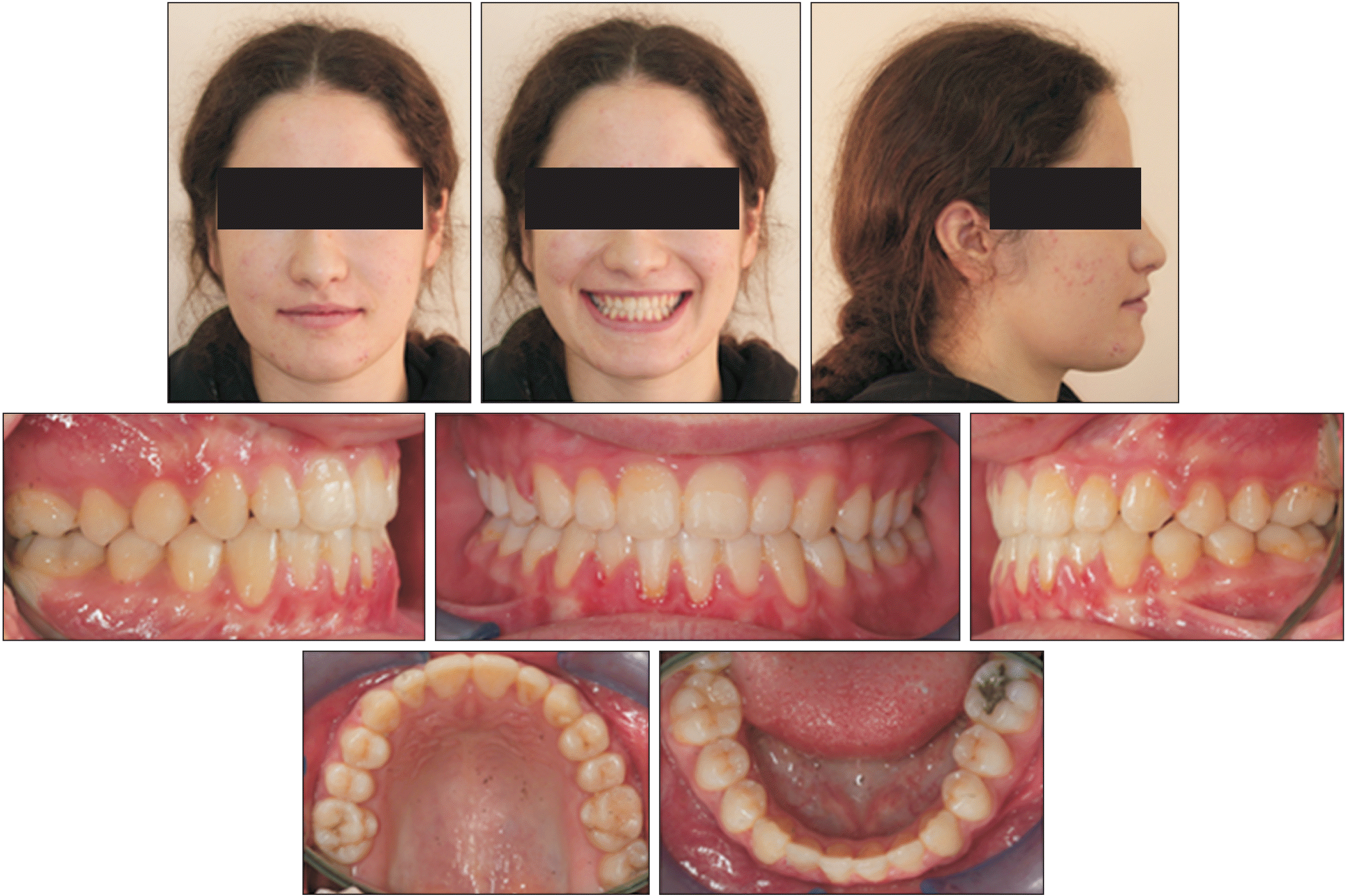
The patient's post-treatment short-term results (after 6 months) (
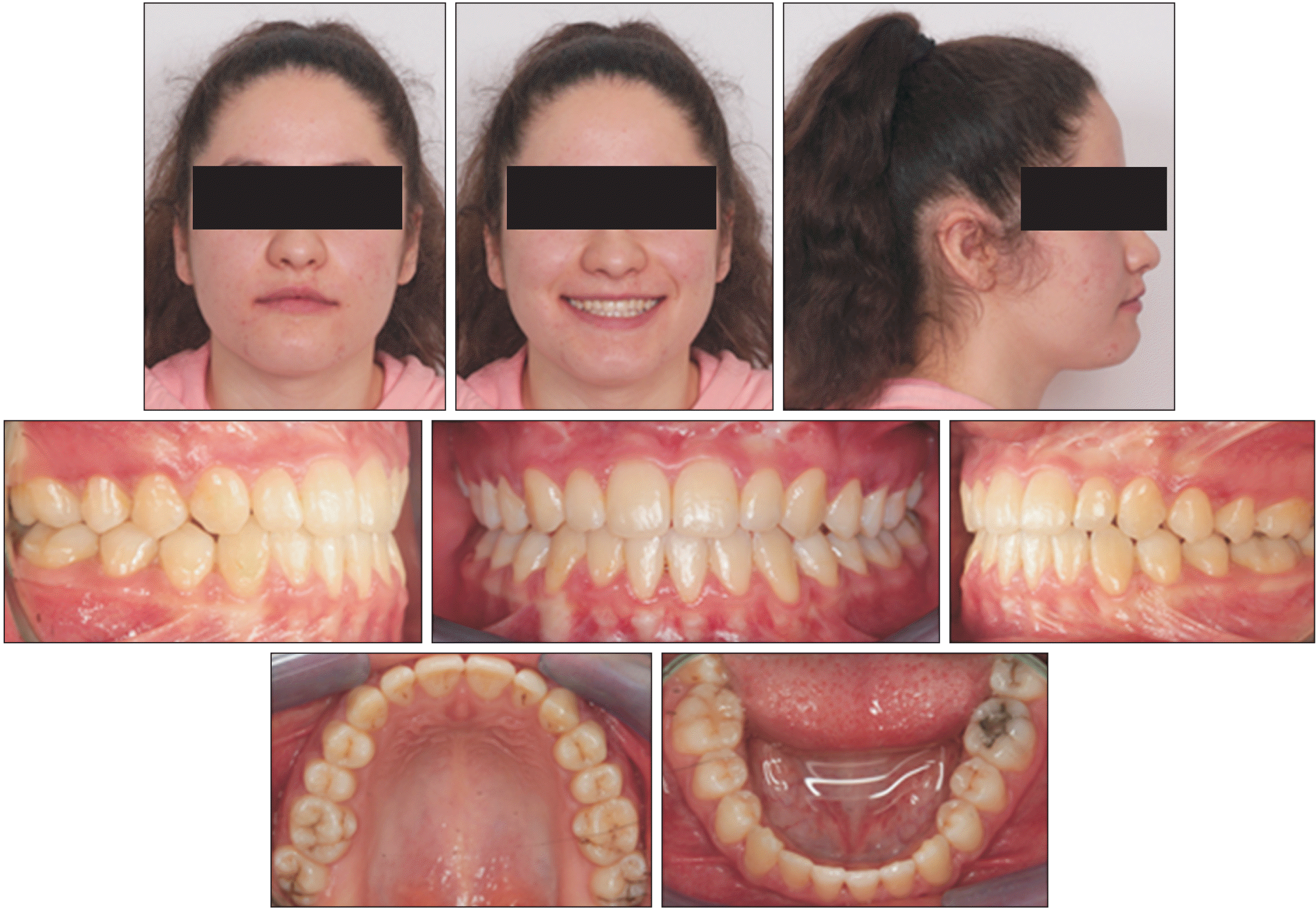
Figure 10) and long-term results (2 years later) also showed a low relapse rate (
Figure 11). Both the profile view and the relationship between the teeth and jaws remained stable (
Figures 12 and
13). The patient did not report any temporomandibular joint pain or discomfort during or after orthodontic treatment. She was satisfied with her occlusion and facial esthetics and showed a perfect fit while putting on the face mask and elastics, which ensured that the treatment plan proceeded as expected.
DISCUSSION
The widespread use of skeletal anchorage units in orthodontic treatments has helped to solve many previously reported treatment-related problems. Direct support from the jaws can prevent unwanted anchorage losses in fixed orthodontic treatments and is aimed to minimize the possible dental effects and increase the skeletal effect in functional and orthopedic treatments. For this purpose, skeletal anchorage units have been frequently used in the treatment of skeletal Class III malocclusions.
6
The literature contains multiple studies reporting treatments such as the use of face masks from plates placed in the zygoma region or Class III elastics between the anchorage units placed in the symphysis region and the zygoma. Maxillary protraction with a face mask from the plates placed in the aperture piriformis region can provide pure skeletal protraction while reducing the dental effect by preventing the dental mesialization of the upper arch. In addition, this method also eliminatesthe potential for posterior rotation of the mandible and the related negative effects, since it allows application of force through the resistance center of the maxilla.
5-7
In this case report, three treatment options were presented to our patient in late adolescence who had completed active growth development. The patient refused dental camouflage treatment because her main complaint was her profile appearance. The patient was also informed about the possibility of an orthognathic surgical procedure and was explained that maxillary protraction with skeletal anchorage could be tried before the operation.
To increase the chance of treatment success, we aimed to mobilize the sutures with the Alt-RAMEC protocol before protraction. Many studies using the Alt-RAMEC protocol in the literature have reported that an average of two-fold more protraction can be achieved in comparison with conventional face mask applications.
8,9 However, studies using both Alt-RAMEC and skeletal anchorage units are very limited in the literature.
10 To our knowledge, the current literature does not include any study in which this method was applied in the late period, making the present case report the first of its kind.
The patient was successfully treated because of her cooperation and because the malocclusion were not very severe. An ideal overjet–overbite relationship was achieved without any protrusion in the upper incisors. To examine the movement of the jaws skeletally, examinations were conducted according to the fixed reference planes. With the application of a skeletal anchorage-supported face mask in the maxilla, 2.8 mm of forward movement was detected, while only a 0.3° increase was observed in the vertical direction of the patient. Most of the overjet change obtained in the light of cephalometric registrations is skeletal, which also ensured post-treatment retention.
There are very limited publications on maxillary protraction treatments in the late period. In some of them, surgical support was also used before protraction. In the study by Yılmaz et al.,
11 corticotomy was performed before protraction in 19 patients with an average age of 13.12 years in the CS4 period. The results of that study showed a successful skeletal change. Nevzatoğlu and Küçükkeleş
12 similarly examined the treatment results of patients with a mean age of 12.54 years who underwent maxillary protraction with corticotomy. Their results showed that although this method is effective in the short term, it was not stable in the long term.
Meazzini et al.
13 examined the short- and long-term effects of late-term maxillary protraction with the Alt-RAMEC protocol. However, both patient groups in their study had a cleft lip and palate, and the oldest patient in their study cohort was 13.2 years old. Ganesh et al.
14 applied maxillary protraction to a 15-year-old male patient in the late period. However, in that case report, although the chronological age of the patient was late, continued growth could be evidenced both in the fact that he was male and in the cephalometric radiographs. Therefore, direct comparisons of the findings of that case with the present case are not possible. Moreover, since Class III elastics were applied from the appliance in the maxilla, a protrusion in the upper incisors in their overlap and the dental effect can be considered to be more effective in the correction of the overjet. In the current case, the overjet was corrected with direct skeletal anchorage-supported protraction. Shi et al.
15 also published a meta-analysis of the applications of skeletal anchorage-assisted protraction in adolescents. Again, in the studies included in that meta-analysis, studies were carried out in individuals aged 11–12 at most.
Park et al.
16 applied camouflage treatment to a 19-year-old patient using an RME and a face mask. They concluded that if the mandibular plane angle of an adult patient with a Class III malocclusion is low, an expander and face mask can be used to allow the mandible to rotate down and back to improve its profile. However, when the patient was examined both cephalometrically and in terms of soft tissue profile, the overjet improved with mesialization in the upper arch and protrusion in the upper incisors (U1-SN: pre, 111.9°; post, 116°). In our case report, the skeletal change was determined to be effective in correction.
Determination of the optimal time to start treatment is one of the critical points for the successful treatment of a Class III malocclusion. Although most studies report that face mask treatment is more effective in patients with primary and early mixed dentition, few studies have indicated that this treatment should be performed at 13–14 years of age.
17,18 This case report demonstrates that miniplates with protraction and an expander appliance can be used to treat early adult patients with a Class III malocclusion. The decision to perform orthopedic treatment in the early period or to wait for the completion of growth development is not an easy one. The advantages of early treatment include minimization of excessive closure of the dentition and mandible during this period, which can yield better facial esthetics and increase the patient's self-confidence. However, the results of this study also indicate that the treatment of Class III malocclusion with the use of miniplates and the Alt-RAMEC protocol may provide a window of opportunity for maxillary protraction and enlargement in patients who are in the late stages of growth.
CONCLUSIONS
Using both Alt-RAMEC and skeletal anchorage, protraction was achieved in the late period, consistent with the findings obtained with early applications in the literature. These findings should be supported by prospective studies with large sample sizes in the future. This approach may be an alternative to orthognathic surgery in compromised cases in the late stages of growth and development.


 PDF
PDF Citation
 Print
Print



 XML Download