Journal List > J Stroke > v.25(1) > 1516081114

Marshall, Liebeskind, III, Edwards, Howard, Meschia, Brott, Lal, Heck, Lanzino, Sangha, Kashyap, Morales, Cotton-Samuel, Rivera, Brickman, and Lazar: Cortical Thinning in High-Grade Asymptomatic Carotid Stenosis

Abstract

Background and Purpose

High-grade carotid artery stenosis may alter hemodynamics in the ipsilateral hemisphere, but consequences of this effect are poorly understood. Cortical thinning is associated with cognitive impairment in dementia, head trauma, demyelination, and stroke. We hypothesized that hemodynamic impairment, as represented by a relative time-to-peak (TTP) delay on MRI in the hemisphere ipsilateral to the stenosis, would be associated with relative cortical thinning in that hemisphere.

Methods

We used baseline MRI data from the NINDS-funded Carotid Revascularization and Medical Management for Asymptomatic Carotid Stenosis–Hemodynamics (CREST-H) study. Dynamic contrast susceptibility MR perfusion-weighted images were post-processed with quantitative perfusion maps using deconvolution of tissue and arterial signals. The protocol derived a hemispheric TTP delay, calculated by subtraction of voxel values in the hemisphere ipsilateral minus those contralateral to the stenosis.

Results

Among 110 consecutive patients enrolled in CREST-H to date, 45 (41%) had TTP delay of at least 0.5 seconds and 9 (8.3%) subjects had TTP delay of at least 2.0 seconds, the maximum delay measured. For every 0.25-second increase in TTP delay above 0.5 seconds, there was a 0.006-mm (6 micron) increase in cortical thickness asymmetry. Across the range of hemodynamic impairment, TTP delay independently predicted relative cortical thinning on the side of stenosis, adjusting for age, sex, hypertension, hemisphere, smoking history, low-density lipoprotein cholesterol, and preexisting infarction (P=0.032).

Conclusions

Our findings suggest that hemodynamic impairment from high-grade asymptomatic carotid stenosis may structurally alter the cortex supplied by the stenotic carotid artery.

Introduction

High-grade carotid artery stenosis may reduce cerebral perfusion in the hemisphere ipsilateral to the stenosis by 8% to 24% [1,2]. Other hemodynamic measures, such as a relative delay in arrival time of contrast material to the hemisphere supplied by the stenotic carotid artery, may be seen in up to 50% of patients [3,4]. Although hemodynamic impairment is associated with increased risk of stroke [5,6] and cognitive dysfunction [7-9], the consequence of these hemodynamic abnormalities on brain structure is not currently known. Cortical thinning is an imaging marker associated with cognitive impairment [10], seen in patients with traumatic brain injury [11], neurodegeneration [12,13], and cerebrovascular disease [14-16]. In this study, we investigated the effect of hemodynamic impairment on cortical thickness in patients with unilateral, high-grade, asymptomatic carotid artery stenosis.
The Carotid Revascularization and Medical Management for Asymptomatic Carotid Stenosis–Hemodynamics (CREST-H) is a National Institute of Neurological Disorders and Stroke (NINDS)-funded clinical investigation, ancillary to the NINDS-funded Carotid Revascularization and Medical Management for Asymptomatic Carotid Stenosis Study (CREST-2) randomized clinical trial [17]. CREST-2 is a pair of outcome-blinded, phase 3 randomized clinical trials to assess treatment differences in stroke and death rates in patients assigned to: (1) carotid endarterectomy (CEA) plus intensive medical management (IMM) versus IMM alone, and (2) carotid artery stenting (CAS) plus IMM versus IMM alone [18]. CREST-H is designed to determine if a subset of CREST-2 patients with high-grade, asymptomatic carotid artery stenosis and hemodynamic impairment would demonstrate cognitive benefit by revascularization of the stenotic carotid artery by CEA or CAS (clinicaltrials.gov NCT03121209). For this report, we used baseline imaging data from the CREST-H study to investigate whether degree of hemodynamic impairment as measured by time-to-peak (TTP) delay in the hemisphere ipsilateral to the stenotic carotid artery correlated with relative cortical thinning in that hemisphere. We hypothesized that patients enrolled in CREST-2 and CREST-H with severe carotid occlusive disease but without recent stroke would show cortical thinning in the hemisphere ipsilateral to the side of the stenosis prior to treatment onset (baseline) if there was hemodynamic impairment in that hemisphere. Because the CREST-H and CREST-2 clinical trials are still ongoing, there are additional imaging and cognitive outcomes data that remain blinded. These will be analyzed for publication at a later date. The present study contains baseline data currently available for analysis.

Methods

Participants

Baseline images were assessed from consecutive subjects randomized into the CREST-2 parent study and enrolled in CREST-H between January 30, 2018 and November 9, 2020. Per the CREST-2 protocol, patients had ≥70% internal carotid artery (ICA) stenosis and no symptomatic stroke or transient ischemic attack (TIA) in the target carotid territory within the last 180 days. Stenosis was defined by a catheter angiogram showing ≥70% stenosis, or a carotid duplex ultrasound showing a Doppler peak systolic velocity ≥230 cm/sec plus one of the following additional findings: end-diastolic velocity ≥100 cm/sec or ICA/CCA (common carotid artery) ratio ≥4.0, or CT angiography or MR angiography demonstrating ≥70% stenosis. Additional eligibility criteria for CREST-H included age 18–86, fluency in English, no contralateral ICA occlusion or stenosis ≥70%, no pre-existing diagnosis of dementia, no history of severe head trauma or current major depression, and education ≥8 years [17]. Written informed consent based on Institutional Review Board (IRB) approval from a central IRB at University of Cincinnati or local site IRB was obtained from all participants, first for CREST-2 and then separately for CREST-H. The current study was approved by the Columbia University Irving Medical Center IRB (AAAR4210), according to the ethical standards of Columbia University under the Common Rule as revised January 21, 2019.

Imaging acquisition

MR images were obtained on 1.5-T and 3-T scanners according to parameters established in the CREST-H study protocol and were processed centrally at the imaging core lab at UCLA Geffen School of Medicine for the perfusion imaging, at the Mayo Clinic at Rochester, MN for the structural imaging, and at Columbia University Irving Medical Center for cortical thickness measurements. The protocol included dynamic contrast susceptibility MR perfusion-weighted images (PWI), T2-weighted fluidattenuated inversion recovery (FLAIR) images to assess infarcts and white matter hyperintensities (WMH), and high-resolution T1-weighted images for cortical thickness measurements. PWI sequences were tailored for each participating site’s standard MRI vendor system field strength. 3-T parameters included: slice thickness 5.0 mm, gap=0 mm, slices=max for repetition time (TR), TR=1,500 ms, echo time=30 ms, field of view=22 cm, frequency=96 Hz, phase=128°, mode=2D. PWI source images were postprocessed with a semi-automated system that computed quantitative perfusion maps using deconvolution of tissue and arterial signals (Olea, Cambridge, MA, USA), yielding standardized data regardless of the acquisition system at each site. The relative TTP delay was represented as a difference TTP (dTTP) calculated by subtraction of voxel values in the hemisphere ipsilateral minus those contralateral to the stenosis, using OleaSphere 3.0 software, with the TTP delay measured in the middle, anterior, and posterior cerebral artery territories. TTP thus reflects any extracranial or intracranial proximal stenosis, as well as variations in circle of Willis collateral anatomy. We used the whole hemisphere to assess hemodynamic asymmetry since ICA stenosis may affect blood flow in the anterior brain regions directly and posterior regions indirectly [2,19]. We chose TTP delay as the hemodynamic measure for this study because of its high sensitivity to interhemispheric asymmetry, reproducibility, and availability across different scanners and field strengths [20]. Arterial Spin Labeling is a means to obtain quantitative perfusion data, but is not widely available and so could not be used in this study. TTP delay in carotid artery disease has been shown to correlate with other perfusion-related measures such as increased mean transit time (MTT) and time to maximum (Tmax) and does not require an arterial input function [3,21]. The computer algorithm binned dTTPs into thresholds of 0.25-second increments up to ≥2 seconds, except for 0–0.5 seconds, the only interval requiring a larger delay for reliable measurements distinguishing asymmetry from no asymmetry by the automated algorithm. To be classified as asymmetrical, a minimum volume of 10 cm3 was required at each level of relative TTP delay. As a result, there were 8 gradations of asymmetry, ranging from no asymmetry (TTP delay=0.0–0.49 seconds) to a TTP delay of ≥2 seconds. Because infarcts may be associated with cortical thinning independent of hemodynamic impairment [14,15], presence and volume of prior ischemic lesions on the ipsilateral and contralateral sides were considered in statistical models. A neuroradiologist blinded to clinical information, side of stenosis and perfusion imaging data, measured (1) cortical and subcortical infarct size and location in the hemisphere ipsilateral and contralateral to the carotid stenosis, including lacunes, and (2) degree of WMH using the Fazekas scale [22]. Infarct burden on the side ipsilateral and contralateral to the stenosis was measured by taking the sum of the largest diameters of each lesion from axial FLAIR slices in that hemisphere. Total infarct volume was derived by summing the infarct burden from each hemisphere. We also a priori defined a binary measure for infarct burden in each hemisphere, setting a threshold of ≥10 mm diameter for cortical infarcts and ≥15 mm for subcortical infarcts, thresholds that would exclude small and punctate infarcts that would be unlikely to contribute to cortical thinning.

Cortical thickness measurement

We used Freesurfer (version 6), a semi-automated, validated MRI analytic package [23] to calculate the average cortical thickness of the ipsilateral versus contralateral hemisphere. For our main analysis we chose to use an asymmetry measure rather than absolute measure in each hemisphere to control for inter-individual differences in cortical thickness, which can be substantial [24] compared with the intra-subject difference between hemispheres, which was our outcome of interest. The arithmetic difference between the mean thickness in the contralateral minus the ipsilateral hemisphere served as a measure of thickness asymmetry. A positive asymmetry value indicated a thinner cortex in the hemisphere ipsilateral to the carotid stenosis. The asymmetry approach would also help control for scan acquisition and scan platform differences.

Statistical analysis

We used Spearman’s correlation to examine the association between TTP delay and cortical thickness asymmetry and adjusted linear regression models with significance level set at α=0.05 for our primary analysis. A linear regression model with cortical thickness asymmetry as the outcome variable was calculated with TTP delay as the primary predictor, adjusting for age, sex, left vs. right hemisphere, hypertension as defined by systolic blood pressure >140 mm Hg or on hypertensive medication for control of blood pressure, current or past smoking history, low-density lipoprotein (LDL) cholesterol, HbA1c, WMH, and ipsilateral and contralateral infarction. Our covariates were chosen from data available in the CREST-2 data set, based on clinical and demographic factors that could contribute to stroke risk, cognitive impairment, and cortical thickness, independently of hemodynamics. All covariates were kept in the fully adjusted model.

Data availability

Data will be made available to all researchers in the academic community subsequent to the publication of the manuscript upon reasonable request.

Results

There were 110 participants included in the study. As shown in Table 1, average age was 70.2±7.8 years, 61% male. Eighty-five percent had hypertension, mean HbA1c=6.1%, mean LDL cholesterol=79.1 mg/dL, 64% were current or prior smokers, and 47% had left sided stenosis. Twenty-four percent of participants reported a history of prior stroke, and 44% had infarctions on structural MR imaging, ranging in size from 3 mm to 74 mm in largest diameter (average=10.5±10 mm). Of the 110 participants, one had inadequate perfusion imaging and two others had incomplete structural imaging, thus 107 were included in the regression analysis. The distribution of TTP delay across the 109 participants with perfusion imaging and cortical thickness measurements is shown in Figure 1. Of these, 64 (59%) had no TTP asymmetry, 45 (41%) had TTP delay of at least 0.5 seconds. With increasing TTP delay thresholds, fewer subjects were represented, with 9 (8.3%) subjects having TTP delay of at least 2.0 seconds. Of note, no subjects included in the study had TTP greater than 4.0 seconds.
Our primary goal was to determine whether there was an association between degree of TTP delay and cortical thickness asymmetry between the ipsilateral and the contralateral hemisphere. Results for this multivariable model are shown in Table 2, which represents the fully adjusted model, including all variables. Our findings demonstrate that TTP delay was an independent predictor of cortical thickness asymmetry (P=0.032), indicating that relative hemodynamic impairment on the side of the stenosis was associated with relative cortical thinning on the ipsilateral side relative to the contralateral side. The relationship between degree of TTP delay and mean cortical thickness asymmetry is illustrated in Figure 2, based on the unadjusted model: the greater the TTP delay, the greater the asymmetry in cortical thickness. For every 0.25 second increase in TTP delay, there is a 0.006 mm (6 micron) increase in cortical thickness asymmetry. The magnitude of the difference in thickness between the 2 hemispheres averaged 0.025 mm (25 microns) at TTP delay of 2 seconds (approximately 1% of mean cortical thickness of the human brain) [25]. The association between TTP delay and cortical thickness asymmetry remained significant in the presence of infarction. As shown in Table 2, hypertension, left vs. right hemisphere and contralateral infarction were also associated with cortical thickness asymmetry. Of note, in all analyses we chose to use the binary variable for infarct because it provided a natural cut-point for interpretation (≥10 mm diameter for cortical infarcts and ≥15 mm for subcortical infarcts). In addition, the distribution of infarct volumes was highly skewed; a majority of patients had no infarct at all (infarct volume=0) in either the ipsilateral hemisphere (n=74, 69%) or the contralateral hemisphere (n=78, 73%). This violates assumptions for linear regression. Using infarct as a continuous variable, which would incorporate the skew, the P-value for TTP was nonsignificant (P=0.074).

Discussion

Cortical thinning, an imaging marker associated with cognitive decline, is found in different brain pathologies, including neurodegenerative diseases, traumatic brain injury, demyelinating disease, and stroke [10-16,26]. It also occurs with normal aging [10]. Associations between cerebral hemodynamic impairment and some of these conditions have been reported. In stroke, cognitive dysfunction has been reported when ipsilateral flow velocities are reduced in the middle cerebral artery [7] or when there is impaired cerebrovascular vasoreactivity [8,9]. The question has remained unsettled, however, whether cerebral hemodynamic impairment can be a contributing factor in cortical thinning. At least one carotid study showed no correlation between degree of stenosis, cortical thickness, and cognition, although no hemodynamic measurement was made in that study [27]. When reduced cerebral blood flow has been identified in the setting of neurodegeneration, cortical flow reduction is thought to be a consequence of reduced metabolism in the thinner cortex [28]. In the current study, we showed that relative cortical thinning was associated with hemispheric TTP delay on the side of asymptomatic high-grade carotid artery stenosis and so it is plausible to infer that the thinning might have been a consequence of chronic hemodynamic impairment.
Vascular cognitive impairment is widely described clinically as a step-wise or progressive condition resulting from accumulated ischemic injury [29,30]. Cerebral hemodynamic failure in patients with high-grade carotid artery stenosis can also impair cognition, even if no overt clinical stroke has occurred. This has been shown with measures of resting cerebral blood flow and metabolism [31], with low middle cerebral artery mean flow velocity [7], and with impaired vasoreactivity [8,9], contributing independently to cognitive decline either directly [32], or as a consequence of silent infarction [33]. Whether cognitive function can improve with restored flow is also unclear. Recent case series showed cognitive improvement after carotid endarterectomy or stenting [34-36], but the hypothesis has never been tested in the context of a randomized clinical trial. We recently found that the baseline cognitive profile of the first 1,000 patients in the CREST-2 trial is diminished relative to control participants from the Reasons for Geographic And Racial Differences in Stroke (REGARDS) study, matched for age, education, and comorbidity [37]. In rodent studies of ischemia, experimental occlusion of large arteries has been reported to produce selective neuronal loss [38] and thinning of the neuropil [39]. It is unknown whether cellular correlates of cortical thinning are the same for human subjects as for animal models. The magnitude of the cortical thickness asymmetry in the current study was quite small and could be consistent with selective cellular loss. For context, the diameter of a human red blood cell is about 8 microns, and the diameter of a human hair is about 70 microns. Nonetheless, the magnitude of thinning we identified in this study is equivalent to about a decade of aging in normal healthy adults, as shown in a high-resolution MRI study of 106 healthy subjects, age 18–93 [40]. Cortical thinning occurred on average at a rate of 0.016 mm (16 microns) per decade [40]. Thus, the ipsilateral cortical thickness we observed in a 75-year-old subject from our study with a TTP delay of 2 seconds would be the equivalent of the thickness we might expect if that subject were 85 years old and had no carotid disease.
Patients in this study were required to be asymptomatic for stroke or TIA within the preceding 6 months, but 26 (24%) reported a history of stroke >6 months before, and 47 (44%) had radiographic evidence of ischemic infarctions on structural MR imaging. Because both cortical [41] and subcortical [14] infarction have been associated with cortical thinning, we tested whether the presence of infarction could explain our findings. Adjusting for ipsilateral and contralateral infarction, the association between TTP delay and cortical asymmetry remained significant, supporting an independent role of hemodynamic impairment in the pathophysiology of cortical thinning. Our data also showed that contralateral infarction was independently associated with cortical thinning asymmetry, with relative thinning in the contralateral hemisphere; that is, there was a negative correlation between contralateral infarct and cortical thickness asymmetry in our model. An interpretation would be that relative cortical thinning in the ipsilateral hemisphere was affected by both hemodynamic impairment and infarction, whereas in the contralateral hemisphere where there was no hemodynamic impairment, the thinning was driven predominantly by infarction. Hemisphere side was also independently associated with cortical thickness asymmetry, presumably due to the left hemisphere being dominant in most patients [40].
Limitations for this study include the small number of participants, although the study had positive findings with 110 subjects. We also did not have information about the duration of hemodynamic impairment in each subject, nor data on directional flow across the circle of Willis to assess collateral status. Finally, we recognize that TTP delay is not equivalent to measuring hypoperfusion by relative cerebral blood flow (rCBF). Much of the information about the impact of hemodynamic impairment has come in the setting of acute stroke. TTP delay of ≥2 seconds is well described in regions with low rCBF in the infarct core and penumbra, and both TTP and MTT have been shown to be sensitive markers of hemodynamic impairment [42,43]. In the chronic setting, correlations between TTP delay and rCBF have been inconsistent [44], although one recent study showed concordant reduced rCBF and delayed TTP in 16 of 18 patients with high-grade unilateral carotid stenosis [45]. TTP itself has also been shown to be highly predictive of subsequent vascular events in carotid stenosis, including for TIA [46], hyperperfusion syndrome after CAS [47], and poor stroke outcomes [21]. Thus both TTP delay and reduced rCBF may be considered part of hemodynamic pathophysiology. Our findings are consistent with a prior study showing significant cortical volume loss after a mean 3.9 years in patients with severe or bilateral high-grade carotid stenosis [48]. Additional information about the evolution of the hemodynamic effect as well as correlations with cognition will be assessed using longitudinal data from this cohort once the clinical trial is completed and cognitive data are fully unblinded. Demonstration of the baseline cognition in the first 1,000 patients in the CREST-2 trial confirmed that this asymptomatic, high-grade stenosis population was a plausible substrate to examine the hemodynamic associations in this study [37].

Conclusions

Our study showed an association between hemodynamic impairment and relative cortical thinning in the hemisphere ipsilateral to asymptomatic high-grade carotid artery stenosis. A causal relationship will require further validation. Although our findings indicated that there are several variables that may contribute to cortical thinning, the hemodynamic association remained significant after adjusting for the presence of other variables including prior infarction. Our findings thus suggest that cortical thinning in this population may be a consequence of chronic hemodynamic impairment. As an imaging marker associated with cognitive decline, identification of cortical thinning may have implications for the management of asymptomatic high-grade carotid artery disease. Whether revascularization can mitigate or reverse cognitive decline is an essential question that remains to be determined and is the primary hypothesis being tested in the ongoing CREST-H study.

Notes

Disclosure

The authors have no financial conflicts of interest.

ACKNOWLEDGMENTS

Grant support was provided from the National Institutes of Health: NINDS R01NS097876, NINDS U01 NS080168, NINDS U01 NS080165, NINDS StrokeNet U01 NS086872, NINDS StrokeNet U24NS107223.

References

1. Ko NU, Achrol AS, Martin AJ, Chopra M, Saloner DA, Higashida RT, et al. Magnetic resonance perfusion tracks 133Xe cerebral blood flow changes after carotid stenting. Stroke. 2005; 36:676–678.
2. Marshall RS, Asllani I, Pavol MA, Cheung YK, Lazar RM. Altered cerebral hemodyamics and cortical thinning in asymptomatic carotid artery stenosis. PLoS One. 2017; 12:e0189727.
3. Teng MM, Cheng HC, Kao YH, Hsu LC, Yeh TC, Hung CS, et al. MR perfusion studies of brain for patients with unilateral carotid stenosis or occlusion: evaluation of maps of “time to peak” and “percentage of baseline at peak”. J Comput Assist Tomogr. 2001; 25:121–125.
4. Kluytmans M, van der Grond J, van Everdingen KJ, Klijn CJ, Kappelle LJ, Viergever MA. Cerebral hemodynamics in relation to patterns of collateral flow. Stroke. 1999; 30:1432–1439.
5. Reinhard M, Hetzel A, Lauk M, Lücking CH. Dynamic cerebral autoregulation testing as a diagnostic tool in patients with carotid artery stenosis. Neurol Res. 2001; 23:55–63.
6. Silvestrini M, Vernieri F, Pasqualetti P, Matteis M, Passarelli F, Troisi E, et al. Impaired cerebral vasoreactivity and risk of stroke in patients with asymptomatic carotid artery stenosis. JAMA. 2000; 283:2122–2127.
7. Marshall RS, Pavol MA, Cheung YK, Asllani I, Lazar RM. Cognitive impairment correlates linearly with mean flow velocity by transcranial doppler below a definable threshold. Cerebrovasc Dis Extra. 2020; 10:21–27.
8. Balestrini S, Perozzi C, Altamura C, Vernieri F, Luzzi S, Bartolini M, et al. Severe carotid stenosis and impaired cerebral hemodynamics can influence cognitive deterioration. Neurology. 2013; 80:2145–2150.
9. Silvestrini M, Paolino I, Vernieri F, Pedone C, Baruffaldi R, Gobbi B, et al. Cerebral hemodynamics and cognitive performance in patients with asymptomatic carotid stenosis. Neurology. 2009; 72:1062–1068.
10. Cheng CP, Cheng ST, Tam CW, Chan WC, Chu WC, Lam LC. Relationship between cortical thickness and neuropsychological performance in normal older adults and those with mild cognitive impairment. Aging Dis. 2018; 9:1020–1030.
11. Kaufmann D, Sollmann N, Kaufmann E, Veggeberg R, Tripodis Y, Wrobel PP, et al. Age at first exposure to tackle football is associated with cortical thickness in former professional American football players. Cereb Cortex. 2021; 31:3426–3434.
12. Fjell AM, Walhovd KB. Structural brain changes in aging: courses, causes and cognitive consequences. Rev Neurosci. 2010; 21:187–221.
13. Seo SW, Ahn J, Yoon U, Im K, Lee JM, Kim ST, et al. Cortical thinning in vascular mild cognitive impairment and vascular dementia of subcortical type. J Neuroimaging. 2010; 20:37–45.
14. Duering M, Righart R, Wollenweber FA, Zietemann V, Gesierich B, Dichgans M. Acute infarcts cause focal thinning in remote cortex via degeneration of connecting fiber tracts. Neurology. 2015; 84:1685–1692.
15. Lambert C, Sam Narean J, Benjamin P, Zeestraten E, Barrick TR, Markus HS. Characterising the grey matter correlates of leukoaraiosis in cerebral small vessel disease. Neuroimage Clin. 2015; 9:194–205.
16. Leritz EC, Salat DH, Williams VJ, Schnyer DM, Rudolph JL, Lipsitz L, et al. Thickness of the human cerebral cortex is associated with metrics of cerebrovascular health in a normative sample of community dwelling older adults. Neuroimage. 2011; 54:2659–2671.
17. Marshall RS, Lazar RM, Liebeskind DS, Connolly ES, Howard G, Lal BK, et al. Carotid revascularization and medical management for asymptomatic carotid stenosis - Hemodynamics (CREST-H): study design and rationale. Int J Stroke. 2018; 13:985–991.
18. Howard VJ, Meschia JF, Lal BK, Turan TN, Roubin GS, Brown RD Jr, et al. Carotid revascularization and medical management for asymptomatic carotid stenosis: protocol of the CREST-2 clinical trials. Int J Stroke. 2017; 12:770–778.
19. Watanabe J, Ogata T, Tsuboi Y, Inoue T. Impact of cerebral large-artery disease and blood flow in the posterior cerebral artery territory on cognitive function. J Neurol Sci. 2019; 402:7–11.
20. Hochberg AR, Young GS. Cerebral perfusion imaging. Semin Neurol. 2012; 32:454–465.
21. Mundiyanapurath S, Ringleb PA, Diatschuk S, Eidel O, Burth S, Floca R, et al. Time-dependent parameter of perfusion imaging as independent predictor of clinical outcome in symptomatic carotid artery stenosis. BMC Neurol. 2016; 16:50.
22. Fazekas F, Chawluk JB, Alavi A, Hurtig HI, Zimmerman RA. MR signal abnormalities at 1.5 T in Alzheimer’s dementia and normal aging. AJR Am J Roentgenol. 1987; 149:351–356.
23. Cardinale F, Chinnici G, Bramerio M, Mai R, Sartori I, Cossu M, et al. Validation of FreeSurfer-estimated brain cortical thickness: comparison with histologic measurements. Neuroinformatics. 2014; 12:535–542.
24. Frangou S, Modabbernia A, Williams SCR, Papachristou E, Doucet GE, Agartz I, et al. Cortical thickness across the lifespan: data from 17,075 healthy individuals aged 3-90 years. Hum Brain Mapp. 2022; 43:431–451.
25. Fischl B, Dale AM. Measuring the thickness of the human cerebral cortex from magnetic resonance images. Proc Natl Acad Sci U S A. 2000; 97:11050–11055.
26. Hwang J, Kim CM, Jeon S, Lee JM, Hong YJ, Roh JH, et al. Prediction of Alzheimer’s disease pathophysiology based on cortical thickness patterns. Alzheimers Dement (Amst). 2016; 2:58–67.
27. Nickel A, Kessner S, Niebuhr A, Schröder J, Malherbe C, Fischer F, et al. Cortical thickness and cognitive performance in asymptomatic unilateral carotid artery stenosis. BMC Cardiovasc Disord. 2019; 19:154.
28. MacDonald ME, Williams RJ, Rajashekar D, Stafford RB, Hanganu A, Sun H, et al. Age-related differences in cerebral blood flow and cortical thickness with an application to age prediction. Neurobiol Aging. 2020; 95:131–142.
29. Hachinski V. World stroke day 2008: “little strokes, big trouble”. Stroke. 2008; 39:2407–2420.
30. Gorelick PB, Scuteri A, Black SE, Decarli C, Greenberg SM, Iadecola C, et al. Vascular contributions to cognitive impairment and dementia: a statement for healthcare professionals from the American Heart Association/American Stroke Association. Stroke. 2011; 42:2672–2713.
31. Tatemichi TK, Desmond DW, Prohovnik I, Eidelberg D. Dementia associated with bilateral carotid occlusions: neuropsychological and haemodynamic course after extracranial to intracranial bypass surgery. J Neurol Neurosurg Psychiatry. 1995; 58:633–636.
32. Marshall RS, Festa JR, Cheung YK, Chen R, Pavol MA, Derdeyn CP, et al. Cerebral hemodynamics and cognitive impairment: baseline data from the RECON trial. Neurology. 2012; 78:250–255.
33. Marshall RS, Rundek T, Sproule DM, Fitzsimmons BF, Schwartz S, Lazar RM. Monitoring of cerebral vasodilatory capacity with transcranial Doppler carbon dioxide inhalation in patients with severe carotid artery disease. Stroke. 2003; 34:945–949.
34. Huang P, He XY, Xu M. Effects of carotid artery stent and carotid endarterectomy on cognitive function in patients with carotid stenosis. Biomed Res Int. 2020; 2020:6634537.
35. Lattanzi S, Carbonari L, Pagliariccio G, Bartolini M, Cagnetti C, Viticchi G, et al. Neurocognitive functioning and cerebrovascular reactivity after carotid endarterectomy. Neurology. 2018; 90:e307–e315.
36. Turowicz A, Czapiga A, Malinowski M, Majcherek J, Litarski A, Janczak D. Carotid revascularization improves cognition in patients with asymptomatic carotid artery stenosis and cognitive decline. Greater improvement in younger patients with more disordered neuropsychological performance. J Stroke Cerebrovasc Dis. 2021; 30:105608.
37. Lazar RM, Wadley VG, Myers T, Jones MR, Heck DV, Clark WM, et al. Baseline cognitive impairment in patients with asymptomatic carotid stenosis in the CREST-2 trial. Stroke. 2021; 52:3855–3863.
38. Hughes JL, Beech JS, Jones PS, Wang D, Menon DK, Aigbirhio FI, et al. Early-stage 11C-flumazenil PET predicts day-14 selective neuronal loss in a rodent model of transient focal cerebral ischemia. J Cereb Blood Flow Metab. 2020; 40:1997–2009.
39. Karl JM, Alaverdashvili M, Cross AR, Whishaw IQ. Thinning, movement, and volume loss of residual cortical tissue occurs after stroke in the adult rat as identified by histological and magnetic resonance imaging analysis. Neuroscience. 2010; 170:123–137.
40. Salat DH, Buckner RL, Snyder AZ, Greve DN, Desikan RS, Busa E, et al. Thinning of the cerebral cortex in aging. Cereb Cortex. 2004; 14:721–730.
41. Kraushar D, Molad J, Hallevi H, Bornstein NM, Ben-Assayag E, Auriel E. Cerebral microinfarcts disruption of remote cortical thickness. J Neurol Sci. 2021; 420:117170.
42. Singer OC, de Rochemont Rdu M, Foerch C, Stengel A, Lanfermann H, Sitzer M, et al. Relation between relative cerebral blood flow, relative cerebral blood volume, and mean transit time in patients with acute ischemic stroke determined by perfusion-weighted MRI. J Cereb Blood Flow Metab. 2003; 23:605–611.
43. Sobesky J, Zaro Weber O, Lehnhardt FG, Hesselmann V, Thiel A, Dohmen C, et al. Which time-to-peak threshold best identifies penumbral flow? A comparison of perfusion-weighted magnetic resonance imaging and positron emission tomography in acute ischemic stroke. Stroke. 2004; 35:2843–2847.
44. Cheng XQ, Tian JM, Zuo CJ, Liu J, Zhang Q, Lu GM. Quantitative perfusion computed tomography measurements of cerebral hemodynamics: correlation with digital subtraction angiography identified primary and secondary cerebral collaterals in internal carotid artery occlusive disease. Eur J Radiol. 2012; 81:1224–1230.
45. Khan AA, Patel J, Desikan S, Chrencik M, Martinez-Delcid J, Caraballo B. Asymptomatic carotid artery stenosis is associated with cerebral hypoperfusion. J Vasc Surg. 2021; 73:1611–1621.e2.
46. Lu J, Li KC, Hua Y. Primary study on imaging in transient ischemic attacks. Chin Med J (Engl). 2005; 118:1812–1816.
47. Chang CH, Chang TY, Chang YJ, Huang KL, Chin SC, Ryu SJ, et al. The role of perfusion computed tomography in the prediction of cerebral hyperperfusion syndrome. PLoS One. 2011; 6:e19886.
48. Muller M, van der Graaf Y, Algra A, Hendrikse J, Mali WP, Geerlings MI; SMART Study Group. Carotid atherosclerosis and progression of brain atrophy: the SMART-MR study. Ann Neurol. 2011; 70:237–244.
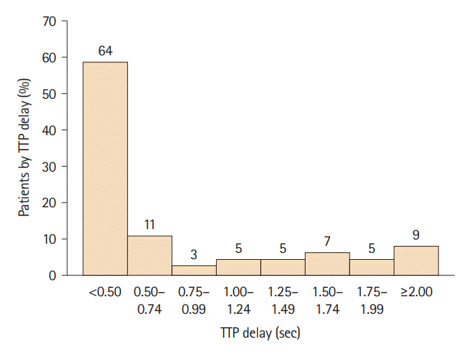
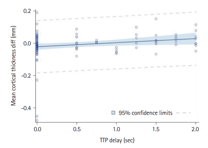
Figure 1.
Histogram showing the distribution of time-to-peak (TTP) delays among 109 subjects, with n=64 (59%) having no delay (0.00–0.49 sec), the remainder having TTP delay ≥0.5 seconds. Nine patients (8.3%) had TTP delay ≥2.0 seconds. Number of subjects in each TTP category is shown above the bars.
jos-2022-02285f1.tif
Figure 2.
Linear regression line showing the relationship between time-to-peak (TTP) delay and cortical thickness asymmetry in the unadjusted model. A positive slope indicates that the greater the TTP delay (greater relative TTP delay in the hemisphere ipsilateral to the carotid stenosis) the greater the relative thinning of the cortex in that hemisphere. Dashed lines indicate the 95% confidence intervals.
jos-2022-02285f2.tif
Table 1.
Demographic, medical, and radiographic characteristics of the study subjects
Characteristic Value (n=110)
Age (yr) 70.2±7.8
Sex
 Male 67 (60.9)
 Female 43 (39.1)
Race
 White 99 (90.0)
 Hispanic 8 (7.3)
 Asian 1 (0.9)
 Native American 1 (0.9)
 Not reported 1 (0.9)
HTN
 Yes 93 (84.5)
 No 17 (15.5)
Smoking
 Never smoked 40 (36.4)
 Current or prior smoker 70 (63.6)
HbA1c (%) (n=109) 6.1±1.20
LDL cholesterol (mg/dL) (n=107) 79.1±32.55
Prior stroke (by report)
 Yes 26 (23.6)
 No 84 (76.4)
iINFARCT (n=108)
 Yes 12 (11.1)
 No 96 (88.9)
cINFARCT (n=108)
 Yes 14 (13.0)
 No 94 (87.0)
Side of stenosis
 Left 51 (46.6)
 Right 59 (53.4)
Fazekas (n=108)
 0 14 (13.0)
 1 10 (9.3)
 2 63 (58.3)
 3 3 (2.8)
 4 18 (16.7)
TTP delay, sec (n=109)
 <0.50 64 (58.7)
 0.50–0.75 11 (10.1)
 0.75–1.00 3 (2.8)
 1.0–1.25 5 (4.6)
 1.25–1.50 5 (4.6)
 1.50–1.75 7 (6.4)
 1.75–2.00 5 (4.6)
 ≥2 9 (8.3)
Cortical thickness mean difference (contra-ipsi) (mm) -0.01±0.08

Values are presented as mean±SD or n (%). All percentages may not total 100% due to rounding.

HTN, hypertension; smoking, current or past history of smoking; LDL, lowdensity lipoprotein; side, left-right hemisphere; iINFARCT, infarct ipsilateral to the stenotic carotid (≥10 mm for cortical, ≥15 mm for subcortical); cINFARCT, infarct contralateral to the stenotic carotid; Fazekas, white matter hyperintensity burden by Fazekas scale; TTP delay, time-to-peak delay in seconds.

Table 2.
Independent predictors of cortical thickness asymmetry by linear regression model
Parameter Estimate SD t P>|t|
Intercept -0.120 0.814 -1.51 0.134
Age 0.001 0.009 0.94 0.348
Sex -0.002 0.155 -0.16 0.873
HTN 0.039 0.196 2.04 0.044
Smok -0.006 0.144 -0.40 0.689
HbA1c 0.002 0.062 0.26 0.794
LDL cholesterol 0.000 0.002 0.71 0.477
Side -0.034 0.144 -2.48 0.015
Fazekas 0.009 0.064 1.45 0.150
iINFARCT 0.042 0.227 1.91 0.059
cINFARCT -0.134 0.237 -5.86 <0.001
TTP delay 0.023 0.103 2.34 0.032

HTN, hypertension; Smok, current or past history of smoking; LDL cholesterol, low-density lipoprotein cholesterol; Side, left-right hemisphere; Fazekas, white matter hyperintensity burden by Fazekas scale; iINFARCT, infarct ipsilateral to the stenotic carotid; cINFARCT, infarct contralateral to the stenotic carotid; TTP delay, time-to-peak delay in seconds.

TOOLS
Similar articles