

 PDF
PDF Citation
 Print
Print


INTRODUCTION
ERCP is performed to diagnose and manage pancreaticobiliary disorders. The guidewire is an essential accessory for ERCP, which facilitates selective cannulation, passage, and dilatation of strictures and supports accessory placement or insertion.1 The guidewires used in ERCP consist of a monofilament core covered with an outer coating. The monofilament core is made of stainless steel, nitinol, or a shape-memory alloy, and the outer sheath is coated with polytetrafluoroethylene, polyurethane, or other polymers. The guidewire tip may be angled, straight, J-shaped, or tapered. In addition, most ERCP guidewires have a hydrophilic coated tip that becomes lubricated when it comes in contact with water. Generally, the length of the guidewire ranges from 150 to 650 cm, and the thickness ranges from 0.018 to 0.038 inches. Guidewires commonly used in ERCP are approximately 450 cm in length and 0.025-0.035 inches in width. The guidewire’s material, tip, length, and thickness determine its stiffness, surface friction, solidity, flexibility, torquability, and hydrophilicity.2 Although rare, guidewires can cause complications, such as perforations,3 knotting,4,5 and fractures.6 Herein, we report a case of guidewire impaction, an unusual complication, during ERCP in our hospital.
CASE REPORT
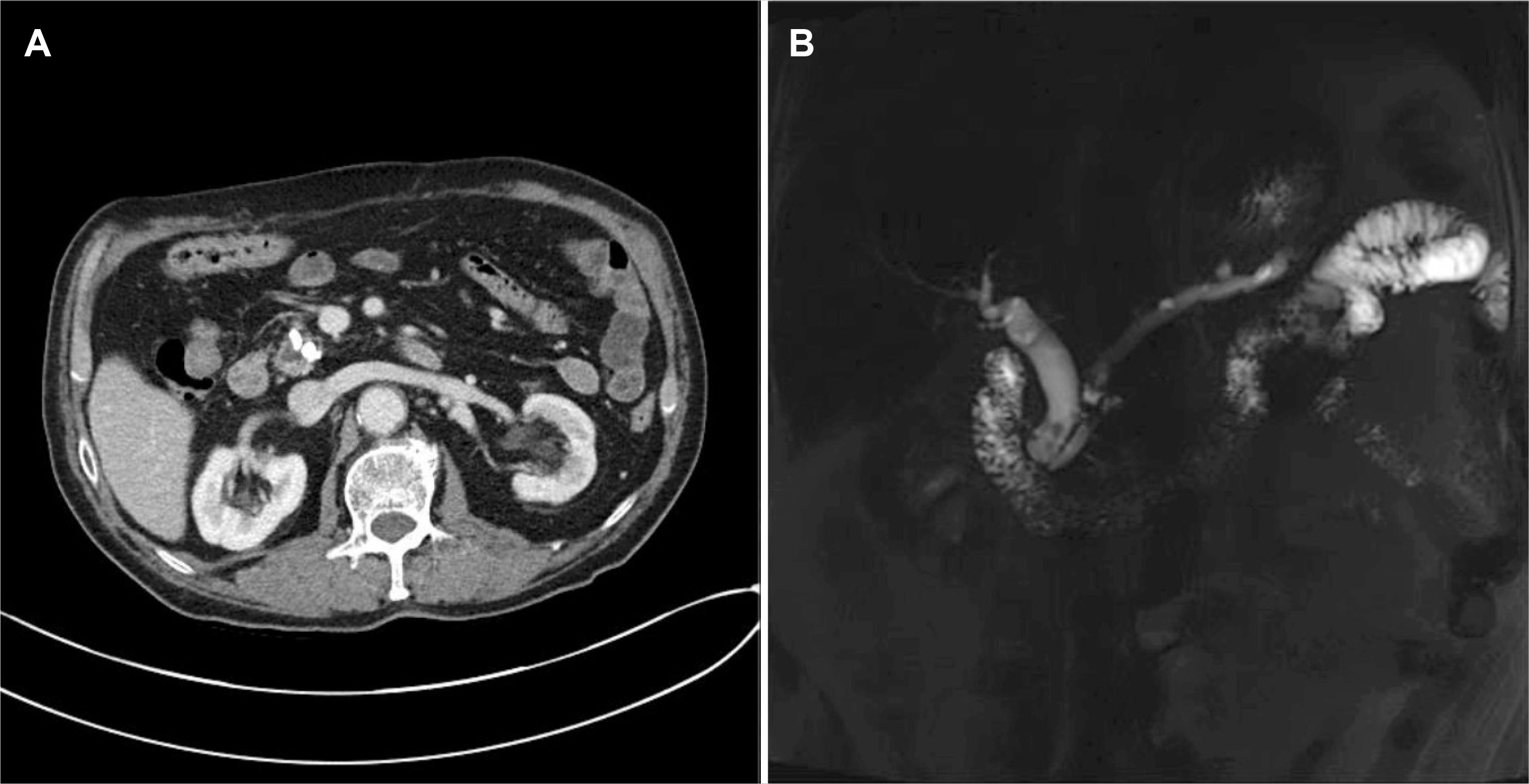
A 73-year-old male with a history of liver transplantation for alcoholic liver cirrhosis was referred to our hospital with abdominal pain. An abdominal ultrasound was performed, which revealed dilatation of the main pancreatic duct. Further, CT and MRCP showed distal common bile duct stones and chronic pancreatitis with dilatation of the main pancreatic duct (maximum, 8 mm) due to pancreaticoliths at the head of the pancreas (Fig. 1). We decided to perform ERCP to remove the common bile duct stones and pancreaticoliths. Accordingly, the ERCP was performed by a junior staff member (H.S.). After duodenal insertion, a guidewire (VisiGlide2®, 0.025 inches, straight type; Olympus, Tokyo, Japan) was passed through the main pancreatic duct. Pancreatography revealed a dilated pancreatic duct with a filling defect, suggesting pancreaticoliths (Fig. 2A). The passage of the guidewire through the pancreaticoliths proved to be difficult. Therefore, we decided to deploy plastic stents for extracorporeal shock wave lithotripsy (ESWL). After endoscopic sphincterotomy, a stent (Zimmon®, 7 Fr, 5 cm, single pigtail; COOK, Limerick, Ireland) was introduced. However, the guidewire was jammed in the stone and could not be removed. The stent was also stuck between the scope and the impacted site, as the guidewire could not be removed (Fig. 2B). It was not possible to remove the impacted guidewire despite using rat tooth forceps. To address this issue, an experienced endoscopist (S.J.B.) was consulted, who inserted another guidewire into the main pancreatic duct to dilate the stenosis. Along the guidewire, a Soehendra® stent retriever (8.5 Fr; COOK Medical, Bloomington, IN, USA) was passed through the stenosis (Fig. 2C). It took approximately 10 minutes to cross the stenosis, and the impacted guidewire was released after the passage of the stent receiver through the stenosis. Finally, the impacted guidewire was removed. After retrieving the impacted guidewire and stent, we deployed another stent (Zimmon®, 7 Fr, 9 cm, single pigtail; COOK) in the main pancreatic duct (Fig. 2D). The patient did not experience any complications. Subsequently, the common bile duct stones were removed by ERCP two days later, and the patient was discharged.
DISCUSSION
In this report, we presented a rare case of guidewire impaction between pancreaticoliths. The impaction of the basket during ERCP is common and expected. However, guidewire impaction during ERCP has been reported in only one case in 1993.7 Similar to the present case, the authors described a guidewire jammed by calculi during an ERCP performed for chronic pancreatitis. In that case, a strong pull resulted in a fracture of the guidewire and a 25-mm long fragment remained in the main pancreatic duct. To address this complication, the patient underwent the Whipple procedure. In the present case, despite the application of a considerable amount of force, the impacted guidewire did not fracture. Therefore, we had to undertake an alternative approach to retrieve the impacted guidewire. The procedure was challenging since the stent was located between the scope and the impaction site. There are various rescue methods for basket impaction, such as extended sphincterotomy, stent insertion, endoscopic mechanical lithotripsy, ESWL, endoscopic laser lithotripsy, transhepatic choledochoscopic lithotripsy, and the use of a second basket.8 However, the methods to resolve guidewire impaction are not well known. In the present case, the impacted guidewire could be retrieved by selective cannulation and dilation of the space next to the impacted guidewire. This is similar to the method used to address basket impaction.
Although reports of complications related to the guidewire used in ERCP are rare, there could be significant complications, such as subcapsular hepatic hematoma,9-11 subcapsular biloma, 12 perforations,3 knotting,4,5 and fracture.6 Subcapsular hepatic hematoma is the most commonly reported guidewire-related complication in ERCP9-11 with an incidence rate of 0.8% to 4%.9 Subcapsular hepatic hematoma is caused by the deep insertion of a guidewire. It is therefore important to check the guidewire tip position during the procedure. Perforation may occur if the guidewire is manipulated excessively or if repeated procedures are performed at the wrong location.3 Knotting may occur if the guidewire is kinked during loop formation.5 A fracture refers to the breakage of the guidewire tip, which may occur when the guidewire is manipulated excessively during torque or if it is pulled at the knot formation stage, and can lead to guidewire retention.6 Accordingly, retained guidewire fragments may lead to infection or abscess formation, and in rare cases, migration of the fragments.13 It is suggested that guidewire retention occurs mostly due to human (34.4%) and leadership-related factor (25.3%). The human factors include inadequate training, credentialing, experience, or supervision of individuals performing the procedure of inserting the catheters and the leadership factors include inadequate policies and procedures or noncompliance with policies and procedures. Other factors are related to communication, assessment, and the physical environment.14 In the present case, the cause of impaction could be attributed to the inexperience of the junior staff member performing the procedure and patient factors, including the large size of the stones and a narrow main pancreatic duct. Furthermore, the joint between the flexible part of the attachment and the hard part of the body of the guidewire is often sharply bent, causing impaction. If the bend could not be further advanced before the stricture, endoscopists should be aware of impaction or parenchyma penetration and should relocate the guidewire using various methods, such as changing the position of the endoscope or using a rotating papillotome. After crossing the stricture, endoscopists should check whether the guidewire has made a smooth loop and can move freely before stent deployment. If the endoscopist wishes to insert a stent when the guidewire has not passed through the stricture, it may be better to gently form a loop with the guidewire in front of the stricture and then insert the stent along it to prevent impaction or parenchyma penetration.15 It is also important to request assistance from experienced senior staff to overcome any challenges faced during the procedure.
In conclusion, although rare, a guidewire might become impacted during ERCP. Therefore, endoscopists should be aware of this issue.

 XML Download