Journal List > Neonatal Med > v.29(3) > 1516080991

Hong, Shin, Choi, Kim, and Kim: Case Series of Isolated Deep Gray Matter Injuries in Preterm Infants

Abstract

Hypoxic-ischemic encephalopathy in neonates is an important cause of brain damage that leads to severe neurological sequelae or death. Brain injury patterns on magnetic resonance imaging (MRI) scans are used to predict neurodevelopmental outcome severity. This case series describes the clinical manifestations and neurologic outcomes of four preterm infants with isolated deep gray matter injuries. Basal ganglia and thalamic lesions were noted without white matter and cerebral cortex lesion on brain MRI. All patients were preterm infants born at less than 33 weeks’ gestation and required resuscitation in the delivery room. All had seizures during the neonatal period requiring anti-seizure medications. Severe neurologic disability was identified in three patients using neurodevelopmental assessment tools. Another patient has not been evaluated with assessment tools yet as he was 2 months’ corrected age, but he was supported by home ventilation via a tracheostomy due to insufficient self-respiration. This case series demonstrates that isolated deep gray matter injuries in preterm infants could predict severe neurodevelopmental outcomes.

서론

신생아기의 저산소성 뇌허혈은 주산기 뇌손상의 중요한 원인이며 저산소허혈뇌증(hypoxicischemic encephalopathy, HIE)으로 진행할 경우 심각한 신경학적 손상이나 사망을 초래하기도 한다. 만삭아에서 HIE의 유병률은 출생아 1,000명당 1–2명 정도이며, 조산아의 경우 출생아 1,000명당 5–9명까지 보고되고 있다[1,2]. 또한 전 세계 연간 신생아 사망 4백만명 중 23%에서 HIE가 관련된 것으로 보고되고 있어[3], 신생아 소생술 프로그램의 개발 및 교육, 위험 인자 및 예후에 관한 연구와 함께, 저체온치료(therapeutic hypothermia)와 같은 예후 개선을 위한 치료 방법에 대한 연구가 활발히 진행되고 있다[4-6].
심부뇌회백질(deep brain gray matter)은 뇌 구조물 중 뇌피질에 위치한 회백질 이외 회백질을 일컫는 말로, 주된 해부학적인 구조물로는 기저핵(basal ganglia), 시상(thalamus)과 시상하부 (hypothalamus)가 있다. 이들의 기능적인 역할에 대해서는 운동과 자세 조절, 보행, 인지 기능에 역할을 하는 것으로 알려졌다[7,8]. 기저핵과 시상의 손상 정도를 크게 경도(mild), 중간 정도(moderate), 그리고 심한 정도(severe)로 구분할 수 있으며, 이에 따라 식이진행, 언어발달, 시력, 발달 지수(developmental quotient) 및 경련의 정도에 차이가 있음이 알려졌다[9]. 또한 신생아 HIE를 진단받은 만삭아를 대상으로 한 이전 연구에 따르면 기저핵과 시상에 병변이 있는 HIE 환자에게서 운동 및 인지기능 장애가 가장 심하게 나타나며 이는 30개월경 뇌성마비로 이행하는 것과도 연관되어 있음이 알려졌다[10]. 하지만, 이러한 심부뇌회백질의 손상은 단독으로 나타나는 경우보다는 뇌백질이나 뇌피질의 회백질 손상과 동반되어 나타나는 경우가 많다.
만삭아에서도 기저핵과 시상하부에 주된 병변이 있는 환자에서 뇌 자기공명영상(magnetic resonance imaging, MRI) 소견상 심한 손상을 나타낸 경우 신경학적인 예후가 불량하다고 보고되었다[9,11,12]. 조산아 중 HIE를 진단받은 환자에게서 뇌 MRI에서 보인 손상 범위에 따른 예후 비교에 대한 연구가 소수 보고되었으나[2,13] 그 수가 만삭아 대상 연구와 비교하여 적으며, 심부뇌회백질 단독 손상이 있는 조산아에 대한 연구는 부족한 상황이다.
이에 연구자들은 본원에서 신생아집중치료실 치료를 받은 환자를 대상으로 심부뇌회백질 단독 손상이 있는 조산아 4명에 대한 임상 양상 및 신경 발달 예후에 대해 보고하고자 한다.

증례

1. 대상

2006년 1월부터 2022년 3월까지 서울대학교 어린이병원 신생아집중치료실에서 치료받은 신생아들을 대상으로 하였다. 출생 체중 1,000 g 미만 혹은 임신나이 29주 미만의 조산아에서 퇴원 전 뇌 MRI를 시행하였으며, 그 외에도 주산기 가사가 의심되거나 출생후 신경학적인 증상이 발생한 환자에서도 뇌 MRI를 시행하였다. 이중 심부뇌회백질 단독 손상이 확인된 신생아 4명에 대해 후향적으로 진료기록 검토하였으며, 신생아집중치료실 입실 이후 입원 시 진료 기록 및 외래 추적관찰 진료기록을 전자의무기록시스템을 통해 수집 및 분석하였다. 본 연구는 기관생명윤리위원회(Institutional Review Board)의 승인을 받았다(IRB No.: 2205-058-1322, 승인 일시: 2022.06.20).

2. 뇌 MRI 촬영

뇌 MRI는 1.5 T MR system (Avanto 1.5T, Siemens Healthcare, Erlangen, Germany), Ingenia Elition X와 3.0 T MR system (SIGNA Premier 3.0T, GE Healthcare, Chicago, IL, USA)으로 시행하였고, 가로면(axial)의 T1-가중영상(T1-weighted conventional spin echo image), T2-가중영상(T2-weighted fast spin echo image), 액체감쇠역전회복(fluid attenuated inversion recovery), 확산강조영상(diffusion weighted Imaging), 겉보기확산계수(apparent diffusion coefficient)를 관찰하였다. 모든 대상아는 호흡이 안정화된 상태에서 진정이 가능한 시기에 뇌 MRI 촬영을 시행하였다. 촬영된 영상은 한 명 이상의 소아영상의학과 전문의가 판독하였다. National Institute of Child Health and Human Development (NICHD) Neonatal Research Network (NRN) score [5]에서는 만삭아에서 발생한 저산소성 허혈성 뇌병변을 분류하고 있다. NICHD-NRN score 0은 정상; 1A는 전두(frontal)와 두정엽(parietal) 피질하 영역의 병변이 있는 경우; 1B는 광범위한 전두, 두정 및 후두(occipital) 피질하 영역에 병변이 있는 경우; 2A는 기저핵, 시상 및 속섬유막(internal capsule)에 병변이 있는 경우; 2B는 기저핵 및 시상병변과 함께 대뇌영역에 병변이 있는 경우; 3은 대뇌 반구의 대대적인 손상이 있는 경우로 정의하고 있다. 금번 연구는 뇌 MRI 판독에서 심부뇌회백질에 속하는 미상핵(caudate nucleus), 렌즈핵(lentiform nucleus)에 주된 손상이 있으면서 표재뇌회백질, 뇌백질 등 손상이 없는 환자, 즉 NICHD-NRN score 2A에 해당하는 환자를 대상으로 하였다. 뇌실내출혈 및 배아기질 출혈은 뇌 MRI에서 확인되지 않더라도 출생 초기에 시행한 뇌초음파에서 발견된 경우를 포함하였다[14].

3. 대상 신생아의 임상 소견

본 연구에서 대상 신생아에 대해 의무기록을 후향적으로 분석하였다. 출생 주수, 산전 검사 결과, 산전 산모 발생 임신 질환, 출생 방법, 출생 사유, 분만 중 태아의 뇌손상을 유발할 수 있는 주요 위험 사건(intrapartum sentinel events), 출생 시 아프가 점수, 신생아 소생술 시행 여부, 출생 체중, 다태아 출생 여부, 임상적으로 확인된 경련(clinical seizure) 여부, 저긴장(hypotonia), 무호흡(apnea) 여부를 확인하였다. 이외 뇌손상 원인, 신생아집중치료실 입원 중 발생한 질환, 출생 후 치료로 승압제 사용 여부, 인공호흡기 사용 여부, 저체온 치료 시행 여부, 신생아집중치료실 입원 기간 등이 포함되었다. 아울러 최근 외래 방문 시 나이, 최근 외래 방문 시 시행한 신경학적인 검진 결과, 최근 시행한 발달검사 결과, 외래 방문 시 진단명, 감각 신경성 난청 여부 등을 수집하였다.

4. 신경학적인 예후평가

신경학적인 평가는 생후 2개월에서 5세까지 마지막 외래 경과 관찰 환자 상태, 신경학적인 검진 및 가장 최근 시행한 객관적인 신경학적 평가 도구인 Gross Motor Function Classification System(GMFCS), 한국 영유아 발달선별검사(Korea Developmental Screening Test for Infants & Children, K-DST)를 바탕으로 시행하였다. 뇌성마비가 있는 경우에 GMFCS 상 level 4 또는 level 5인 경우는 중증, level 2 또는 level 3은 중등도, level 1은 경도 장애로 구분하였다[11].

5. 대상 환자의 임상적 특징

총 4명의 환자가 심부뇌회백질 단독 손상을 보이는 것으로 확인되었다. 모든 환자는 출생 시 조산아였으며, 모두 남아였다. 임신나이 29주부터 32주에 출생한 신생아였으며, 출생체중은 1,250 g부터 2,400 g이었다. 신생아집중치료실 입원 이후 모든 환자에서 경련이 확인되어 항경련제 복용을 시작하였으며, 3명의 환자에서 명확한 저긴장이 있었다(Table 1). 2명의 환자가 신생아집중치료실 입원 중 미숙아 망막병증(retinopathy of prematurity)으로 수술적인 치료를 받았으며, 2명의 환자가 총정맥영양공급(total parenteral nutrition)으로 인한 담즙정체증이 확인되었다. 환자 C의 경우 신생아집중치료실 입실 5일경까지 크레아티닌 상승이 지속된 급성 신손상이 확인되었으나 신대체요법 등의 치료 없이 호전되었다(Table 2). 네 명의 환자에서 심각한 심장질환이나 선천성 유전질환, 대사질환, 신경계 질환 등은 확인되지 않았다.

6. 산모 및 주산기 특징

1명의 산모에서 식이만으로 조절되는 임신성 당뇨(gestational diabetes)가 있었으며, 이외 임신으로 인한 부작용 있는 산모는 없었 다. 1명의 산모가 다태아 분만이었으며, 환자를 제외한 다른 쌍둥이는 정상 신경 발달을 보였다. 출생 전 3명의 환자에게서 태아심박수 감소가 확인되었으며, 태아심박수 감소의 원인은 각각 조기진통, 제대염전(cord entanglement), 산모의 심한 폐렴으로 인한 심폐소생술 시행 등이었다. 다른 한 명의 환자는 태반조기박리로 출산예정일보다 이르게 응급제왕절개술을 시행 받았다. 모든 신생아가 응급 제왕절개술을 통해 분만하였다. 1분 Apgar 점수는 모두 3점 이하로 확인되었으며, 5분 Apgar 점수는 4명 중 3명이 4점 이하였다. 대상환자 모두 분만장에서 기관내삽관을 시행 받았으며, 3명이 분만장에서 심장마사지 및 승압제 투약을 받았다. 신생아집중치료실 입실 이후에 저혈압으로 인해 승압제 사용이 지속적으로 필요했던 대상아는 1명이었다(Table 2).

7. 뇌 MRI 촬영 결과 및 신경 발달 평가

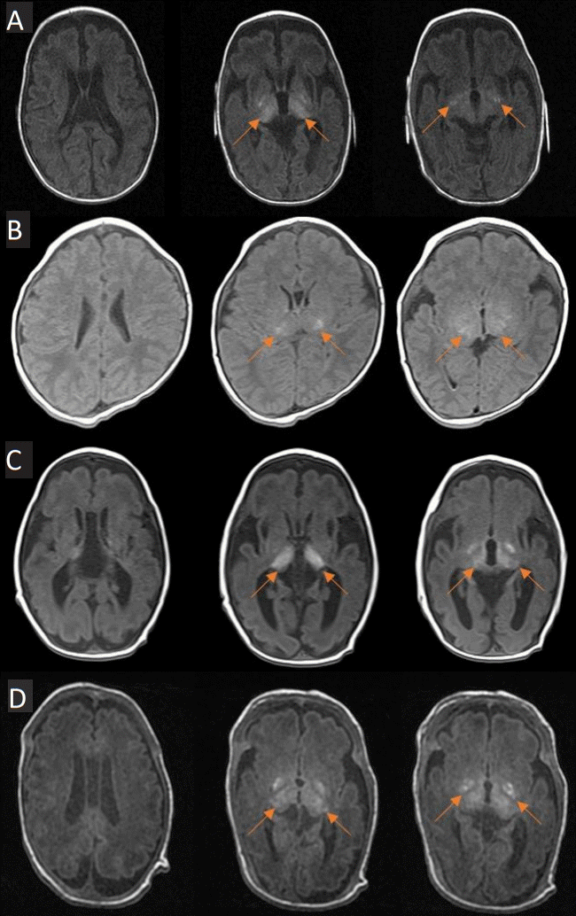
대상 환자들은 생후 23일 이후부터 3개월 이내에 첫 뇌 MRI 촬영을 시행하였다. 특징적인 소견으로는 T1 protocol에서 양측 기저핵과 시상에 대칭적으로 고신호강도가 확인되었으며(Figure 1), 기저핵과 시상의 손상 외에 환자 A에서는 대뇌섬 겉질밑조직(insula subcortex), 환자 C에서는 해마(hippocampus)에 손상이 의심되는 소견이 있었으나 점상(spotty) 양상으로 병변의 크기는 크지 않았다(Table 2).
첫 뇌 MRI 시행 이후 3명의 환자에서 뇌 MRI 추적관찰을 시행하였다. 환자 A는 2세경 항경련제 조절을 위해 입원 시 추적검사를 시행하였고, T2 protocol에서 양측 기저핵과 시상 부위뿐 아니라 신생아기에는 저명하지 않던 양측 시상 옆 백질과 뇌실 옆 백질에도 대칭적으로 고신호강도가 확인되었다. 환자 C는 생후 10개월경 조절되지 않는 경련으로 타병원 입원하였고, 당시 입원기간 중 10분간 심폐소생술 시행 후 본원으로 전원이 되었다. 당시 MRI 추적검사 소견에서 이전 광범위한 뇌 위축 소견과 함께, 양측 뇌 반구 전반적인 급성 저산소 허혈 손상 소견도 함께 확인되었다. 환자 D는 신생아중환자실 퇴원 전 추적관찰을 위한 뇌 MRI 시행하였고, 기존에 있던 창백핵 담창구(globus pallidus)와 시상 병변이 덜 두드려져 보이는 소견 이외에 추가적으로 확인된 병변은 없었다.
4명 중 2명이 GMFCS를 이용한 신경학적인 평가를 각각 교정 월령 11개월, 만 5세에 시행하였으며, 모두 level 4 이상으로 심각한 뇌병변 장애가 확인되었다. 환자 C는 교정 1개월부터 만 4세까지 차례로 시행한 K-DST에서 모두 0점을 기록하였으며, 경피적 내시경 위조루관을 이용한 영양 섭취 중이다. 또한 지속적인 근 긴장 및 경직과 간헐적인 근 경직 조절 불능으로 항경련제와 더불어 baclofen, diazepam, clonazepam을 투약하고 있다. 환자 D는 현재 교정연령 2개월로 객관적인 신경학적 평가를 진행하지 못했으나, 신생아집중치료실 퇴원 시 자가호흡으로는 산소포화도 유지가 불충분하여 기관절개술 후 가정용 인공호흡기 적용하며 퇴원 진행하였다(Table 2).

고찰

뇌 MRI는 뇌손상의 위치 및 정도를 영상자료를 통해 직관적으로 확인할 수 있는 장점이 있으며, 뇌 MRI 소견이 환자의 신경학적인 예후와도 연관되어 있음이 잘 알려졌다[9,15]. 금번 보고에서는 뇌손상이 의심되는 환자에 대해 시행한 뇌 MRI에서 심부뇌회백질 단독 손상, 특히 기저핵과 시상에 국한된 손상이 있는 조산아 4례의 발달 상태를 기술하였다. 조산아에서 HIE가 있는 경우에 만삭아와 다르게 심부뇌회백질의 손상이 잘 발생할 수 있으며, 사망이나 심한 신경학적 손상과 연관성이 있는 것으로 보고된바 있다[16]. Okereafor 등[16]에 따르면 기저핵 및 시상의 손상과 함께 뇌백질 손상이 동반된 경우엔 뇌성마비 혹은 사망과 같이 예후가 불량하였으나 시상에 국한된 뇌손상이 확인된 조산아 2명의 경우 발달지연 정도의 양호한 예후를 보였다. 또한 Logitharajah 등[13]에 따르면 뇌간 손상과 동반한 심한 기저핵, 시상 손상이 있는 경우엔 예후가 불량하였으나, 경도 또는 중등도의 기저핵, 시상 단독 손상이 있는 3명의 조산아의 경우는 양호한 예후를 보였다. 하지만, 최근 국내 보고에서는 기저핵과 시상 손상에 국한된 병변을 보인 만삭아 4명 모두 불량한 신경학적 예후를 보였다[17]. 본 증례에서 보고한 4명의 조산아 모두 가장 최근 추적관찰 시 확인한 임상 양상, 신경학적인 검진, 발달 평가를 종합하였을 때 불량한 신경학적 예후를 보임이 확인되었다. 이번 연구는 심부뇌회백질 단독 손상을 보인 조산아를 보고한 연구로, 기존 만삭아에서 불량한 예후를 보인 국내 사례와 그 예후가 다르지 않음을 보여 주었다. 하지만 앞서 언급한 기존 외국 사례와는 차이가 있는 결과로 향후 대규모 연구를 통해 심뇌회백질 단독 손상이 있는 경우의 유병률과 예후에 대한 명확한 정립이 필요하겠다.
혈관 취약성(vascular fragility), 미숙한 혈뇌장벽기능(blood-Brain barrier function), 발달 단계에 있는 신경아교세포(developing neuroglial cell), 희소돌기아교세포의 전구세포와 희소돌기아교세포(oligodendroglial progenitor and the immature oligodendrocytes), 선택적 취약성(selective vulnerability) 등은 조산아에서 저산소성 허혈과 연관 있는 요인으로 알려졌다[18]. 조산아에서 분만 중 태아의 뇌손상을 유발할 수 있는 주요 위험 사건 등으로 주산기 동안 심한 저혈압이 발생할 경우 미성숙한 뇌 영역 중 가장 대사 활동이 활발한 시상, 뇌간, 소뇌가 허혈성 손상에 취약한 부분으로 알려졌다[19]. 조산아에서 심부뇌회백질 영역이 손상되는 기전에 대한 정립은 되어있지 않으나, 조산아에서 단일광자 단층촬영(single photon emission computed tomography)을 이용하여뇌 영상을 확인하였을 때, 뇌피질의 2배, 뇌백질의 4배가량의 높은 혈액 흐름이 기저핵 부분에 분포하는 것이 밝혀졌다[20].
신생아 HIE 환자에서 심부뇌회백질 손상은 비교적 흔하게 확인되는 병변이나, 이 증례보고에서는 극소조산아에 해당하는 환자 2명을 포함한 환자 4명의 조산아에서 심부뇌회백질 단독 손상을 보이는 환자의 예후에 대해 보고 하였다. 이를 통해 조산아에서 심한 뇌실내출혈이나 뇌백질연화증이 없는 경우에도 단독으로 심부뇌회백질이 발견되는 경우 불량한 예후를 예측하는 데 활용될 수 있을 것이다.

Notes

Ethical statement

This study was approved by the Institutional Review Board of Seoul National University Hospital (IRB No.: 2205-058-1322, approval date: June 20, 2022). Written informed consent by the patients was waived due to the retrospective nature of our study.

Conflicts of interest

No potential conflict of interest relevant to this article was reported.

Author contributions

Conception or design: S.H.S.

Acquisition, analysis, or interpretation of data: K.T.H., Y.H.C., E.K.K., H.S.K., S.H.S.

Drafting the work or revising: K.T.H., S.H.S.

Final approval of the manuscript: All authors read and approved the final manuscript.

Funding

None

ACKNOWLEDGMENTS

None

REFERENCES

1. Schmidt JW, Walsh WF. Hypoxic-ischemic encephalopathy in preterm infants. J Neonatal Perinat Med. 2010; 3:277–84.
2. Chalak LF, Rollins N, Morriss MC, Brion LP, Heyne R, Sanchez PJ. Perinatal acidosis and hypoxic-ischemic encephalopathy in preterm infants of 33 to 35 weeks’ gestation. J Pediatr. 2012; 160:388–94.
3. Lawn JE, Cousens S, Zupan J; Lancet Neonatal Survival Steering Team. 4 Million neonatal deaths: when? where? why? Lancet. 2005; 365:891–900.
4. Shankaran S, Laptook AR, McDonald SA, Hintz SR, Barnes PD, Das A, et al. Acute perinatal sentinel events, neonatal brain injury pattern, and outcome of infants undergoing a trial of hypothermia for neonatal hypoxic-ischemic encephalopathy. J Pediatr. 2017; 180:275–8.
5. Shankaran S, Barnes PD, Hintz SR, Laptook AR, Zaterka-Baxter KM, McDonald SA, et al. Brain injury following trial of hypothermia for neonatal hypoxic-ischaemic encephalopathy. Arch Dis Child Fetal Neonatal Ed. 2012; 97:F398–404.
6. Jacobs SE, Berg M, Hunt R, Tarnow-Mordi WO, Inder TE, Davis PG. Cooling for newborns with hypoxic ischaemic encephalopathy. Cochrane Database Syst Rev. 2013; 2013:CD003311.
7. Utter AA, Basso MA. The basal ganglia: an overview of circuits and function. Neurosci Biobehav Rev. 2008; 32:333–42.
8. Nelson AB, Kreitzer AC. Reassessing models of basal ganglia function and dysfunction. Annu Rev Neurosci. 2014; 37:117–35.
9. Martinez-Biarge M, Diez-Sebastian J, Rutherford MA, Cowan FM. Outcomes after central grey matter injury in term perinatal hypoxic-ischaemic encephalopathy. Early Hum Dev. 2010; 86:675–82.
10. Miller SP, Ramaswamy V, Michelson D, Barkovich AJ, Holshouser B, Wycliffe N, et al. Patterns of brain injury in term neonatal encephalopathy. J Pediatr. 2005; 146:453–60.
11. Martinez-Biarge M, Diez-Sebastian J, Kapellou O, Gindner D, Allsop JM, Rutherford MA, et al. Predicting motor outcome and death in term hypoxic-ischemic encephalopathy. Neurology. 2011; 76:2055–61.
12. Rutherford MA, Pennock JM, Counsell SJ, Mercuri E, Cowan FM, Dubowitz LM, et al. Abnormal magnetic resonance signal in the internal capsule predicts poor neurodevelopmental outcome in infants with hypoxic-ischemic encephalopathy. Pediatrics. 1998; 102(2 Pt 1):323–8.
13. Logitharajah P, Rutherford MA, Cowan FM. Hypoxic-ischemic encephalopathy in preterm infants: antecedent factors, brain imaging, and outcome. Pediatr Res. 2009; 66:222–9.
14. Inder TE, Perlman JM, Volpe JJ. Preterm intraventricular hemorrhage/posthemorrhagic hydrocephalus. In : Volpe JJ, Inder TE, Darras BT, de Vries LS, du Plessis AJ, Neil J, editors. Volpe's Neurology of the Newborn. 6th ed. Philadelphia: Elsevier;2018. p. 637–98.e21.
15. Martinez-Biarge M, Bregant T, Wusthoff CJ, Chew AT, Diez-Sebastian J, Rutherford MA, et al. White matter and cortical injury in hypoxic-ischemic encephalopathy: antecedent factors and 2-year outcome. J Pediatr. 2012; 161:799–807.
16. Okereafor A, Allsop J, Counsell SJ, Fitzpatrick J, Azzopardi D, Rutherford MA, et al. Patterns of brain injury in neonates exposed to perinatal sentinel events. Pediatrics. 2008; 121:906–14.
17. Jeong SY, Choi SY, Chang YP, Lee YS. Neurodevelopmental outcomes according to brain injury patterns in neonates with postasphyxial hypoxic ischemic encephalopathy. Neonatal Med. 2017; 24:32–9.
18. Gopagondanahalli KR, Li J, Fahey MC, Hunt RW, Jenkin G, Miller SL, et al. Preterm hypoxic-ischemic encephalopathy. Front Pediatr. 2016; 4:114.
19. Chao CP, Zaleski CG, Patton AC. Neonatal hypoxic-ischemic encephalopathy: multimodality imaging findings. Radiographics. 2006; 26 Suppl 1:S159–72.
20. Borch K, Greisen G. Blood flow distribution in the normal human preterm brain. Pediatr Res. 1998; 43:28–33.

Figure 1.
Transverse T1-weighted brain magnetic resonance imaging findings of all patients. (A) Images of patient A taken at a post-menstrual age (PMA) of 38+0 weeks. (B) Images of patient B taken at a PMA of 40+2 weeks. (C) Images of patient C taken at a PMA of 34+2 weeks. (D) Images of patient D taken at a PMA of 34+2 weeks. The arrows indicate T1 high signal intensity lesions in the deep gray matter.
nm-2022-29-3-117f1.tif
Table 1.
Perinatal Characteristics and Clinical Case Presentations
GA (wk) Birth weight (g) Sex Outborn Multiple birth Seizure Hypotonia Perinatal problem FHRP Mode of delivery Apgar score Resuscitation at delivery room Cord BGA (pH/BE) Diagnosis during NICU care
A 32+3 1,250 M Yes No + NA Placental abruption Normal EmCS 0-3-6 Intubation (+) NA HIE, ROP stage 3, 2’ASD, mild BPD
CM (+)
B 29+3 1,510 M Yes No + + PTL Deceleration EmCS 0-1-4 Intubation (+) NA HIE, recurrent aspiration pneumonia, CRE sepsis, TPN-induced cholestasis, ROP stage 2
CM (+)
C 31+0 1,690 M No Yes* + + Cord entanglement Deceleration EmCS 3-7 Intubation (+) 7.33/–3.3 HIE, AKI, ROP stage 1, GMH
CM (–)
D 29+0 1.36 M Yes No + + GDM, pneumonia due to COVID-19 infection Deceleration due to maternal cardiac arrest EmCS 3-4 Intubation(+) 7.316/–0.5 HIE, recurrent aspiration pneumonia, ROP stage 3, TPN induced cholestasis
CM (+)

ROP stage: 1-demarcation line, 2-ridge, 3-ridge with extra retinal fibrovascular proliferation, 4-subtotal retinal detachment, 5-total retinal detachment.

* No significant injury in co-twin.

Abbreviations: GA, gestational age; FHRP, fetal heart rate pattern; BGA, blood gas analysis; BE, base excess; NICU, neonatal intensive care unit; NA, not available; EmCS, emergency Cesarean section; CM, cardiac massage; HIE, hypoxic ischemic-encephalopathy; ROP, retinopathy of prematurity; ASD, atrial septal defect; BPD, bronchopulmonary dysplasia; PTL, preterm labor; CRE, carbapenem-resistant Enterobacteriaceae; TPN, total parenteral nutrition; AKI, acute kidney injury; GMH, germinal matrix hemorrhage; GDM gestational diabetes; COVID-19, coronavirus disease 2019.

Table 2.
Radiologic Findings and Neurodevelopmental Outcomes of Preterm Infants with Deep Gray Matter Injuries
Age at brain MRI PMA at initial brain MRI Reading NICHD-NRN score IVH Age of last OPD FU Neurologic developmental outcome
A 1 month 38+0 Symmetric hyperintense both thalamus T1WIs 2A 3-year-old GMFCS level 5 (at CA 10 months), epilepsy
Relatively symmetric T2 high/T1 low lesions at both BG (mainly globus pallidus) and insula subcortex CP, severe disability
B 2 months 40+2 T1 high SI lesions at bilateral thalamus 2A 5-year-old GMFCS level 5 (at 5-year-old)
CP, severe disability
C 23 days 34+2 Symmetric T1 high signal intensity in the bilateral thalamus, hippocampus and parahippocampal gyrus 2A +, Grade 1 5-year-old K-DST: all zero (until 4- yearold), epilepsy, tube feeding through PEG tube
CP, severe disability
D 1 month 34+2 Bilateral symmetric T1 high signal lesion at the globus pallidus and thalamus 2A CA 2 months Tracheostomy with home ventilator apply

Abbreviations: MRI, magnetic resonance imaging; PMA, post menstrual age; NICHD-NRN, National Institute of Child Health and Human Development Neonatal Research Network; IVH, intraventricular hemorrhage; OPD, out-patient department; FU, follow-up; T1WI, T1-weighted image; BG, basal ganglia; SI, signal intensity; GMFCS, Gross Motor Function Classification System; CP, cerebral palsy; K-DST, Korean Developmental Screening Test for Infants & Children; PEG, percutaneous endoscopic gastrostomy; CA, corrected age.

TOOLS
Similar articles