

 PDF
PDF Citation
 Print
Print


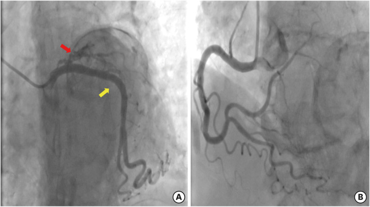
A 91-year-old woman with a history of myocardial infarction (MI) 36 years ago presented with aggravating dyspnea for one week. Since her diagnosis of MI, she has been prescribed guideline-directed medical therapy (GDMT), including an anti-platelet agent, beta-blocker, renin-angiotensin-aldosterone system inhibitor (RAASi), nitrate, and statin. Chest radiography revealed cardiomegaly and pulmonary edema with pleural effusion. There was a large, well-demarcated, spherical calcified mass (Figure 1A). Computed tomography showed an extensive, curvilinear myocardial calcification along with the apical left ventricle (LV) aneurysm (Figure 1B). Transthoracic echocardiography showed dilated LV with apical aneurysm complicated by severely decreased LV systolic function (LV ejection fraction=19%) (Supplementary Videos 1, 2, 3). Coronary angiography performed 10 years before presentation showed the chronic total occlusion at the ostium of the left anterior descending artery with an absence of other epicardial coronary artery stenoses (Figure 2). The patient was diagnosed with acute on chronic systolic heart failure. After intensive medical care with diuresis and inotropic therapy, the patient’s condition improved. She was discharged with medication adjustment, changing RAASi into an angiotensin receptor-neprilysin inhibitor and adding a sodium-glucose cotransporter-2 inhibitor.
A massive myocardial calcification is a rare sequel of extensive MI,1) associated with poor prognosis by causing complications such as chronic heart failure and arrhythmia.2) We report a rare case of a long-term survivor of MI with extensive myocardial calcification. The patient described here had not received revascularization but maintained GDMT for the management of post-MI heart failure.
We obtained written informed consent from the patient.


 XML Download