Journal List > Acute Crit Care > v.37(4) > 1516080751

Huh, Choi, Jo, Huh, Hong, Shim, Lim, and Koh: Incidence and risk factors associated with progression to severe pneumonia among adults with non-severe Legionella pneumonia

Abstract

Background

Legionella species are important causative organisms of severe pneumonia. However, data are limited on predictors of progression to severe Legionella pneumonia (LP). Therefore, the risk factors for LP progression from non-severe to the severe form were investigated in the present study.

Methods

This was a retrospective cohort study that included adult LP patients admitted to a 2,700-bed referral center between January 2005 and December 2019.

Results

A total of 155 patients were identified during the study period; 58 patients (37.4%) initially presented with severe pneumonia and 97 (62.6%) patients with non-severe pneumonia. Among the 97 patients, 28 (28.9%) developed severe pneumonia during hospitalization and 69 patients (71.1%) recovered without progression to severe pneumonia. Multivariate logistic regression analysis showed platelet count ≤150,000/mm3 (odds ratio [OR], 2.923; 95% confidence interval [CI], 1.100–8.105; P=0.034) and delayed antibiotic treatment >1 day (OR, 3.092; 95% CI, 1.167–8.727; P=0.026) were significant independent factors associated with progression to severe pneumonia.

Conclusions

A low platelet count and delayed antibiotic treatment were significantly associated with the progression of non-severe LP to severe LP.

INTRODUCTION

Legionella species (spp.) are small Gram-negative bacilli usually found in natural aqueous environments [1,2]. The bacteria can cause respiratory infection in humans and are an important causative organism of pneumonia [3,4]. Legionella pneumonia (LP) accounts for 2.4%–12.5% of community-acquired pneumonia (CAP) cases [5,6]. In the last decade, the incidence of LP has rapidly increased worldwide [7-9]. The number of cases reported to the Korea Centers for Disease Control and Prevention was 6 in 2006 in contrast to 501 in 2019 [10]. The reasons for such a sharp increase include widespread nucleic acid amplification, urinary antigen testing, and the introduction of mandatory reporting systems for the disease in several countries [11-13]. Furthermore, an aging population, infrastructure, and changes in the global climate have been suggested as factors contributing to this increase [14].
In prior studies, 20.7%–78.6% of LP patients were shown to initially have or eventually progress to severe pneumonia requiring intensive care unit (ICU) admission [15-18]. Despite the growing importance of LP in public health, the issues associated with the progression of LP have been addressed in only a few studies. Therefore, in the present study, the risk factors associated with the progression of non-severe LP to severe LP in adults were investigated.

MATERIALS AND METHODS

The study protocol was approved by the Institutional Review Board of Asan Medical Center (No. 2021-2424). The requirement for informed consent was waived due to the retrospective nature of the analysis.

Study Population and Data Collection

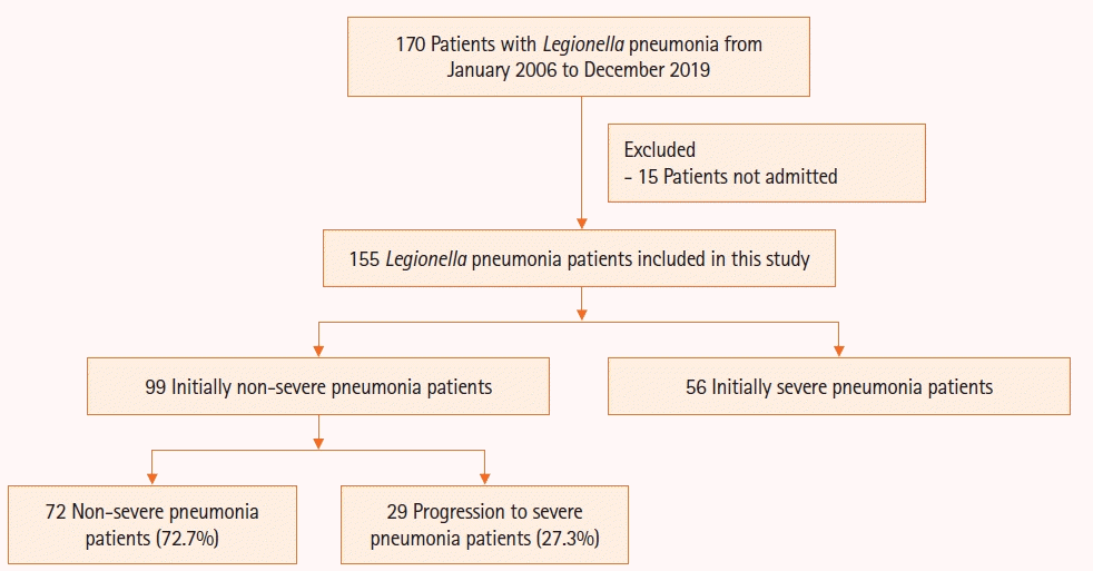
Patients whose clinical specimens yielded positive results for Legionella spp. between January 2005 and December 2019 at Asan Medical Center, a 2,700-bed tertiary hospital, were screened for this study. Using the clinical microbiology electronic database of the hospital, 170 LP patients >18 years of age were identified, their medical records were reviewed, and patients treated in-hospital for LP were selected. Finally, a total of 155 patients were included in the present study (Figure 1). Data on demographics, underlying diseases or conditions, clinical manifestations, laboratory and radiologic findings, pathogens, treatment, and outcomes were retrospectively collected from the electronic records. Survival data were obtained from the National Health Insurance records.

Definitions

A LP case was defined as a patient with pneumonia whose specimens yielded microbiologic evidence of Legionella spp. The diagnosis of pneumonia was confirmed if the patient presented with new or increased cough, sputum, fever or hypothermia, abnormal white blood cell count, or elevated C-reactive protein as well as new pulmonary infiltration on radiologic examination. Legionella spp. were detected based on serology (type-specific immunofluorescence antibody assay conducted at the Seoul Research Institute of Public Health and Environment), BinaxNOW urinary antigen testing (Abbott Diagnostic Medical, Lake Forrest, CA, USA), PCR with the AmpliSens Legionella pneumophila-FRT PCR kit (InterLabService Ltd., Moscow, Russia), Seeplex PneumoBacter ACE Detection assay (Seegene, Seoul, Korea), or culture of respiratory tract specimens using buffered charcoal yeast extract agar (bioMérieux, Marcy-l'Étoile, France).
Severe pneumonia was defined as septic shock with the need for vasopressor administration, respiratory failure requiring mechanical ventilation, as defined in the American Thoracic Society and Infectious Disease Society of America’s guidelines [19], or PaO2/FiO2 (partial pressure of arterial oxygen/fraction of inspired oxygen) ratio <200. The criteria for clinical stability were temperature ≤37.8℃, heart rate ≤100 beats/min, respiratory rate ≤24 breaths/min, systolic blood pressure ≥90 mm Hg, arterial oxygen saturation ≥ 90% or pO2 ≥60 mm Hg on room air, ability to maintain oral intake, and normal mental status [20].
Adequate antibiotic treatment was defined as the use of macrolides or fluoroquinolones. Although other classes of empirical antibiotics were also frequently administered, they were not categorized as “adequate” due to their lack of efficacy against LP. The macrolides used in this study were clarithromycin and azithromycin. The fluoroquinolones included ciprofloxacin, levofloxacin, and moxifloxacin. Chronic lung diseases included chronic obstructive pulmonary disease, interstitial lung disease, bronchiectasis, emphysema, asthma, and chronic bronchitis.

Statistical Analysis

Continuous variables were compared using Student t-test or Mann-Whitney test as appropriate. Categorical variables were compared using chi-square test or Fisher’s exact test. The relationship among risk factors for progression to severe pneumonia was evaluated using logistic regression analysis. Factors with P<0.1 in univariate analysis were selected for multivariate analysis. Statistical analysis was performed using R version 3.6.0 (R Foundation for Statistical Computing, Vienna, Austria). All tests of significance were two-sided, and P<0.05 was considered statistically significant (two-tailed).

RESULTS

Baseline Characteristics

The baseline characteristics of the 155 included patients are presented in Table 1. The mean number of patients per year increased from 5.2 per year between 2005 and 2009 to 12.9 per year between 2010 and 2019. The mean age was 64.9 years and 76.1% of patients were male. CAP was the most common (45.2%) and 34.8% and 20.0% of patients had healthcare-associated pneumonia and hospital-acquired pneumonia, respectively. A total of 89 patients (57.4%) were immunocompromised. The most common symptom was fever (67.1%), followed by sputum (63.9%) and cough (62.6%). A total of 22 patients (14.2%) had altered mental status. At LP diagnosis, the median serum sodium concentration was 134.0 mmol/L and the median C-reactive protein level was 16.8 mg/dl. The median leukocyte and platelet counts were 9,200/mm3 and 174,000/mm3, respectively. A total of 65 patients had thrombocytopenia (<150,000/mm3). Radiologic examination revealed bilateral lung involvement in 58.7% of patients and pleural effusion in 16.1% of patients.

Treatment and Clinical Outcomes of LP

During hospitalization, most patients (98.7%) were treated with macrolides, fluoroquinolones, or both for LP. Fluoroquinolone therapy, without macrolide was administered to 102 patients (65.8%) and macrolide therapy without fluoroquinolone was administered to 11 patients (7.1%). A combination of both was administered to 40 patients (25.8%). At presentation, 58 patients (37.4%) had severe pneumonia. Among the 97 patients without severe pneumonia (62.6%, 97/155), 28 patients (28.9%, 28/97) progressed to severe pneumonia and 69 patients (71.1%, 69/97) recovered without deterioration (Figure 1). At presentation, leukocyte and platelet counts were lower in the progressed group than in the non-progressed group; however, other baseline characteristics were comparable (Table 2). Clinical stability was achieved in 60 patients and the median time since the presentation of LP was 6 days (interquartile range, 4–11 days). The 90-day mortality rates of patients with non-severe LP, initially severe LP, and severe LP after progression were 17.4% (12/69), 53.4% (31/58), and 75.0% (21/28), respectively (P=0.001).

Risk Factors for Progression to Severe Pneumonia in LP

Table 3 shows analysis of the risk factors for progression to severe pneumonia for the 97 patients with initially non-severe LA. In univariate analysis, platelet count ≤150,000/mm3 (odds ratio [OR], 3.30; 95% confidence interval [CI], 1.34–8.43; P=0.010) and delayed antibiotic treatment >1 day (OR, 2.75; 95% CI, 1.11–7.10; P=0.032) were significant factors associated with the development of severe pneumonia during hospitalization. In addition, immunocompromised state (OR, 2.35; 95% CI, 0.93–6.38; P=0.078) and bilateral involvement on radiologic examination (OR, 2.13; 95% CI, 0.88–5.35; P=0.098) were included for multivariate analysis. Multivariate analysis identified platelet count ≤150,000/mm3 (adjusted OR [aOR], 2.92; 95% CI, 1.10–8.11; P=0.034) and delayed antibiotic treatment >1 day (aOR, 3.09; 95% CI, 1.17–8.73; P=0.026) as independent risk factors for severe disease in LP.

DISCUSSION

In the present study, approximately one-third of patients initially had severe LP at the time of hospital presentation. Among the remaining two-thirds of patients with non-severe LP, one-quarter progressed to severe pneumonia during hospitalization. Risk factors associated with the development of severe pneumonia were low platelet count and delayed antibiotic treatment. Overall, the death rate was high among patients who progressed to severe LP.
The high percentage of severe pneumonia among patients with LP in this study is comparable to the findings in several previous studies. In a prospective study conducted in Spain, among 30 hospitalized LP patients, 50% had respiratory failure [15]. The outcomes of 14 LP patients were reported in a Canadian study and 78.6% (11/14) required ICU admission [16]. However, conflicting results have also been reported. In a large prospective French study, 27.4% (30/540) of 540 community-acquired LP patients required ICU care [17]. In a study in Italy, 20.7% (24/116) of 116 patients retrospectively analyzed required ICU admission [18]. These discrepancies may be attributed to differences in the patient population and number of participants. The present study was relatively large and performed at a tertiary care hospital. The inclusion of immunocompromised patients may have contributed to the relatively high rate of severe LP.
A low platelet count has been associated with the progression of non-severe LP. In a recent prospective cohort study involving 250 hospitalized CAP patients, both ICU admission rate and the rate of mechanical ventilation were higher in the thrombocytopenia group (<100,000/mm3) [21]. In another retrospective cohort study involving 500 hospitalized patients with CAP, thrombocytopenia was significantly associated with death [22]. The present study results are in agreement with these studies, indicating a low platelet count might be predictive of non-severe LP progressing to severe LP. Platelets play various roles in the immune response; they can recognize pathogenic bacteria or toxins, induce the acute phase response, and promote the innate immune cell response [23]. Recently, streptococcal M1 protein was shown to activate platelets, resulting in the acquisition of C1q on the surface of the platelets and increase apoptosis and phagocytosis [24]. A similar mechanism for enhancing complement activity may be present in Legionella spp. The L. pneumophila lipopolysaccharide activates the classical complement pathway [25]. In a study of samples from infected patients, an activation of platelets and thrombocytopenia was observed [26]. In addition, L. pneumophila is an intracellular organism that utilizes complement receptors for entry into monocytes for replication [27]. A decreased platelet count may indirectly indicate susceptibility to Legionella infection.
In the present study, delayed antibiotic treatment >1 day was a risk factor for progression to severe pneumonia, which is consistent with a recent Italian study [10]. The investigators showed macrolide/levofloxacin administration within 24 hours of admission was associated with fewer transfers to the ICU (OR, 0.20; 95% CI, 0.05–0.73). In a Taiwanese study, the ICU admission rate was also reportedly higher in patients who received delayed treatment compared with subjects who received timely antibiotic treatment (68.7% vs. 31.2%) [28]. The importance of administering antibiotics in a timely manner has also been demonstrated for all-cause pneumonia [29]. Our finding reemphasizes the importance of adequate early therapy for non-severe LP as well as severe LP.
The present study had several limitations. First, this was a retrospective study performed at a single referral center. Approximately half the patients were immunocompromised. Due to the possibility of selection bias of the study population, the results may not be generalizable to other patient populations. Second, patients who were not hospitalized were excluded from the analysis which may have been a source of bias. However, the number of excluded patients was relatively small. In conclusion, severe pneumonia frequently occurs among patients with LP and the mortality rate is high. The risk of progression to severe pneumonia is higher among patients with subnormal platelet counts and delayed antibiotic treatment >1 day. Thus, that early administration of antibiotics against Legionella spp. may be beneficial for LP patients, especially those with low platelet counts.

HIGHLIGHTS

▪ Among 155 patients with Legionella pneumonia at presentation, 58 patients (37.4%) had severe pneumonia and 97 patients (62.6%) had non-severe pneumonia; 28 of the 97 patients (28.9%) progressed to severe pneumonia.
▪ Platelet count ≤150,000/mm3 and delayed antibiotic treatment >1 day were factors associated with progression to severe pneumonia in initially non-severe Legionella pneumonia patients.

Notes

CONFLICT OF INTEREST

No potential conflict of interest relevant to this article was reported.

AUTHOR CONTRIBUTIONS

Conceptualization: JYH, SHC, YK. Data curation: JYH, SHC, KWJ, TSS. Formal analysis: JYH, SHC. Methodology: JYH, SHC, YK. Project administration: JWH, SBH, CML. Visualization: JYH, SHC. Writing–original draft: JYH, SHC. Writing–review & editing: JYH, SHC, YK.

REFERENCES

1. Diederen BM. Legionella spp. and Legionnaires’ disease. J Infect. 2008; 56:1–12.
crossref
2. Cunha BA, Burillo A, Bouza E. Legionnaires’ disease. Lancet. 2016; 387:376–85.
crossref
3. Carratalà J, Garcia-Vidal C. An update on Legionella. Curr Opin Infect Dis. 2010; 23:152–7.
crossref
4. Morgan AJ, Glossop AJ. Severe community-acquired pneumonia. BJA Educ. 2016; 16:167–72.
crossref
5. Chong YP, Jung KS, Lee KH, Kim MN, Moon SM, Park S, et al. The bacterial etiology of community-acquired pneumonia in Korea: a nationwide prospective multicenter study. Infect Chemother. 2010; 42:397–403.
crossref
6. Sopena N, Sabrià M, Pedro-Botet ML, Manterola JM, Matas L, Domínguez J, et al. Prospective study of community-acquired pneumonia of bacterial etiology in adults. Eur J Clin Microbiol Infect Dis. 1999; 18:852–8.
crossref
7. Centers for Disease Control. Notice to readers: final 2014 reports of nationally notifiable infectious diseases. MMWR Morb Mortal Wkly Rep. 2015; 64:1019.
8. European Centre for Disease Prevention and Control. Legionnaires’ disease: annual epidemiological report for 2019. Stockholm: European Centre for Disease Prevention and Control; 2019.
9. Fukushima S, Hagiya H, Otsuka Y, Koyama T, Otsuka F. Trends in the incidence and mortality of legionellosis in Japan: a nationwide observational study, 1999-2017. Sci Rep. 2021; 11:7246.
crossref
10. Korea Disease Control and Prevention Agency. Legionellosis [Internet]. Cheongju: Korea Disease Control and Prevention Agency; 2020 [cited 2022 Sep 1]. Available from: https://www.kdca.go.kr/npt/biz/npp/ist/simple/simplePdStatsMain.do.
11. Cassell K, Thomas-Lopez D, Kjelsø C, Uldum S. Provincial trends in Legionnaires’ disease are not explained by population structure in Denmark, 2015 to 2018. Euro Surveill. 2021; 26:2000036.
crossref
12. Diederen BM, Kluytmans JA, Vandenbroucke-Grauls CM, Peeters MF. Utility of real-time PCR for diagnosis of Legionnaires’ disease in routine clinical practice. J Clin Microbiol. 2008; 46:671–7.
crossref
13. Han BS, Lee MJ, Kwon YH, Lee WC. A comparative study of the epidemiological aspects of Legionnaires’ disease: outbreaks in Korea and Japan, 2010 - 2014. J Clin Med Res. 2017; 9:67–70.
crossref
14. Rubin R. Why are Legionnaires disease diagnoses becoming more common in the United States? JAMA. 2018; 319:1753–4.
crossref
15. Falcó V, Fernández de Sevilla T, Alegre J, Ferrer A, Martínez Vázquez JM. Legionella pneumophila: a cause of severe community-acquired pneumonia. Chest. 1991; 100:1007–11.
16. Cargnelli S, Powis J, Tsang JL. Legionella pneumonia in the Niagara region, Ontario, Canada: a case series. J Med Case Rep. 2016; 10:336.
crossref
17. Chidiac C, Che D, Pires-Cronenberger S, Jarraud S, Campèse C, Bissery A, et al. Factors associated with hospital mortality in community-acquired legionellosis in France. Eur Respir J. 2012; 39:963–70.
crossref
18. Falcone M, Russo A, Tiseo G, Cesaretti M, Guarracino F, Menichetti F. Predictors of intensive care unit admission in patients with Legionella pneumonia: role of the time to appropriate antibiotic therapy. Infection. 2021; 49:321–5.
crossref
19. Metlay JP, Waterer GW, Long AC, Anzueto A, Brozek J, Crothers K, et al. Diagnosis and treatment of adults with community-acquired pneumonia: an official clinical practice guideline of the American Thoracic Society and Infectious Diseases Society of America. Am J Respir Crit Care Med. 2019; 200:e45–67.
crossref
20. Mandell LA, Wunderink RG, Anzueto A, Bartlett JG, Campbell GD, Dean NC, et al. Infectious Diseases Society of America/American Thoracic Society consensus guidelines on the management of community-acquired pneumonia in adults. Clin Infect Dis. 2007; 44(Suppl 2):S27–72.
crossref
21. Ghoneim AH, Mohammad MA, Elghamrawy MA, Embarak S. Platelet count as a predictor of outcome of hospitalized patients with community-acquired pneumonia at Zagazig University Hospitals, Egypt. Egypt J Bronchol. 2020; 14:11.
crossref
22. Mirsaeidi M, Peyrani P, Aliberti S, Filardo G, Bordon J, Blasi F, et al. Thrombocytopenia and thrombocytosis at time of hospitalization predict mortality in patients with community-acquired pneumonia. Chest. 2010; 137:416–20.
crossref
23. Eriksson O, Mohlin C, Nilsson B, Ekdahl KN. The human platelet as an innate immune cell: interactions between activated platelets and the complement system. Front Immunol. 2019; 10:1590.
crossref
24. Palm F, Sjöholm K, Malmström J, Shannon O. Complement activation occurs at the surface of platelets activated by Streptococcal M1 protein and this results in phagocytosis of platelets. J Immunol. 2019; 202:503–13.
crossref
25. Mintz CS, Schultz DR, Arnold PI, Johnson W. Legionella pneumophila lipopolysaccharide activates the classical complement pathway. Infect Immun. 1992; 60:2769–76.
crossref
26. Larsson A, Nilsson B, Eriksson M. Thrombocytopenia and platelet microvesicle formation caused by Legionella pneumophila infection. Thromb Res. 1999; 96:391–7.
crossref
27. Chauhan D, Shames SR. Pathogenicity and virulence of Legionella: intracellular replication and host response. Virulence. 2021; 12:1122–44.
crossref
28. Kao WF, Wang JT, Sheng WH, Chen YC. Community-acquired Legionnaires’ disease at a medical center in northern Taiwan. J Microbiol Immunol Infect. 2019; 52:465–70.
crossref
29. Zasowski EJ, Bassetti M, Blasi F, Goossens H, Rello J, Sotgiu G, et al. A systematic review of the effect of delayed appropriate antibiotic treatment on the outcomes of patients with severe bacterial infections. Chest. 2020; 158:929–38.
crossref

Figure 1.
Flowchart of study findings.
acc-2022-00521f1.tif
Table 1.
Baseline characteristics of 155 Legionella pneumonia patients included in the study
Characteristics Value
Number of patients 155
Age (yr) 64.9±12.9
Male 118 (76.1)
Body mass index (kg/m2) 22.2±3.4
Current smoker 19 (12.3)
Category of pneumonia
 Community-acquired pneumonia 70 (45.2)
 Healthcare-associated pneumonia 54 (34.8)
 Hospital-acquired pneumonia 31 (20.0)
Comorbidity
 Immunocompromised statea) 89 (57.4)
 Diabetes mellitus 39 (25.2)
 Chronic lung disease 30 (19.4)
  Chronic obstructive pulmonary disease 12 (40.0)
  Interstitial lung disease 8 (26.7)
  Bronchiectasis 5 (16.7)
  Emphysema 3 (10.0)
  Chronic bronchitis 1 (3.3)
  Asthma 1 (3.3)
 Chronic kidney disease 26 (16.8)
 Chronic heart disease 18 (11.6)
 Liver cirrhosis 13 (8.4)
Clinical finding
 Fever 104 (67.1)
 Sputum 99 (63.9)
 Cough 97 (62.6)
 Dyspnea 80 (51.6)
 Altered mental status 22 (14.2)
 Chest discomfort 17 (11.0)
 Diarrhea 17 (11.0)
 Headache 5 (6.9)
Laboratory finding
 Leukocytes (/mm3) 9,200 (4,100–13,500)
 Platelets (×103/mm3) 174.0 (91.5–238.5)
 Blood urea nitrogen (mg/dl) 24.0 (17.5–37.5)
 Sodium (mmol/L) 134.0 (130.0–137.0)
 Lactate dehydrogenase (U/L) 362.0 (255.0–481.0)
 C-reactive protein (mg/dl) 16.8 (8.3–27.2)
 PaO2/FiO2 ratio 276.2 (206.7–342.8)
Radiologic finding
 Multilobar involvement 104 (67.1)
 Bilateral involvement 91 (58.7)
 Pleural effusion 25 (16.1)
Pneumonia severity
 CURB-65 score 2 (1–2)

Values are presented as mean±standard deviation, number (%), or median (interquartile range).

PaO2: partial pressure of arterial oxygen; FiO2: fraction of inspired oxygen; CURB-65: confusion, uremia, blood pressure, age ≥65 years.

a) An immunocompromised state was defined if one of the following criteria were met: (1) receiving immunosuppressants daily, including corticosteroids; (2) infection with human immunodeficiency virus; (3) receiving solid organ or hematopoietic stem cell transplantation; (4) receiving chemotherapy for underlying malignancy during the previous 6 months; and (5) presence of other underlying immunodeficiency disorders.

Table 2.
Baseline characteristics of Legionella pneumonia patients who progressed or did not progress to severe pneumonia
Characteristics Progressed Non-progressed P-value
Number of patients 28 69 -
Age (yr) 66.0±11.2 63.0±14.2 0.327
Male 24 (85.7) 49 (71.0) 0.207
Body mass index (kg/m2) 23.5±3.4 22.2±3.4 0.098
Current smoker 2 (7.1) 10 (14.5) 0.512
Immunocompromised statea) 20 (71.4) 34 (51.5) 0.119
Chronic lung disease 5 (17.9) 15 (21.7) 0.880
Leukocytes (/mm3) 7,850 (3,150–10,775) 9,800 (6,500–15,100) 0.040
Platelets (×103/mm3) 102 (44.5–180.5) 193 (131–269) <0.001
Protein (mg/dl) 5.9±0.9 6.3±1.0 0.361
Albumin (mg/dl) 2.5±0.6 2.8±0.6 0.203
Sodium (mmol/L) 134.5 (131.8–136.2) 134.0 (130.0–137.0) 0.984
Blood urea nitrogen (mg/dl) 20.5 (18.0–29.0) 22 (22.0–34.0) 0.837
PaO2/FiO2 ratio <300 12 (42.9) 18 (26.1) 0.169
Bilateral involvement 17 (60.7) 29 (42.0) 0.148
Pleural effusion 2 (7.1) 10 (14.5) 0.512
Fluoroquinolone use 24 (85.7) 60 (87.0) >0.999
Macrolide use 10 (35.7) 24 (34.8) >0.999
Both fluoroquinolone and macrolide use 15 (21.7) 8 (28.6) 0.650
CURB-65 score 2 (1–2) 1 (1–2) 0.236

Values are presented as mean±standard deviation, number (%), or median (interquartile range).

PaO2: partial pressure of arterial oxygen; FiO2: fraction of inspired oxygen; CURB-65: confusion, uremia, blood pressure, age ≥65 years.

a) An immunocompromised state was defined if one of the following criteria were met: (1) receiving immunosuppressants daily, including corticosteroids; (2) infection with human immunodeficiency virus; (3) receiving solid organ or hematopoietic stem cell transplantation; (4) receiving chemotherapy for underlying malignancy during the previous 6 months; and (5) presence of other underlying immunodeficiency disorders.

Table 3.
Risk factors for progression to severe pneumonia among 97 patients who were initially presented with non-severe Legionella pneumonia
Risk factor Univariate analysis
Multivariate analysis
OR 95% CI P-value Adjusted OR 95% CI P-value
CURB-65 score 1.311 0.782–8.231 0.308
Body mass index 1.121 0.982–1.296 0.101
Current smoker 0.454 0.067–1.877 0.329
Chronic lung disease 0.783 0.233–2.293 0.669
Immunocompromised state 2.353 0.934–6.377 0.078 2.104 0.767–6.150 0.157
Leukocytes <4,000/mm3 2.250 0.817–6.179 0.115
Platelets ≤150,000/mm3 3.302 1.344–8.427 0.010 2.923 1.100–8.105 0.034
PaO2/FiO2 ratio ≤300 2.125 0.839–5.365 0.109
Bilateral involvement on CXR 2.132 0.879–5.345 0.098 2.081 0.785–5.776 0.147
Delayed antibiotic treatment >1 day 2.746 1.109–7.103 0.032 3.092 1.167–8.727 0.026
Fluoroquinolone use 0.900 0.265–3.574 0.871
Macrolide use 1.042 0.406–2.583 0.931
Both fluoroquinolone and macrolide use 1.440 0.512–3.864 0.475

OR: odds ratio; CI: confidence interval; CURB-65: confusion, uremia, blood pressure, age ≥65 years; PaO2: partial pressure of oxygen; FiO2: fraction of inspired oxygen; CXR: chest X-ray.

TOOLS
Similar articles