This article has been
cited by other articles in ScienceCentral.
Abstract
The implant prosthesis of anterior maxilla requires careful consideration in planning. In order to satisfy both esthetic and functional needs of a patient, fusion of intra-oral scan in Cone-beam computed tomography (CBCT) and facial scan can be considered. Bony structures and soft tissues captured in CBCT and occlusal surfaces of intra oral scan were incorporated into personal characteristics from facial scan. The patient had insufficient buccal bone on maxillary anterior area. The maxillary implants could not be placed on the most ideal position. However, the “top down” approach completed by computer-generated arranging of teeth in implant planning and surgery with surgical guide resulted in esthetically and functionally satisfying result regardless of the limitation. Careful diagnosis with digital technique and the usage of surgical guide resulted in successful surgery and esthetic restoration. The temporary fixed prostheses were designed, restored and evaluated. The patient was not satisfied with the first design of temporary prosthesis, which showed uneven space distribution between teeth due to the position of maxillary implant. The design was modified by changing proximal emergence contours and line angle to alter the perceived since of incisors. The patient was satisfied with the new design of provisional restoration. A digital occlusion analyzer (Arcus Digma II, KaVo, Leutkirch, Germany) was used to measure inherent condylar guidance and anterior guidance of a patient to provide a definitive prosthesis.
Go to :

초록
성공적인 상악전치부의 임플란트 식립은 많은 고려를 필요로 한다. 환자를 만족시키며 기능적이며 심미적인 보철수복을 위해서는, 치료에 앞서 정확한 진단이 선행되어야 하며 이는 구내스캔, 콘빔시티, 그리고 안면스캔을 중첩함으로 시도해 볼 수 있다. 구내스캔을 통해 얻어지는 환자의 교합양상과 콘빔시티에서 얻어지는 환자의 치조골의 양상 및 질, 그리고 안면스캔에서 얻어지는 환자의 특성과 구외평가를 어우르는 진단은 보다 성공적인 보철수복을 위한 발판이 될 수 있다. 다양한 정보의 중첩을 통해, 보철적으로 이상적인 치관의 위치를 우선순위에 두지만, 환자의 생물학적 한계를 포용하는 “탑다운”형식의 임플란트 진단은, 이를 고려한 서지컬 가이드를 3D 프린팅하고, 수술에 사용함으로써 구현되었다. 환자의 치조골 소실로 인한 임플란트 식립위치의 한계점은, 치아의 선각과 착시효과를 이용한 보철수복을 통하여 극복하려고 노력하였다. 이후, 임시수복물을 사용하고 환자가 가장 편하다고 느끼는 보철물을 장착한 뒤 디지털 교합장비(Arcus Digma II)를 이용하여 전방유도와 하악의 운동을 측정하여 이를 최종보철물에 적용하였다.
Go to :

Keywords: implant, CAD, CAM, virtual patient, dental esthetic, intraoral scan
색인어: 임플란트, CAD, CAM, 가상환자, 심미수복, 구내스캔
Introduction
Restoring a patient’s esthetic and functional smile has long been a goal for dentists especially in maxillary anterior teeth. In many cases, esthetics is a primary consideration for patients since it not only affects dental aesthetics, but also affects the facial aspect as well. The longing for geometric restoration is frequently achieved with “Golden proportion” proposed by Levin
1 in crown and bridge. Yet in many cases of implant prosthesis, patients’ degree and site of alveolar bone resorption limits the placement of implant and further inhibits prosthesis restoration that follows the “Golden proportion”. This constraint can be surmounted with the concept of prosthetic-driven implant placement as known as “top-down approach” aided by the rapid development of digital workflow.
2
Diagnosis of placement of implants starts with a thorough preoperative planning of prosthesis, which integrates the biological, functional and esthetic requirements of each patient. Accurate transfer of diagnosis into implant placement can be supported by static surgical guide as Jung et al. stated.
3
Unideal placement of endosseous implant due to alveolar resorption was resolved by appropriate line angles, used as a tool to create a visual illusion for geometric restoration.
Go to :

Case Report
A 86-year-old male presented to dental hospital without maxillary anterior tooth. His anterior teeth were extracted due to chronic periodontitis (
Fig. 1). He wanted implant prosthodontic restoration with natural lip support. Both right and left sides of posterior teeth had cusp to fossa occlusion, denoting a stable contact relationship (
Fig. 2).
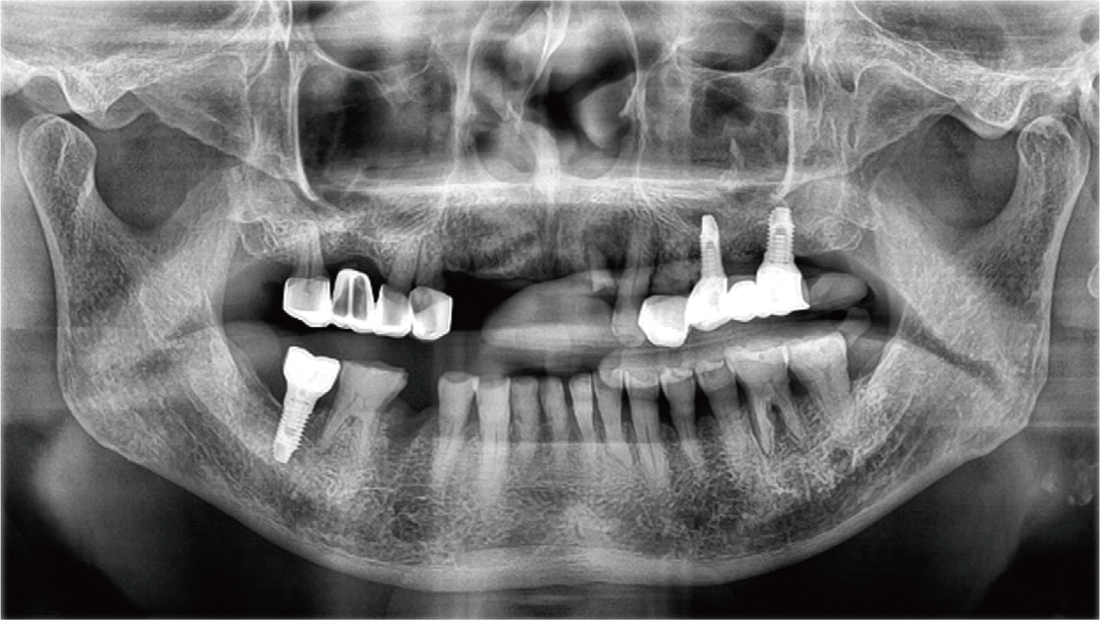 | Fig. 1Initial panoramic radiograph. 
|
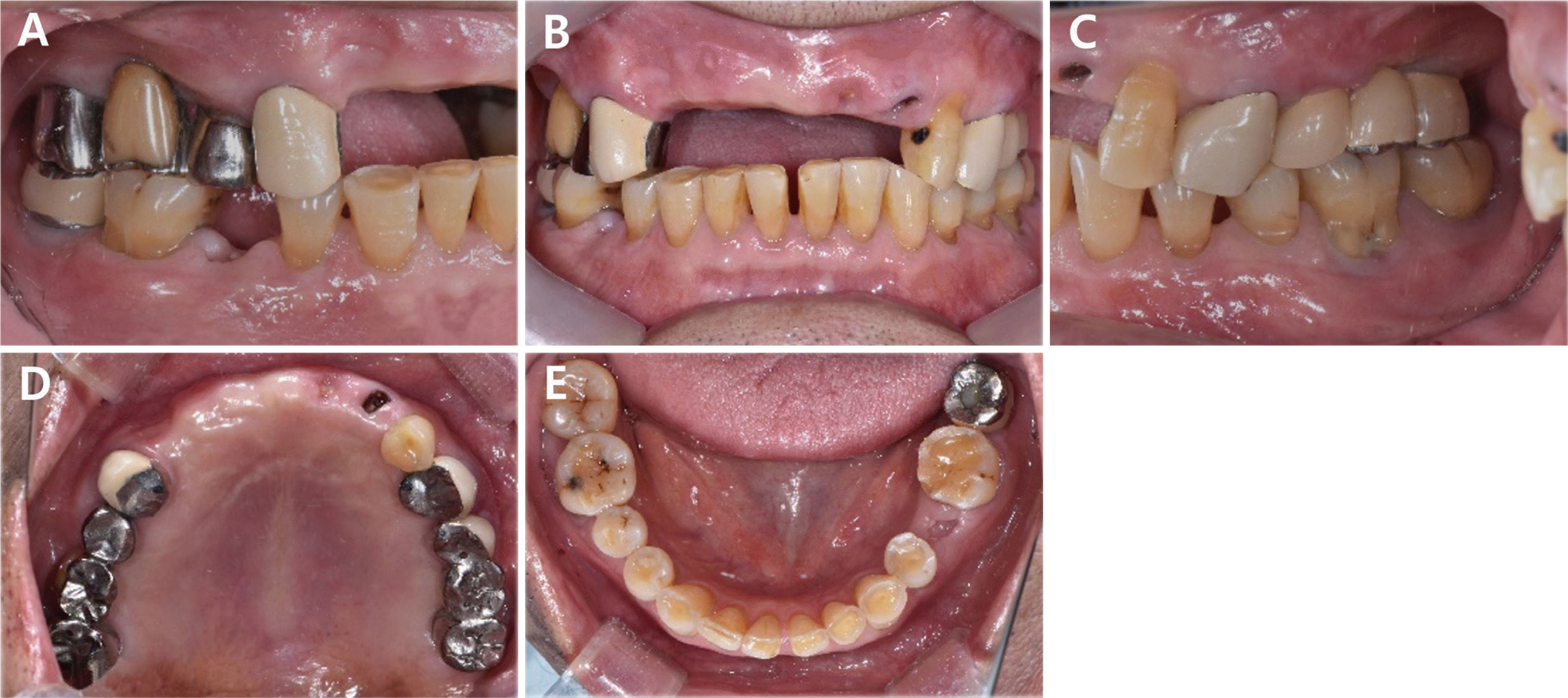 | Fig. 2Initial intraoral photographs. (A) Right lateral view, (B) Frontal view, (C) Left lateral view, (D) Maxillary occlusal view, (E) Mandibular occlusal view. 
|
Data retrieved from an intraoral scanner (Trios4, Copenhagen, Denmark), CBCT, and facial scanner were superimposed. Missing teeth were designed on computer aided design (CAD) and static surgical guide was created on virtual wax-up (
Fig. 3,
4). Due to the alveolar bone resorption especially on the left maxillary lateral incisor area, implant fixtures were not expected to be placed on the most ideal place prosthetically.
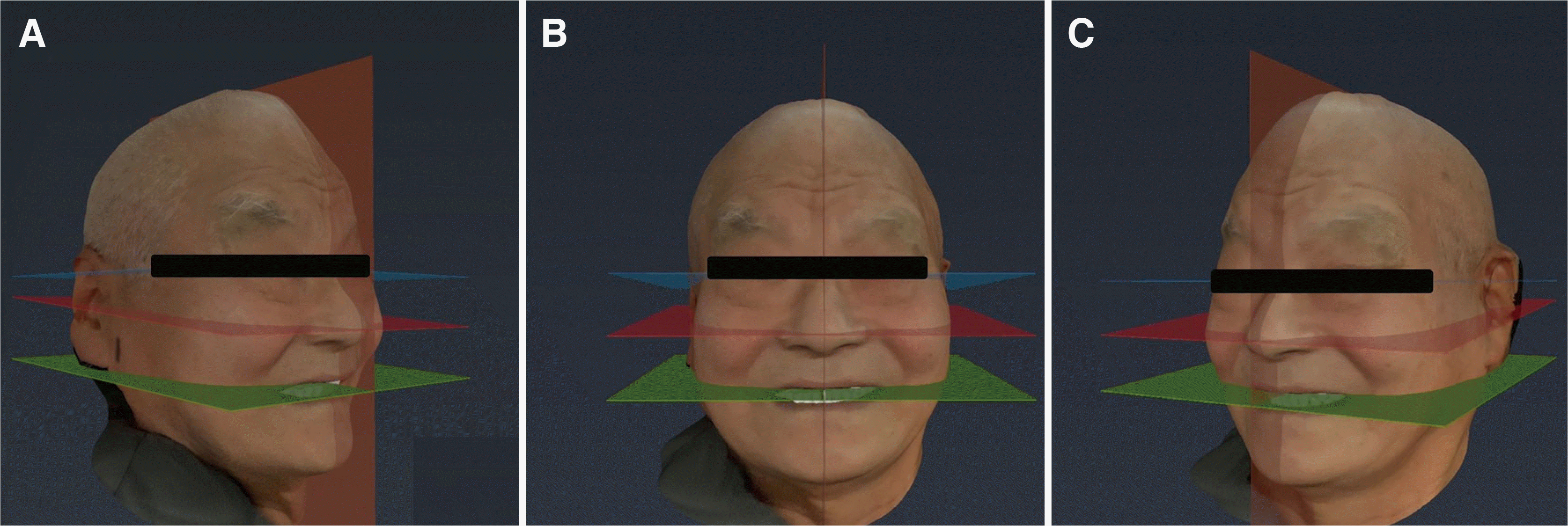 | Fig. 3Digital facebow transfer. (A) Digital facebow: right view, (B) Digital facebow: front view, (C) Digital facebow: left view. 
|
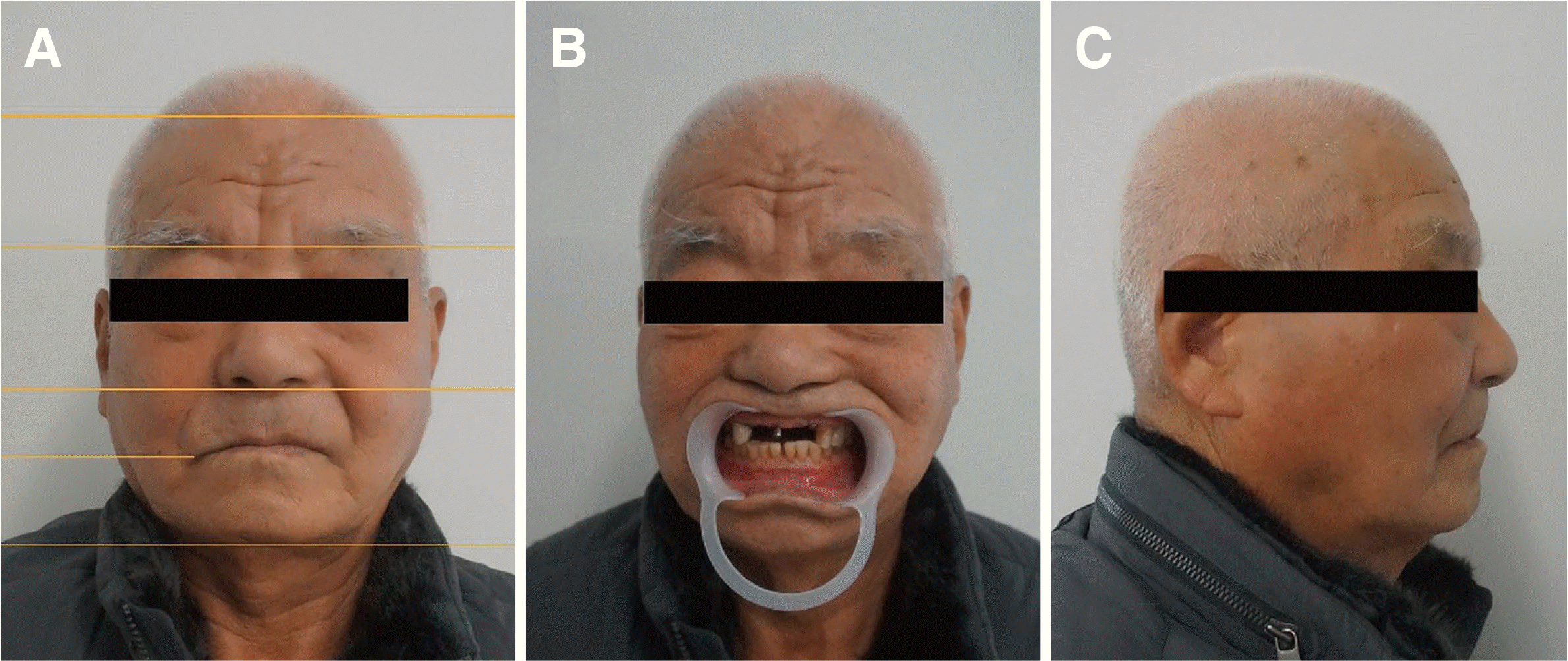 | Fig. 4Extra-oral photographs for esthetic analysis. (A) Front view, showing hairline, glabella, subnasale, lip line, and menton, (B) Front view with lip retractor, (C) Lateral view. 
|
From extra-oral photograph, inter-pupillary line was checked to be parallel. Commissural line was right slanted, occlusal plane was left slanted. Facial profile was concave due to missing maxillary anterior teeth. Menton point deviation was to the right. Nasofrontal angle was 147°, Nasolabial angle was 120°, Nasomental angle was 15°, lip protrusion with Richetts E-line was -4.5 mm (Upper lip), and -1.5 mm (lower lip) (
Fig. 5).
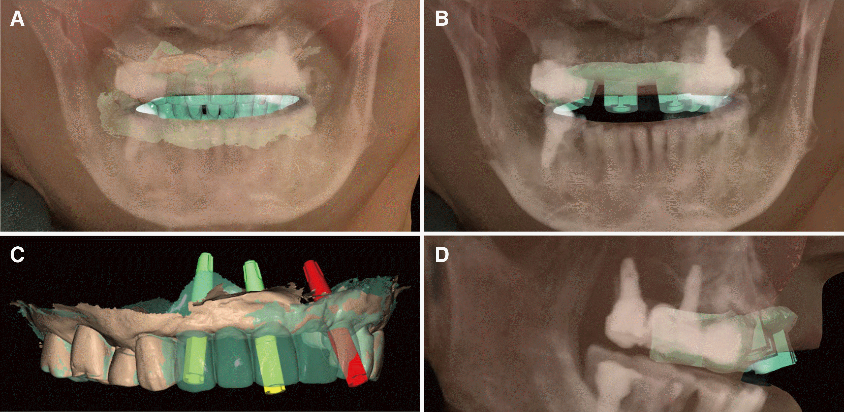 | Fig. 5Design of Surgical Guide with facial scan, CBCT (Dicom file), and intra oral scan. (A) Digital wax up on CAD software and virtual implant placement, (B) Front view: Superimposition of wax up, intra oral scan, CBCT (dicom file), and facial scan, (C) Digital wax up of maxillary anterior area and placement of implant fixture, (D) Lateral view: superimposition of surgical guide, intra oral scan, CBCT (dicom file), a facial scan. 
|
Impression taking of implants were taken with scanbodies and intraoral scanner (Trios4)(
Fig. 6). Interim prosthesis was designed on CAD program (Exocad, Darmstadt, Germany). Since the patient had insufficient buccal bone on Left Maxillary lateral incisor, the implant was placed slightly posterior than the ideal location. Maxillary central incisor could be designed symmetrically, yet the lateral incisors could not be done due to the placement of implant. Sesemann defined a line angle as the line that two planes intersect.
4 By using specific vertical and horizontal line angles on anterior teeth, visual perception of teeth morphology can be affected. Chiche and Pinault wrote of moving line angle to create visual illusion in order to affect the perceived morphology of tooth.
5 Bilaterally symmetrical maxillary first incisors were prioritized in designing 5 unit maxillary prosthesis.
 | Fig. 6Impression taking with intraoral scan. (A) Digital impression taking with scanbody, (B) Intra oral scan of mandible. 
|
Due to the placement of implant, Maxillary lateral incisors on both sides did not have equal horizontal space to be restored. Right maxillary lateral incisor had narrower space than the left side, horizontally. Therefore, line angles were moved laterally and facial embrasures were decreased on right lateral incisor. Furthermore, horizontal lines and ridges were added. The left maxillary lateral incisor was modified to be seen narrower, by adding vertical lines and flatten cervical convexity. Right maxillary canine implant prosthesis had wider space than the left maxillary canine. Since the mesial half of the canine is only visible from the frontal view when patient smiles, smaller space for mesial half was allowed and wider space for the distal half was allotted. From the front view, the reflective surface of right maxillary canine prosthesis seemed similar in size and shape to the left maxillary canine (
Fig. 7). Maxillary canine plays a critical point in creating a pleasing smile as Bhuvaneswaran stated,
6 right maxillary canine prosthesis was designed so that it not only depicts personal characteristics of a patient but also supports frontal muscles and determine buccal corridor and canine guidance as well.
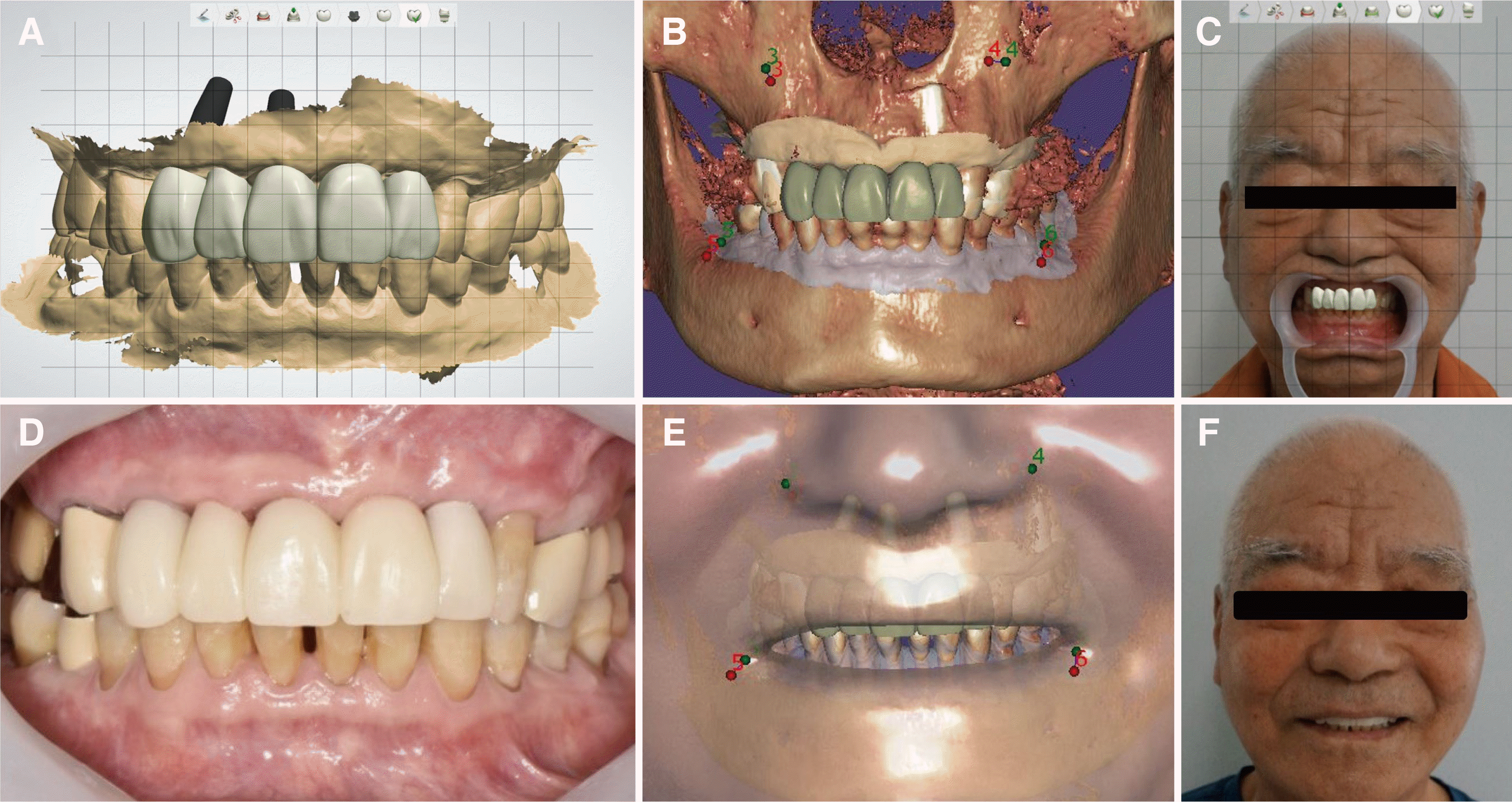 | Fig. 7Provisional restoration. (A) CAD design of provisional restoration, (B) Superimposition of Provisional restoration, intra oral scan and CBCT (dicom file), (C) Virtual patient with Provisional restoration, (D) Superimposition of Provisional restoration, intra oral scan, CBCT (dicom file), and facial scan, (E) Intraoral photo of provisional restoration, (F) Extraoral photo of a patient with provisional restoration. 
|
Final titanium abutment and provisional prosthesis made of PMMA (Polymethylmethacrylate) was proceeded before the final restoration. This enabled gingival modeling and further allowed dentist to determine the anterior guidance and esthetic requirement of definitive prosthesis. When the patient was fully satisfied with the newly given anterior guidance, a digital occlusion analyzer (Arcus Digma II, KaVo, Leutkirch, Germany) was used to measure Bennett’s angle, the horizontal condylar inclination, anterior guidance, and ISS (Immediate side shift) (
Fig. 8).
7,8 This measurement was then sent to the dental lab studio, and was applied when making final Zirconia prosthesis in CAD software.
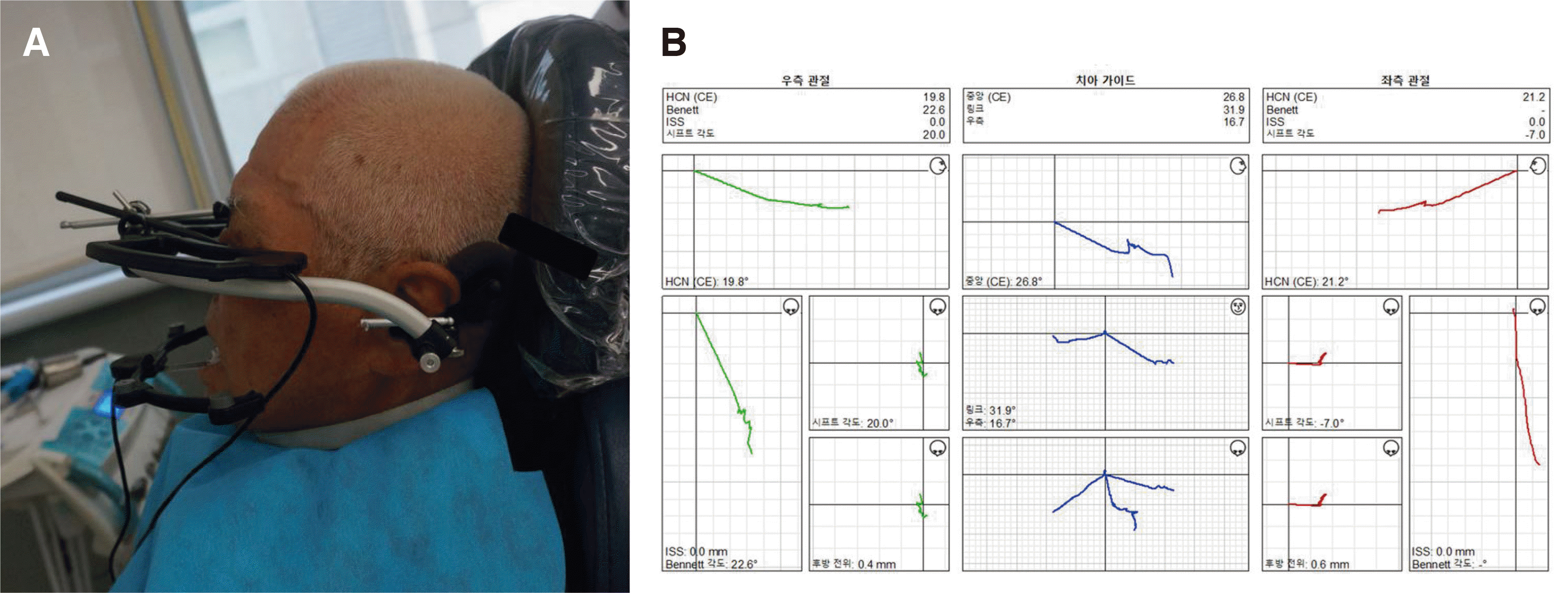 | Fig. 8A digital occlusion analyzer (Arcus Digma II, KaVo, Leutkirch, Germany). (A) A patient with Arcus digma II, (B) Arcus digma II result. 
|
Definitive prosthesis was fabricated with few changes in design such as narrowing the width of right maxillary canine to balance left maxillary canine and a couple changes in line angles to meet the patient’s aesthetic expectation (
Fig. 9). The patient was fully satisfied with the prosthesis and felt comfortable with it.
 | Fig. 9Final restoration. (A) Intraoral photo: Front view, (B) Intraoral photo: Occlusal view. 
|
Go to :

Discussion
The ultimate goal of esthetic dentistry is a combination of art and science, regenerating patients’ genuine smile which looks and functions naturally. The synchronized appliance of technical and creative skills further enhances themselves with an aid of digital dentistry. By virtualize a patient with facial scanner, a lab worker can add the artistic part of esthetic dentistry. By superimposing skeletal information of a patient given in CBCT with facial scan and intra-oral scan, the accuracy of implant planning can be increased. No doubt, digital dentistry took prosthetic treatment a big step forward by analyzing patients’ condylar guidance and apply it into prosthesis. Further, digital dentistry has shortened lab time for prosthetic procedure with the use of CAD and CAM.
However, digital dentistry still has its own draw backs. As Leung
9 stated, by using a voxel size of 0.38 mm at 2 mA, CBCT alveolar bone height can be measured to an accuracy of about 0.6 mm. With this difference, computer generated surgical guide may not fully reflect patients’ alveolar bone loss and may result in implantation on prosthetically unideal place.
Transferring intraoral scan in relation to face is an important step in creating esthetic result. With markers on patient’s face or with digital facebow as Li et al stated,
10 facial scanner could have increased its accuracy when superimposing. If possible, a digital facebow with virtual articulator with mandibular movement analyzer could have increased the accuracy of facial scan.
Final limitation of this case is that the measurement of the patient’s anterior guidance was done with the provisional prosthesis. It would have been the best if the patient visited dental hospital with his own anterior teeth. So that his anterior guidance of natural teeth could be measured and applied to the final prosthesis. Visiting dental hospital with missing anterior teeth, however, adjusting provisional prosthesis until the patient feels comfortable then measuring the anterior guidance, condylar inclination and ISS with a digital occlusion analyzer (Arcus Digma II, KaVo) was the best could be done.
Go to :

Conclusion
This case describes a digital workflow of implant which combines intraoral scan, CBCT data, and a 3-dimensional facial scan to predict the outcome of prosthodontic treatment of anterior implant restoration. Implant planning that prioritizes prosthetic aspect was applied to the patient by using a 3d printed surgical guide. This further resulted in esthetic prosthesis with a visual aid supported by creating a virtual patient. The usage of digital technique supplemented its quality by transferring the patient’s condylar and anterior guidance on a digital articulator (Arcus Digma II, KaVo). If it were not digital, the patient could not have gotten a definitive prosthesis which encompasses skeletal, occlusal, and personal characteristics of a patient. Were it otherwise, or with conventional analysis and steps, it would have taken more time and energy to carry out the procedure with much less certainly of the outcome.
Go to :


 PDF
PDF Citation
 Print
Print


 XML Download