This article has been
cited by other articles in ScienceCentral.
Abstract
Purpose
This study aimed to evaluate the accuracy and clinical usability of an identification model using deep learning for 79 dental implant types.
Materials and Methods
A total of 45396 implant fixture images were collected through panoramic radiographs of patients who received implant treatment from 2001 to 2020 at 30 dental clinics. The collected implant images were 79 types from 18 manufacturers. EfficientNet and Meta Pseudo Labels algorithms were used. For EfficientNet, EfficientNet-B0 and EfficientNet-B4 were used as submodels. For Meta Pseudo Labels, two models were applied according to the widen factor. Top 1 accuracy was measured for EfficientNet and top 1 and top 5 accuracy for Meta Pseudo Labels were measured.
Results
EfficientNet-B0 and EfficientNet-B4 showed top 1 accuracy of 89.4. Meta Pseudo Labels 1 showed top 1 accuracy of 87.96, and Meta pseudo labels 2 with increased widen factor showed 88.35. In Top5 Accuracy, the score of Meta Pseudo Labels 1 was 97.90, which was 0.11% higher than 97.79 of Meta Pseudo Labels 2.
Conclusion
All four deep learning algorithms used for implant identification in this study showed close to 90% accuracy. In order to increase the clinical applicability of deep learning for implant identification, it will be necessary to collect a wider amount of data and develop a fine-tuned algorithm for implant identification.
Go to :

초록
목적
본 연구는 79종의 치과 임플란트에 대해 딥러닝을 이용한 식별 모델의 정확도와 임상적 유용성을 평가하는 것을 목적으로 하였다.
연구 재료 및 방법
2001년부터 2020년까지 30개 치과에서 임플란트 치료를 받은 환자들의 파노라마 방사선 사진에서 총 45396개의 임플란트 고정체 이미지를 수집했다. 수집된 임플란트 이미지는 18개 제조사의 79개 유형이었다. 모델 학습을 위해 EfficientNet 및 Meta Pseudo Labels 알고리즘이 사용되었다. EfficientNet은 EfficientNet-B0 및 EfficientNet-B4가 하위 모델로 사용되었으며, Meta Pseudo Labels는 확장 계수에 따라 두 가지 모델을 적용했다. EfficientNet에 대해 Top 1 정확도를 측정하고 Meta Pseudo Labels에 대해 Top 1 및 Top 5 정확도를 측정하였다.
결과
EfficientNet-B0 및 EfficientNet-B4는 89.4의 Top 1 정확도를 보였다. Meta Pseudo Labels 1은 87.96의 Top 1 정확도를 보였고, 확장 계수가 증가한 Meta Pseudo Labels 2는 88.35를 나타냈다. Top 5 정확도에서 Meta Pseudo Labels 1의 점수는 97.90으로 Meta Pseudo Labels 2의 97.79보다 0.11% 높았다.
결론
본 연구에서 임플란트 식별에 사용된 4가지 딥러닝 알고리즘은 모두 90%에 가까운 정확도를 보였다. 임플란트 식별을 위한 딥러닝의 임상적 적용 가능성을 높이려면 더 많은 데이터를 수집하고 임플란트에 적합한 미세 조정 알고리즘의 개발이 필요하다.
Go to :

Keywords: dental implants, artificial intelligence, deep learning, convolutional neural networks
색인어: 치과 임플란트, 딥러닝, 인공지능, 합성곱 신경망
Introduction
Since the concept of osseointegration was introduced, dental implants have been considered a successful treatment option for partially or completely edentulous patients.
1 It has been reported that dental implants have a high success rate regardless of the type of prosthesis, such as fixed or removable, and their application range and use are gradually increasing.
2-4 With the successful introduction of dental implants, numerous types of dental implants are being produced by different manufacturers, and the number continues to grow.
5
Although dental implants have a high success rate, various surgical or prosthetic complications exist.
6-8 Surgical complications include osseointegration failure and peri-implantitis, and prosthetic complications include fracture of the upper abutment or prosthesis, screw loosening, and screw fracture. When these complications occur, in many cases, the existing abutment and upper prosthesis must be removed, and in particular, when prosthetic complications occur, remake of prosthesis is sometimes necessary. In this case, it is essential for clinicians to accurately identify the type of implant, as there are many manufacturers and each implant is often incompatible.
Radiography is usually used to identify the type of implant.
9,10 For this, it was necessary for clinicians to understand the features of the fixture (internal or external hexagonal, thread shape, tapering degree, etc.) shown in radiographs. However, this conventional identification method has important limitations. Since there are so many types of implants currently in use, it is almost impossible for even an experienced clinician to identify the features of all implants. Therefore, it was difficult to accurately identify the type of implant, unless it is an implant that clinicians mainly use or have experience with.
Artificial intelligence, a concept that has recently been widely used throughout society, is one of the subfields of computer science that attempts to implement computer systems to perform tasks that require human learning ability, reasoning ability, and perceptual ability. Deep learning, a field of artificial intelligence, is machine learning based on a multi-layered neural network, and it is a technique to build a high-level abstraction model from a large amount of data.
11 Recently, studies to confirm the usefulness of deep learning in the field of dentistry are increasing.
12 In particular, studies have been made on implant identification using deep learning image classification.
13-16 In these studies, it was reported that a high level of accuracy was achieved when various deep learning algorithms were used for the identification. However, previous studies have a limitation in that they targeted several types of implants. Clinicians are actually exposed to a much wider variety of implants. Considering that the accuracy of the deep learning model decreases as the number of classes increases, it is necessary to confirm whether deep learning can show a high level of accuracy even with more types of implants.
Therefore, in this study, deep learning model was trained on 79 types of implants, and its performance was analyzed to confirm the applicability of deep learning for identification of various types of implants.
Go to :

Materials and Methods
Data collection
This study was approved by the institutional review board (No.P01-202105-21-009). A total of 45396 implant fixture images were collected through panoramic radiographs of patients who received implant treatment from 2001 to 2020 at 30 dental clinics. Implant images were labeled as one of 79 types of implants from 18 manufacturers according to medical records. The implant manufacturers and systems used in this study are in
Table 1.
Table 1
The manufacturer and system of the implant and the number of images collected for model learning
|
Manufacturer |
System |
Images |
Manufacturer |
System |
Images |
Manufacturer |
System |
Images |
|
Bicon |
260-345-305 |
53 |
Neobiotech |
BIS3508A |
56 |
Point Implant |
POF3008 |
32 |
|
BioHorizons |
IITSS510D |
17 |
BIS4007A |
195 |
POF3008Q |
19 |
|
LITSS410D |
40 |
EB310 |
27 |
POF3008QNP |
111 |
|
SLITSS312D |
20 |
EB407 |
1441 |
POF4007 |
269 |
|
BIOMET 3i |
FNT485 |
402 |
EB3513A |
179 |
POF4007Q |
946 |
|
FNT585 |
104 |
EB4513A |
2867 |
POF4007QNP |
1299 |
|
FNT685 |
53 |
EBI507 |
1022 |
Straumann |
21.2408 |
92 |
|
FOS485 |
49 |
EBI5010A |
128 |
21.3308 |
188 |
|
OSS410 |
198 |
IS33508A |
38 |
21.3508 |
45 |
|
OSS510 |
48 |
Nobel Biocare |
32114 |
217 |
043.054S |
215 |
|
Biotem |
ASTFA4011 |
26 |
32186 |
24 |
043.131S |
94 |
|
Dentis |
DS2FM3708S |
447 |
32199 |
25 |
Thommen Medical |
4.13.243 |
215 |
|
DSFM3708S |
279 |
32200 |
146 |
4.13.900 |
297 |
|
DSSOFR5210S |
191 |
32205 |
367 |
Zimmer Dental |
TSVB8 |
83 |
|
Dentium |
FX3408 |
1251 |
32212 |
403 |
TSVH8 |
848 |
|
FX3610SW |
353 |
37609 |
331 |
|
|
|
|
FXI3608 |
118 |
Osstem Implant |
AUS3M3508S |
507 |
|
|
|
|
Dentsply Sirona |
26-2431 |
232 |
GS2W4511R01 |
175 |
|
|
|
|
24951 |
6827 |
GS3S4011R |
484 |
|
|
|
|
24982 |
1731 |
MSD3008S20 |
24 |
|
|
|
|
Dio Implant |
FTT5015S |
18 |
MSN2508S25 |
47 |
|
|
|
|
SFN3808 |
1023 |
MSP25103R |
38 |
|
|
|
|
SFN3808H |
148 |
MST18104 |
20 |
|
|
|
|
UF(ll)4513S |
1701 |
SS2R4007S18 |
44 |
|
|
|
|
UF(ll)N3315S |
706 |
SS2R4008S28 |
20 |
|
|
|
|
Hi ossen Implant |
ET3M3508S |
438 |
TS3M3008C |
290 |
|
|
|
|
IBS Implant |
451M4009 |
104 |
TS3M3008S |
28 |
|
|
|
|
551M5007 |
262 |
TS3M3508A |
4285 |
|
|
|
|
MegaGen Implant |
EF4011P |
280 |
TS3M3508C |
3265 |
|
|
|
|
IF4010C |
51 |
TS3M3508H |
115 |
|
|
|
|
|
|
TS3M3508S |
1572 |
|
|
|
|
|
|
US3M3508S |
1187 |
|
|
|
|
|
|
US3R411R |
71 |
|
|
|
|
|
|
US4R4007S |
3835 |
|
|
|

Model learning
Regions of interest were cropped and extracted from the panoramic radiographs. Implant images of the maxilla were vertically flipped, and when the fixture was tilted more than 45 degrees, it was manually corrected to be parallel to the vertical axis. Data augmentation was performed on the image to improve the accuracy of the model. For data augmentation, randomly resizing and cropping were performed for each image. Next, the brightness and contrast of the image were randomly changed. Finally, horizontally flipping was performed.
EfficientNet and Meta Pseudo Labels algorithms were used for model learning. EfficientNet is a deep learning architecture and scaling method that uses compound scaling techniques to uniformly scale all dimensions of depth, width, and resolution.
17 Meta Pseudo Labels is a semi-supervised learning method that uses a teacher network to generate pseudo labels on unlabeled data to teach a student network. In EfficientNet, 80% of the images were used for training, and 20% were used for validation. In Meta Pseudo Labels, 80% were used for training, of which 20% were fine tuning, 20% were unlabeled, and 60% were labeled. The remaining 20% were used for validation. For EfficientNet, EfficientNet-B0 and EfficientNet-B4 were used as submodels, and for Meta Pseudo Labels, two models were applied according to the widen factor. The settings of the four algorithms used in this study are as follows (
Table 2).
Table 2
EfficientNet and Meta Pseudo Labels algorithms
|
Algorithm |
Depth, widen factor |
Epoch |
Time (hour) |
|
EfficientNet-B0 |
- |
200 |
3 |
|
EfficientNet-B4 |
- |
300 |
33 |
|
Meta Pseudo Labels 1 |
28, 2 |
300,000 |
32 |
|
Meta Pseudo Labels 2 |
28, 8 |
300,000 |
52 |

Model performance evaluation
The accuracy [(TP +TN) /( TP + FP + FN + TN)] used to evaluate the trained model. TP is a true positive, TN is a true negative, FP is a false positive, and FN is a false negative. According to the default setting of each algorithm, top 1 accuracy was measured in EfficientNet and top 1 and top 5 accuracy in Meta Pseudo Labels were measured. The top 1 accuracy refers to the ratio that the nearest class was predicted and the answer was correct. The top 5 accuracy refers to the ratio in which the five nearest classes were predicted and the answer was in them.
Go to :

Results
The performances of the trained models are presented in
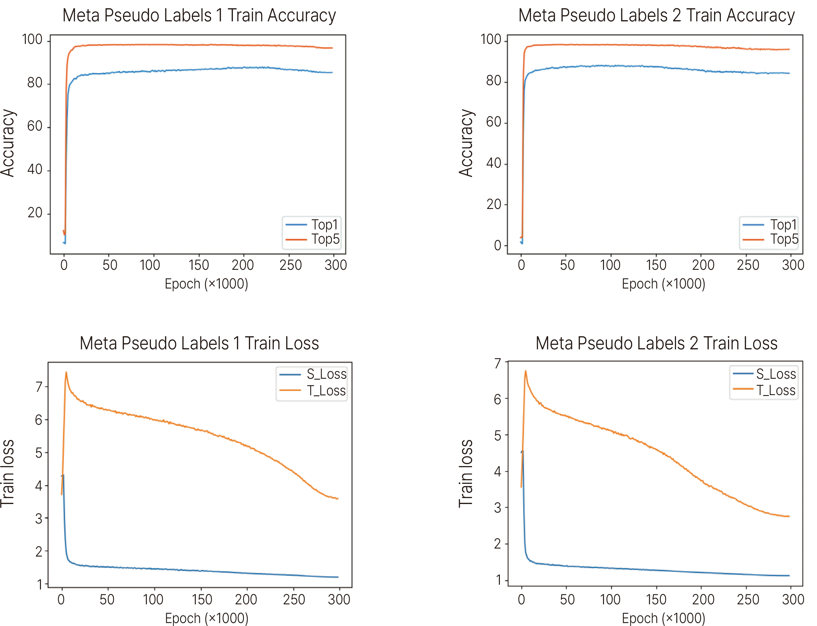
Table 3. EfficientNet-B0 and EfficientNet-B4 showed top 1 accuracy of 89.4. Meta Pseudo Labels 1 showed top 1 accuracy of 87.96, and Meta pseudo labels 2 with increased widen factor showed 88.35. In Top 5 Accuracy, the score of Meta Pseudo Labels 1 was 97.90, which was 0.11% higher than 97.79 of Meta Pseudo Labels 2. Accuracy graphs of train and validation for EfficientNet are shown in
Fig. 1. The top 1 accuracy and top 5 accuracy graphs for Metal Pseudo Labels are shown in
Fig. 2.
 | Fig. 1Accuracy and train loss results for EfficientNet-B0 and EfficientNet-B4 algorithms. The value of validation means top 1 accuracy. 
|
 | Fig. 2Top 1 and Top 5 accuracy and train loss results of Meta Pseudo Labels algorithms. Meta Pseudo Labels 2 is a setting with an increased widen factor. 
|
Table 3
Performance of implant identification models according to algorithm
|
EfficientNet Meta |
Pseudo Labels |
|
EfficientNet-B0 |
EfficientNet-B4 |
Meta Pseudo Labels 1 |
Meta Pseudo Labels 2 |
|
Top 1 accuracy |
89.4 |
89.4 |
87.96 |
88.35 |
|
Top 5 accuracy |
- |
- |
97.90 |
97.79 |

Go to :

Discussion
In this study, we evaluated how accurately the model using deep learning can identify 79 types of implants. According to the study results, the deep learning model identified implants with nearly 90% accuracy, confirming the usefulness of the deep learning model in implant classification.
The clinical importance of the identification of implants has been steadily mentioned since implant treatment became popular. In order to overcome the limitations of the traditionally used radiographic method, various methods have been introduced. Implant Recognition Software is a program that classifies implants according to various characteristics, and when the user selects the characteristics, the type of implant corresponding to the condition is presented.
18 There are also commercialized websites that use a similar method to identify implants. However, the disadvantage of this method is that the clinician has to check the characteristics of the implant one by one, which can be difficult, especially for inexperienced clinicians. In contrast, identification of implants using artificial intelligence has the advantage that the accuracy does not depend on the clinician.
In previous studies using deep learning, it was reported that the accuracy of implant identification was between 0.51 and 0.98.13-16 Although this is high accuracy, it has a limitation in that the types of implants to be studied are limited. Therefore, in this study, data were collected from implants from various manufacturers to increase the clinical applicability of the model. The accuracy of this study is somewhat lower than that of previous studies. This is because there are many kinds of classes for model building. In an image classification model using deep learning, it is generally reported that the accuracy decreases as the number of class increases.
19 To overcome this, it is necessary to collect a sufficient number of images for each class. In the present study, the minimum number of images obtained per class was 17, which may be insufficient for accurate model training, and this will degrade the overall accuracy of the model. In particular, considering that there are many similar types of implants, it is necessary to have high top 1 accuracy so that the type can be accurately identified in a clinical situation. For this, after classifying implants according to design, more images of implant systems with similar shapes should be collected.
In comparison between algorithms, EfficientNet algorithm showed higher accuracy than Meta Pseudo Labels. According to a study that used various algorithms for implant classification, the Resnet algorithm showed the highest accuracy.
16 Algorithms for image classification are very diverse and are being developed at the present time, and research on the strengths and weaknesses of each algorithm continues. Therefore, in order to find the optimal algorithm for classifying implants, it is necessary to train and evaluate models using various algorithms for many types of implants.
Identifying implant images using deep learning is a method of learning the features of each implant type. For this, it is important that the unique features of the implant are clearly revealed on the radiograph. The accuracy of the model can be improved if things like the shape and size of the threads and the taper of the fixtures become clearer. Panoramic radiographs were used in this study. In the case of panoramic radiographs, there is a disadvantage that overlapping of anatomical structures may occur depending on the posture of the patient.
20,21 Therefore, to solve these shortcomings, it will be necessary to utilize standardized periapical radiography or computed tomography.
The limitation of this study is that although various types of implant data were collected, it is much less than the types of implants used in clinical practice. In order to use deep learning-based implant identification in actual clinical practice, more extensive data on implants manufactured in Korea and abroad are needed. In addition, the accuracy was lower than that of the previous studies. A sufficient number of images for each type are required, and a process that can analyze the various features of the implant in more detail is needed. Finally, since data were collected from several dental clinics, the contrast, angle, and resolution of radiographs varied, which may have acted as a factor lowering the accuracy of the model.
Go to :

Conclusion
Deep learning algorithms for identifying 79 dental implant types showed high accuracy close to 90%. It will be necessary to collect a wider amount of data and develop a fine-tuned algorithm for implant identification to increase the clinical applicability.
Go to :

Acknowledgements
This research was financially supported by the Ministry of Science and ICT of Korea through the Institute of Information and Communications Technology Planning and Evaluation in 2021 (No.20210012430012002, Setting up Big data of dental case and Developing dental implant identification system based on Deep learning technology).
Go to :

References
7. Annibali S, Ripari M, LA Monaca G, Tonoli F, Cristalli MP. 2008; Local complications in dental implant surgery: prevention and treatment. Oral Implantol. 1:21–33. PMID:
23285333. PMCID:
PMC3476500.
Go to :


