Abstract
Cleft lip lower-lip deformity is a secondary deformity in patients who underwent primary cheiloplasty of the upper lip, characterized by an enlarged and anteriorly rotated lower lip. In these cases, soft-tissue imbalances remain even after skeletal correction with orthognathic surgery, and additional soft tissue treatment is required for lip harmony and esthetic facial balance in CLP (cleft lip palate) patients. This study describes three cases of transverse myomucosal excision of the lower lip for correction of cleft lip lower-lip deformity to restore facial esthetic balance. Each patient underwent orthognathic surgery, rhinoplasty, or upper lip revision cheiloplasty according to condition. Postoperatively, volume of the lower lip decreased and lip harmony was improved in all three patients. The surgeon should fully understand the anatomical structure around the lips and be able to evaluate overall harmony of the soft tissue. When a lower lip deformity is present, careful surgical planning and execution are important for each patient.
Patients with cleft lip and palate (CLP) have a typical facial profile, characterized by midface depression due to maxillary growth deficiency and disharmony between the upper and lower lips. Unlike patients without CLP, the lateral esthetic balance of patients with CLP may not be well restored after orthognathic surgery, which may be because of upper-lip tightness caused by the primary cleft lip repair and multiple revision operations1,2. Deformities of the midfacial complex, particularly the nose and upper lip, are thought to be the main contributors to esthetic impairment in patients with CLP3-5. Similar to the upper lip, there are also typical soft tissue changes to the lower lip in patients who underwent primary cheiloplasty of the upper lip. The lower lip can be hypertrophied, superiorly displaced, and anteriorly rotated, and these alterations are termed, “cleft lip lower-lip deformity”6. Lip disharmony is accentuated by this lower-lip redundancy and may be one of the most noticeable and esthetically impaired parts of CLP deformity7.
To restore the esthetic balance of the lateral facial contour, it is necessary to identify and correct the factors contributing to facial imbalance. Because many other factors may contribute to soft tissue imbalance in patients with CLP1, one surgical method alone is insufficient to correct lip disharmony. Skeletal abnormalities may appear as soft tissue contours overlying the facial bone, and there are also alterations in the characteristics of the soft tissue itself, such as the elasticity or thickness of the lips3. Although orthognathic surgery can resolve skeletal imbalances, soft tissue imbalances do not significantly improve1 and can be accentuated after surgery8. Therefore, managing lip disharmony in patients with CLP only with orthognathic surgery is difficult, and soft tissue management must also be performed to restore the esthetic balance of the lateral facial contour6.
Although cleft lip lower-lip deformity is recognized clinically6, few studies have been conducted to objectively analyze the characteristics of this deformity, and the management of this condition has received scant attention. This study presents three cases of surgical treatment for volume reduction and inversion of cleft lip lower-lip deformity. The patients had been referred for oral and maxillofacial surgery, such as bimaxillary orthognathic surgery or iliac bone graft, and a lower-lip deformity was observed in all cases. We discuss our experience with lower-lip reduction cheiloplasty and surgical techniques performed to harmonize the upper lip with the lower lip. Institutional Review Board (IRB) approval was granted at Seoul National University Dental Hospital (IRB No. ERI22037).
A 30-year-old man with unilateral CLP was referred to the Department of Oral and Maxillofacial Surgery, Seoul National University Dental Hospital, for orthognathic surgery. The patient had a history of primary cheiloplasty at the age of three months and underwent palatoplasty at the age of 17 months. On clinical examination and radiological study, maxillary retrusion and mandibular protrusion were observed as skeletal problems and a protruding and everted lower lip was observed as a soft tissue problem.(Fig. 1, left column) Therefore, bimaxillary orthognathic surgery and lower-lip reduction cheiloplasty were planned and were performed simultaneously.
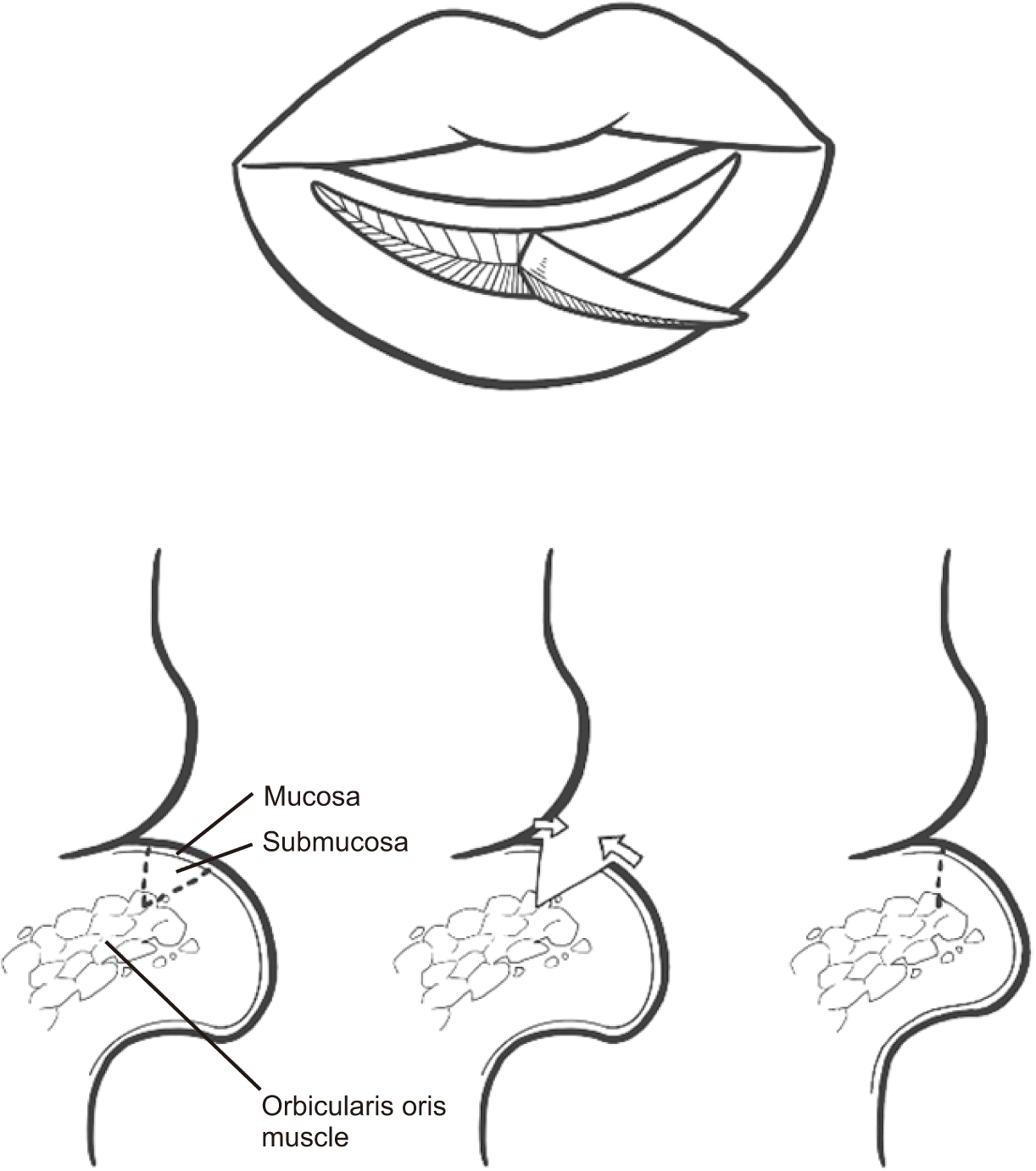
General anesthesia was administered via nasotracheal intubation because bimaxillary orthognathic surgery was scheduled simultaneously. Bimaxillary orthognathic surgery using Le Fort I osteotomy and bilateral sagittal split ramus osteotomy was performed first, according to the standard procedure. After correcting the skeletal disproportion, lip reduction cheiloplasty using transverse myomucosal excision was performed. Preoperative markings were made posterior to the wet/dry border to mark the area for excess tissue removal.(Fig. 2. A) The shape of the marking was a curved ellipse, with both ends stopping a few millimeters from the commissure. After marking the outline, a slightly beveled incision was made along the outline using a #15 blade.(Fig. 2. B) The mucous membrane was excised and removed, including the submucosal tissue and part of the underlying orbicularis oris muscle layer. The muscle layer was closed first, followed by the submucosal tissue, and finally the mucosal layer.(Fig. 2. C) No surgical treatment was performed on the upper lip. After the operation, the lower lip volume was decreased and the most anterior point of the lower lip was moved posteriorly.(Fig. 1, right column)
A 22-year-old man with bilateral CLP had undergone primary cheiloplasty at the age of four months, palatoplasty at the age of 13 months, and bimaxillary orthognathic surgery at the age of 21 years. The patient complained of esthetic dissatisfaction; therefore, lip reduction cheiloplasty and rhinoplasty were planned to improve lateral facial contour. On extraoral examination, both the upper and lower lips were thick and protruded. Additionally, the texture and color of the upper-lip tubercle were different from those in the surrounding area.(Fig. 3, left column) The patient wanted to remove the abnormal area of the tubercle and improve the balance of the lips.
General anesthesia was administered via orotracheal intubation because rhinoplasty was scheduled concurrently. In the upper lip, the excision margin on the tubercle area was set, and a W-flap was designed on both sides.(Fig. 4. A) An incision was made along the marking, and triangular flaps were raised on opposite sides. The mucosa from the submucosal structures within the planned margin was excised (Fig. 4. B), and elevated triangular flaps were transposed and sutured.(Fig. 4. C) Through this method, the abnormal area of the tubercle was removed, and simultaneously, three-dimensional volume reduction and vertical height reduction were achieved. In the lower lip, the wet vermilion was excised, removing the myomucosal tissue to reduce the volume.(Fig. 4. D, 4. E) Postoperatively, the thickness of both lips was reduced, and the overall color of the upper vermilion blended naturally.(Fig. 3, right column)
A 19-year-old man with bilateral CLP visited our facility to improve lip disharmony. The patient had undergone primary cheiloplasty at the age of three months, palatoplasty at the age of 12 months, and alveolorrhaphy with iliac bone grafting at the age of 10 years. On extraoral examination, an exposed portion of the wet vermilion of both lips was presented. The upper-lip vermilion border from the Glogau-Klein point to the oral commissure was concave, and the vermilion height of the upper lip was asymmetric.(Fig. 5, left column) Therefore, lower-lip reduction cheiloplasty and upper-lip cheiloplasty were planned with rhinoplasty.
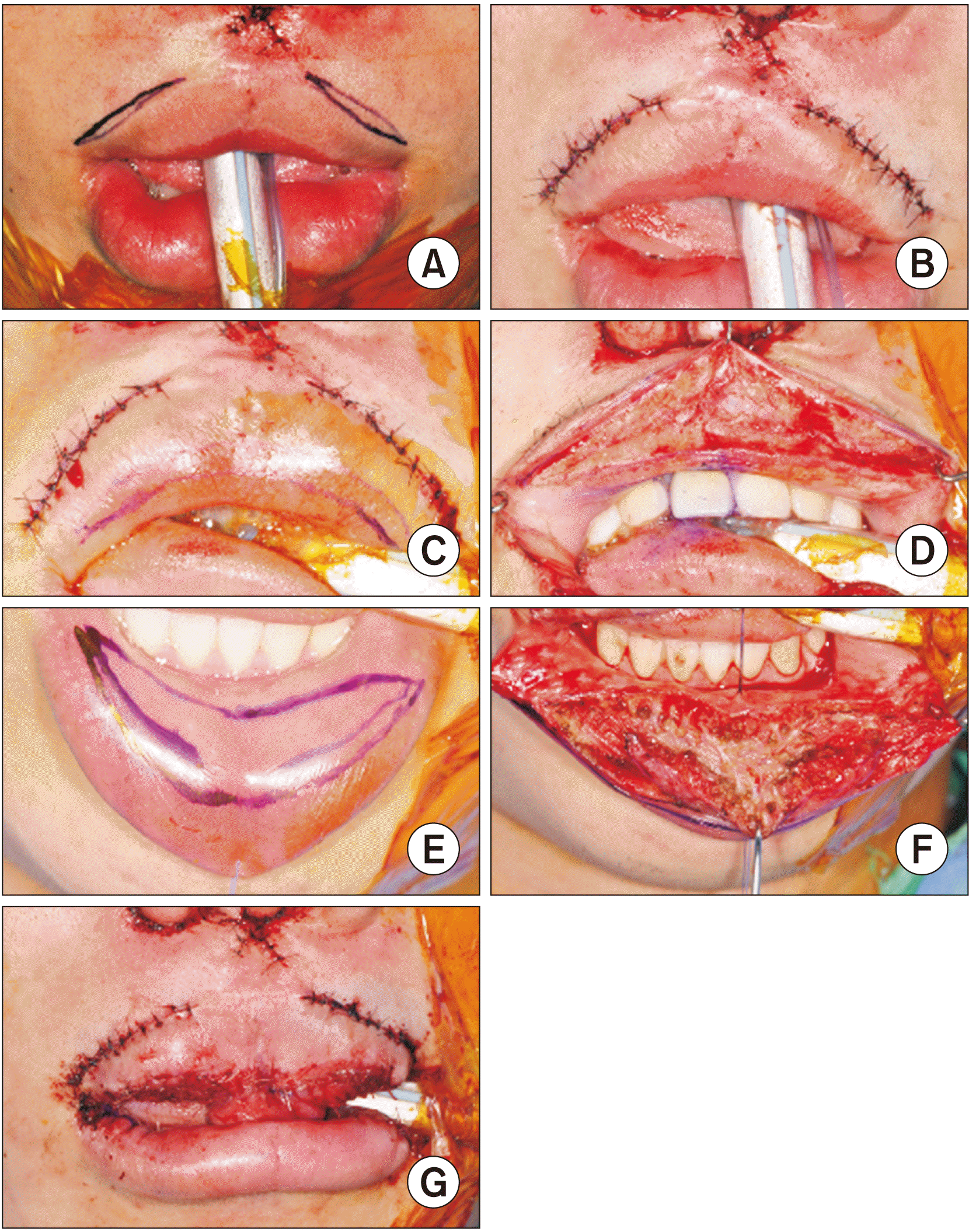
General anesthesia was administered via orotracheal intubation because rhinoplasty was scheduled simultaneously. To improve the contour of the upper-lip vermilion border, lateral vermilion advancement was performed. The incision lines were placed on the skin above the white roll on both sides.(Fig. 6. A, 6. B) The exposed portion of the wet vermilion was removed by myomucosal resection.(Fig. 6. C, 6. D) The lower lip was inverted by direct closure after transverse myomucosal excision posterior to the wet/dry border.(Fig. 6. E-G) Postoperatively, the exposed wet vermilion in the resting state was decreased, and the balance of the upper and lower lips was improved.(Fig. 5, right column)
The residual scar is characteristic of patients with repaired upper-lip cleft; however, when observed clinically, secondary deformities appears in the lower lip as well. This lower-lip dysmorphology, which causes greater disproportion between the lips, may be one of the most visible deformities in patients with CLP7. The cleft lip lower-lip deformity, first described by Pensler and Mulliken6, is characterized by an enlarged and anteriorly rotated lower lip with inferomedial displacement of the commissures.
Several hypotheses can be suggested about the mechanism of development of lower-lip deformity in patients with CLP. The first hypothesis is that the lower lip protrudes relative to the tight and retrusive upper lip1. However, not only protruding, but also significantly thicker and more outwardly rotated lower lips were observed in the cleft group of a previous study8. Another hypothesis is that the upper lip is horizontally contracted because of scar tissue after labial repair, which causes the relatively long lower lip to bulge outward9. This is supported by other studies, which showed that patients with CLP have significantly narrower mouths than people without CLP8-10; however, this may not explain why the lower lip is significantly thicker in patients with bilateral CLP compared with the not significant mouth width differences between patients with bilateral CLP and those with unilateral CLP. The final hypothesis is that strain on the musculature in the lower lip causes hypertrophy in the lower lip and increased lip thickness. The muscular strain in the lower lip might be due to malocclusion secondary to discrepancy between the maxilla and mandible6. However, this does not explain the absence of thickened, everted lower-lip deformity in patients without CLP but with skeletal discrepancy. Finally, it has been suggested that the excessive strain and hyperfunction of the lower-lip musculature compensate for the poor function of the orbicularis oris in the upper lip8.
Although no single hypothesis can fully explain the mechanism, several studies indicated that hyperfunction of the orbicularis oris in the lower lip compensates for the insufficient function of the upper lip and is an important contributor to development of cleft lip lower-lip deformity1,6,8. It is possible that the hyperfunction of the musculature around the lower lip, such as the mentalis and depressor labii inferioris, caused the lower lip to be anteriorly rotated.
To correct lower-lip deformity and improve lip disharmony, a treatment plan should be established considering the skeletal relationship between the maxilla and mandible and the anatomy of the soft tissue surrounding the lips. Patients with CLP have skeletal disharmony due to deficiency of growth and development in the midfacial complex2, and orthognathic surgery is one of a sequence of treatments for managing the functional and esthetic problems of skeletal discrepancy. A principle for correcting lip deformities is that the skeletal imbalance should be addressed before performing soft tissue reconstruction6. In this study, for patients with skeletal discrepancy, which is an indication for orthognathic surgery, orthognathic surgery was performed first, followed by soft tissue management. Soft tissue imbalance remained after skeletal correction with orthognathic surgery, and lip disharmony in patients with CLP was not resolved and may even have been accentuated after orthognathic surgery1,8. In summary, unlike patients without CLP, lip disharmony of patients with cleft-related dentofacial deformities cannot be managed with orthognathic surgery alone, and additional soft tissue treatments are required to achieve lip harmony and esthetic facial balance.
The surgical procedures that can correct cleft lip lower-lip deformity include the cross-lip flap procedure and transverse mucosal excision, including underlying tissue1,6,11. Representative methods for cross-lip flap, which involve transposition of the lower lip to the upper lip, are the Abbe flap and the vermilion switch flap11,12, and other methods, such as island flap13 and bi-winged myomucosal switch flap14. The appropriate type of cross-lip flap can be determined by the extent and location of the deficiency in the upper lip6, and it can be tailored according to the case. The Abbe flap can provide a full-thickness composite flap from the central part of the lower lip to the upper lip and is particularly appropriate for tight upper lips, short prolabium, or lack of a philtral column14. When the patient has an acceptable prolabium and appropriate philtrum and Cupid’s bow, other cross-lip flaps, such as Kawamoto’s vermilion switch flap12 and bi-winged myomucosal switch flap14, may be preferred over the Abbe flap. The cross-lip flap procedure reduces the volume of the lower lip while adding soft tissue to the upper lip to achieve harmony between the lips14. However, these methods require a two-stage surgical procedure to divide the pedicle after 1 to 2 weeks and cause discomfort to patients. One-stage methods applying the concept of island flaps have been described in some studies13-15; however, indications should be carefully considered because dissection of the vascular pedicle is a delicate technique. Therefore, such factors should be considered for patients with cleft lip lower-lip deformity accompanied by severe lip disharmony6,14.
Alternatively, transverse myomucosal excision can remove as much excess soft tissue as desired, and it is an intuitive and simple operative procedure compared with cross-lip flap. A wedge-shaped excision of some of the deeper tissue including submucosa and orbicularis oris muscle allows greater volume reduction and greater inversion16.(Fig. 7) The basic premise of setting the incision lines is to make a mark at or posterior to the wet/dry border because the anterior extent of the incision will be the location of the suture line and the final scar. Placing the incision too far forward can make the scar more visible and remove the excess vermilion. In contrast, placing it too far posteriorly may have less effect on inversion of the lip16. Therefore, setting the appropriate resection location according to patient is necessary. In our cases, we preferred transverse myomucosal excision to reduce the soft tissue volume and invert the lower lip for the following reasons: (1) the transverse scar is inconspicuous and can be hidden behind the wet/dry border, (2) the procedure is not complicated, and (3) the recovery period is short17.
While cleft lip lower-lip deformity has been recognized clinically, few studies have been conducted on its characteristics or the factors contributing to its severity. Furthermore, the enlarged lower lip is recognized by clinicians as a donor site for transplanting soft tissue into a tight and thin upper lip, rather than being recognized as an object for correction. However, cleft lip lower-lip deformity contributes significantly to lip disharmony and impaired facial esthetics in patients with CLP. It must be corrected to restore soft tissue balance.
To reduce the severity of cleft lip lower-lip deformity, it is necessary to prevent secondary deformities of the lower-lip musculature by restoring the function of the orbicularis oris as much as possible during primary upper-lip repair. For deformities already present, the contributing factors should be identified and resolved. After skeletal and soft tissue analyses, if necessary, skeletal discrepancies should be corrected first. Next, soft tissue management suitable for each patient must be planned and performed. To provide the most appropriate surgical treatment to improve aesthetic lip harmony in patients with CLP, the surgeon should fully understand the anatomy surrounding the lips, evaluate the overall harmony of soft tissue, and select the most appropriate cheiloplasty design and method.
Notes
Authors’ Contributions
S.H.H. participated in the study design and helped to draft the manuscript. C.Y.K. wrote manuscript and J.Y.C. operated patients and guided manuscript design. All authors read and approved the final manuscript.
References
1. Park JS, Koh KS, Choi JW. 2015; Lower lip deformity in patients with cleft and non-cleft class III malocclusion before and after orthognathic surgery. J Craniomaxillofac Surg. 43:1638–42. https://doi.org/10.1016/j.jcms.2015.07.029. DOI: 10.1016/j.jcms.2015.07.029. PMID: 26315274.
2. Yilmaz H, Demirkaya AA. Gülşen A, editor. 2020. Orthognathic surgery in cleft lip and palate patients. Current treatment of cleft lip and palate. IntechOpen;London: p. 63–82. DOI: 10.5772/intechopen.89556.
3. Smahel Z, Polivková H, Skvarilová B, Horák I. 1992; Configuration of facial profile in adults with cleft lip with or without cleft palate. Acta Chir Plast. 34:190–203.
4. Semb G, Brattström V, Mølsted K, Prahl-Andersen B, Zuurbier P, Rumsey N, et al. 2005; The Eurocleft study: intercenter study of treatment outcome in patients with complete cleft lip and palate. Part 4: relationship among treatment outcome, patient/parent satisfaction, and the burden of care. Cleft Palate Craniofac J. 42:83–92. https://doi.org/10.1597/02-119.4.1. DOI: 10.1597/02-119.4.1. PMID: 15643921.
5. Ferrari Júnior FM, Ayub PV, Capelozza Filho L, Pereira Lauris JR, Garib DG. 2015; Esthetic evaluation of the facial profile in rehabilitated adults with complete bilateral cleft lip and palate. J Oral Maxillofac Surg. 73:169.e1–6. https://doi.org/10.1016/j.joms.2014.09.012. DOI: 10.1016/j.joms.2014.09.012. PMID: 25511967.
6. Pensler JM, Mulliken JB. 1988; The cleft lip lower-lip deformity. Plast Reconstr Surg. 82:602–10. https://doi.org/10.1097/00006534-198810000-00007. DOI: 10.1097/00006534-198810000-00007. PMID: 3420181.
7. Brown JB, McDowell F. 1941; Secondary repair of cleft lips and their nasal deformities. Ann Surg. 114:101–17. https://doi.org/10.1097/00000658-194107000-00012. DOI: 10.1097/00000658-194107000-00012. PMID: 17857848. PMCID: PMC1385792.
8. Langeveld M, Bruun RA, Koudstaal MJ, Padwa BL. 2019; Etiology of cleft lip lower lip deformity: use of an objective analysis to measure severity. Cleft Palate Craniofac J. 56:1333–9. https://doi.org/10.1177/1055665619853373. DOI: 10.1177/1055665619853373. PMID: 31610716.
9. Zhu NW, Senewiratne S, Pigott RW. 1994; Lip posture and mouth width in children with unilateral cleft lip. Br J Plast Surg. 47:301–5. https://doi.org/10.1016/0007-1226(94)90086-8. DOI: 10.1016/0007-1226(94)90086-8. PMID: 8087366.
10. Duffy S, Noar JH, Evans RD, Sanders R. 2000; Three-dimensional analysis of the child cleft face. Cleft Palate Craniofac J. 37:137–44. https://doi.org/10.1597/1545-1569_2000_037_0137_tdaotc_2.3.co_2. DOI: 10.1597/1545-1569_2000_037_0137_tdaotc_2.3.co_2. PMID: 10749054.
11. Oyama T, Yoshimura Y, Onoda M, Hosokawa K. 2010; One-stage vermilion switch flap procedure for the correction of thin lips in patients with bilateral cleft lips. J Plast Reconstr Aesthet Surg. 63:e248–52. https://doi.org/10.1016/j.bjps.2009.06.004. DOI: 10.1016/j.bjps.2009.06.004. PMID: 19577526.
12. Kawamoto HK Jr. 1979; Correction of major defects of the vermilion with a cross-lip vermilion flap. Plast Reconstr Surg. 64:315–8. https://doi.org/10.1097/00006534-197909000-00005. DOI: 10.1097/00006534-197909000-00005. PMID: 472041.
13. Wintsch K, Ortiz Monasterio F. 1990; Contour correction of the vermilion of the upper lip by island flap of the lower lip. Plast Reconstr Surg. 86:768–72. https://doi.org/10.1097/00006534-199010000-00031. DOI: 10.1097/00006534-199010000-00031. PMID: 2217596.
14. Chung KH, Lo LJ. 2017; A new method for reconstruction of vermilion deficiency in cleft lip deformity: the bi-winged myomucosa switch flap. Plast Reconstr Surg. 140:1251–5. https://doi.org/10.1097/PRS.0000000000003889. DOI: 10.1097/PRS.0000000000003889. PMID: 28820844.
15. Ohtsuka H. 1985; One-stage lip-switch operation. Plast Reconstr Surg. 76:613–5. https://doi.org/10.1097/00006534-198510000-00025. DOI: 10.1097/00006534-198510000-00025. PMID: 4034780.
16. Niamtu J 3rd. 2010; Lip reduction surgery (reduction cheiloplasty). Facial Plast Surg Clin North Am. 18:79–97. https://doi.org/10.1016/j.fsc.2009.11.007. DOI: 10.1016/j.fsc.2009.11.007. PMID: 20206092.
17. Anvar BA, Evans BCD, Evans GRD. 2007; Lip reconstruction. Plast Reconstr Surg. 120:57e–64e. https://doi.org/10.1097/01.prs.0000278056.41753.ce. DOI: 10.1097/01.prs.0000278056.41753.ce. PMID: 17805106.
Fig. 1
Surgical procedure of transverse myomucosal excision. A. Marking of incision lines in the shape of a curved ellipse. B. Excision including the submucosal tissue and part of the underlying orbicularis oris. C. Direct closure.
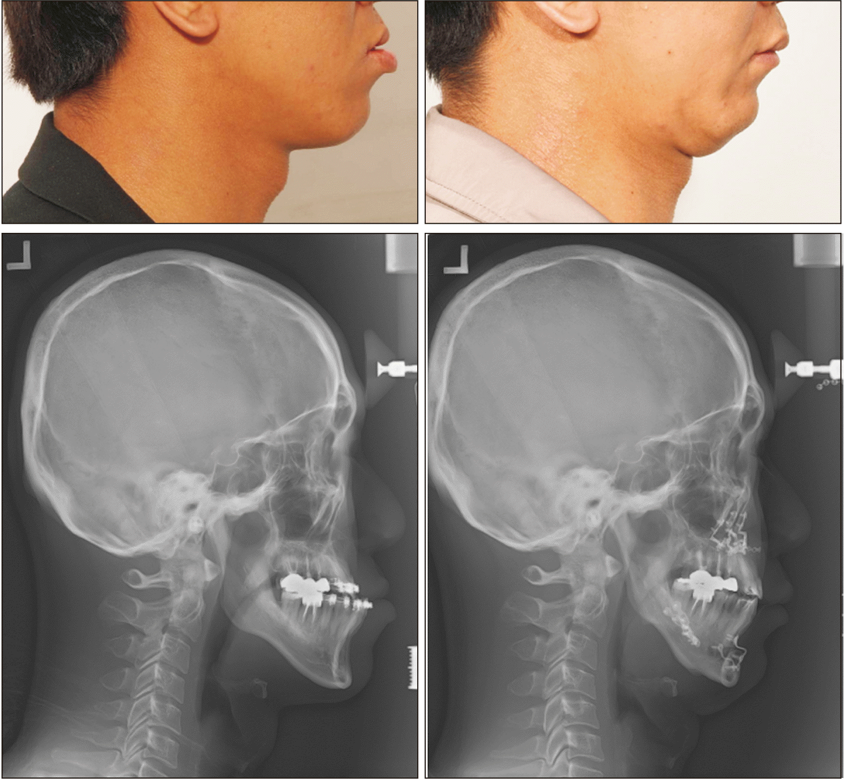
Fig. 2
A case of lower lip reduction using transverse mucosal resection and bimaxillary orthognathic surgery. (Left column) Preoperative clinical photo and lateral cephalogram. (Right column) Postoperative clinical photo and lateral cephalogram.

Fig. 3
A case of lower lip reduction using transverse myomucosal excision, upper lip cheiloplasty, and rhinoplasty. (Left column) Preoperative clinical photos and lateral cephalogram. (Right column) Postoperative clinical photos and lateral cephalogram.
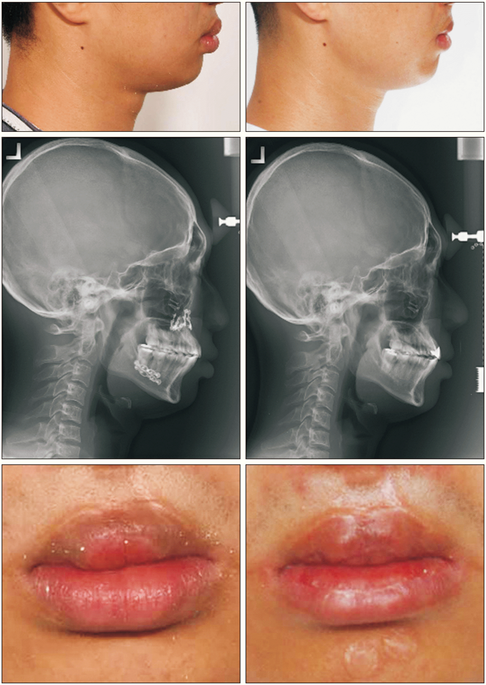
Fig. 4
Surgical procedures of transverse myomucosal excision of the lower lip and W-plasty of the upper lip. A. Marking of incision lines on the upper lip. B. Excision on the tubercle area and elevation of triangular flaps. C. Transposition and suturing of the flaps. D. Marking of incision lines on the lower lip in the shape of an ellipse. E. Direct closure.
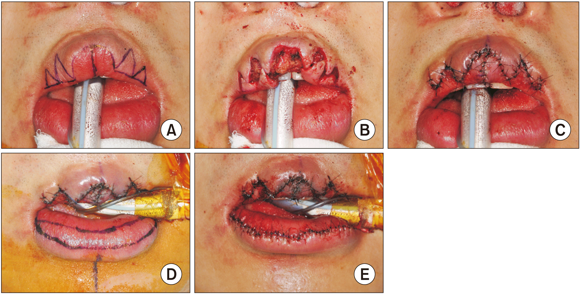
Fig. 5
A case of lower lip reduction using transverse myomucosal excision, upper lip cheiloplasty, and rhinoplasty. (Left column) Preoperative clinical photos. (Right column) Postoperative clinical photos.
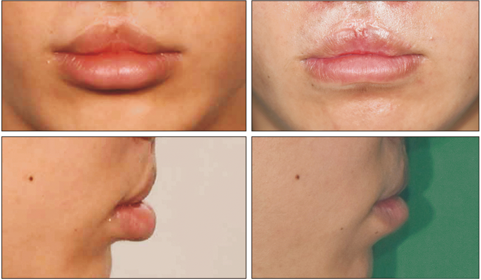
Fig. 6
Surgical procedures of transverse myomucosal excision of both lips and lateral vermilion advancement of the upper lip. A. Marking of incision lines on skin above the white roll. B. Excision and direct closure of skin. C. Marking of incision lines on the upper lip. D. Mucosa, submucosal tissue, and part of the underlying muscle were excised. E. Marking of incision lines on the lower lip in the shape of an ellipse. F. Excision including the submucosal tissue and part of the underlying muscle. G. Direct closure.


 PDF
PDF Citation
 Print
Print


 XML Download