

 PDF
PDF Citation
 Print
Print


I. Introduction
Extraction is one of the most common surgeries in dentistry, performed for various dental problems with different indications1-3. It is considered a traumatic procedure and results in immediate loss of the supporting alveolar bone and soft tissues around the extracted tooth, due to the fact that tooth extraction has followed the same protocols of achieving exodontia for the last several decades with either forceps or newly developed instruments4,5. In all protocols, tooth mobilization is achieved by severing Sharpey’s fibers from the alveolar bone and exerting luxating force by forceps or newer extraction devices, resulting in tearing of periodontal ligament (PDL) fibers and alteration of the supporting bone surrounding the socket5,6.
Although performed under local anesthesia (LA), soreness and pain due to tooth extraction can last for several days7,8. The literature suggests that postoperative pain and use of analgesics following dental extraction are related to the amount of tissue trauma created during extraction and the length of the operation9-11. These problems may be avoided through recent atraumatic extraction techniques that preserve bone and gingival architecture, reduce postoperative pain and discomfort, and allow future or immediate dental implant placement7,12.
The use of devices or instruments such as periotome and piezotome as aids in atraumatic surgical procedures has been reported in many studies4,12-14. Periotomes function by the mechanism of “wedging’’ and “severing’’ to facilitate tooth removal. They are composed of very thin metallic blades that are gently wedged into the PDL space in an orderly circumferential manner. In addition to minimally invasive luxation, the periotome blade severs Sharpey’s fibers that anchor the tooth within the socket. When the majority of Sharpey’s fibers has been separated from the root surface, rotational movements permit easy extraction of the tooth with minimal pressure. This reduces potential trauma to the adjacent bone and supporting gingival structure4,15. Another novel technique is the use of piezoelectric devices, which can overcome the drawbacks related to the conventional rotating hand piece1. The mechanism of action of the piezotome is based on the ability of certain ceramics and crystals to deform when an electric current is passed across them, resulting in micro vibrations at an ultrasonic frequency16. This aids in accurate cutting of hard tissues and protection of soft tissues including nerves and blood vessels, less vibration and noise, and a better view of the surgical field17.
As there is limited literature regarding comparative assessment of periotome and piezotome as aids in atraumatic extraction, the present randomized controlled trial was designed to compare and evaluate the effectiveness of periotome and piezotome as aids for atraumatic extraction and its sequelae.
II. Materials and Methods
1. Study participants and design
A single-blinded randomized controlled trial with concurrent parallel study design was carried out to investigate the effectiveness of periotome and piezotome as aides for atraumatic extraction, to improve operative duration, presence or absence of gingival laceration, operative and postoperative pain and discomfort, and intake of analgesics following extraction. The study sample comprised 48 teeth equally allocated to two study groups by random allocation, from participants aged 19-62 years, selected based on certain inclusion and exclusion criteria among patients who attended Qassim University Dental Clinics, Saudi Arabia, between September 2020 and March 2021. The inclusion criteria were grade I or II according to the American Society of Anesthesiologists (ASA) Physical Status Classification System18, Miller’s grade I or II mobility19, and sound roots requiring simple extraction or extraction and immediate implant placement. The exclusion criteria were grade III-VI according to ASA18, teeth with mobility >grade II19, and teeth indicated for surgical extraction. Written informed consent was obtained from all study participants after explaining the purpose, procedures, and possible complications of the trial. Ethical approval was obtained from the Ethics Committee of the Dental Research Centre of the College of Dentistry, Qassim University (No. EA/m-2019-3013). The trial was registered at https://clinicaltrials.gov with Identification Number-NCT04915443.
A pilot study was conducted on 10 participants to assess the feasibility, sample size, and familiarization with the clinical procedures. Based on the results obtained, 24 teeth samples were allocated to each of periotome and piezotome groups using a mean difference of 3.48, standard deviation of 3.7346, 90% power, and a significance level of 5%. Randomization was performed by randomly allocating each sample to one of the two study groups in a 1:1 ratio using a phone toss app. Since the study was single blinded, the participants were unaware about which group they were assigned. No attrition was reported from the final selected sample.
2. Procedure
Dental and medical histories were obtained from all patients. Prior to starting the procedure, each sample tooth was assessed clinically by visual inspection and mobility grading and radiographically by orthopantomograms and periapical radiographs. The tooth extraction procedure for all patients in both groups followed an aseptic surgical protocol. All procedures were performed by a single skilled investigator under LA using Scandicaine 2% speciale (mepivacaine hydrochloride 0.020 g/mL with adrenaline 0.010 mg/mL).
A periosteal elevator was used for both groups to perform an initial soft tissue detachment from the tooth and to test the efficiency of the LA. A timer was started once the periotome or piezotome tip touched the tooth.
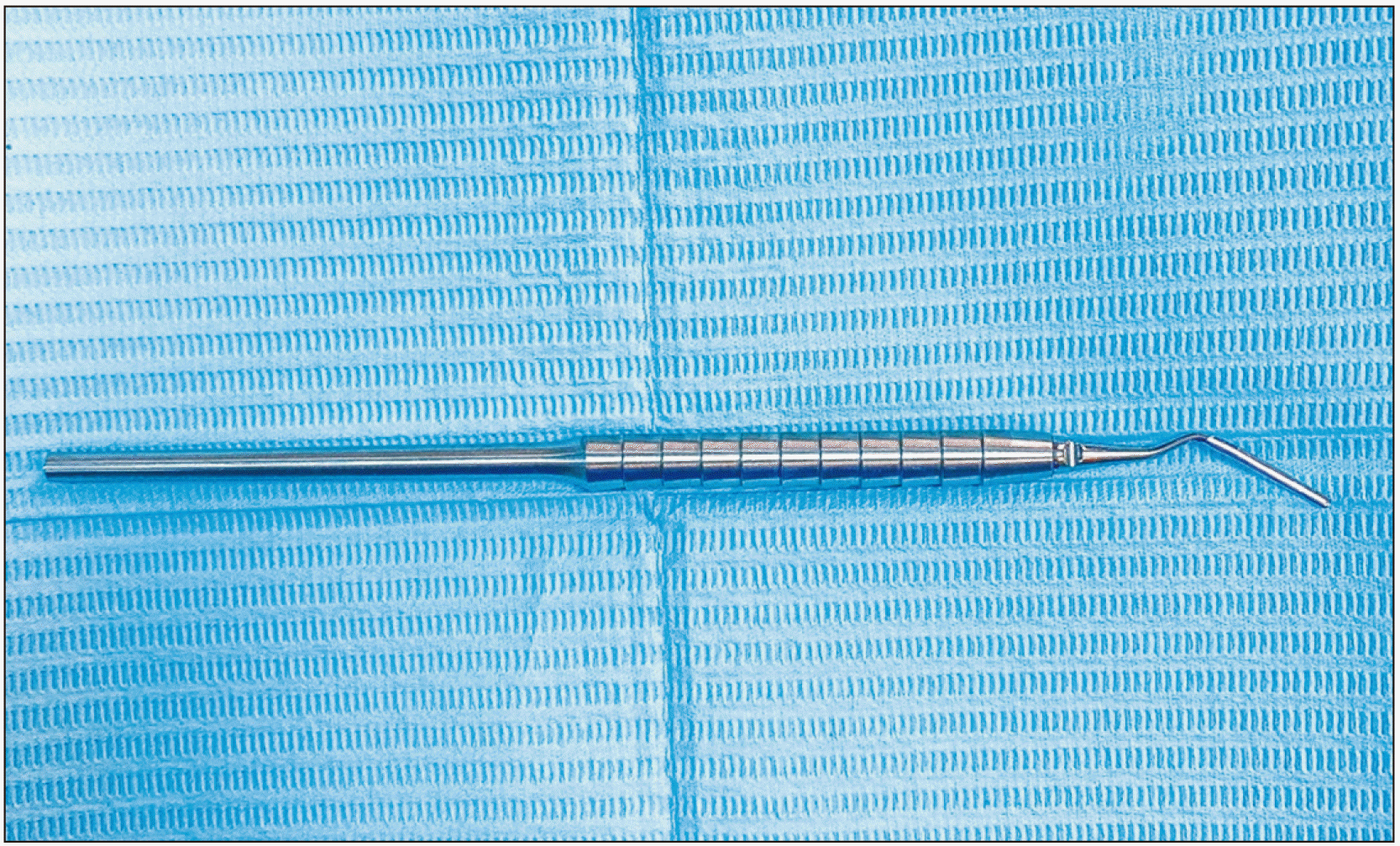
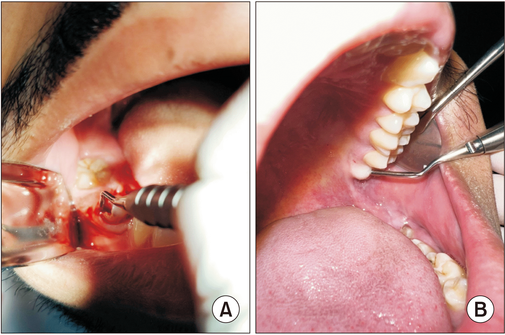
Periotomes numbered as H.ZEPF 26.182.13 and 26.182.11 (Fig. 1, 2) were used in all periotome group extraction procedures. Following initial detachment, the periotome was inserted between the root surface and the bone parallel to the long axis of the root and pushed apically to the maximum possible depth to severe the PDL fibers and was left for 10-15 seconds to allow biomechanical creep. This process was repeated at multiple points of entry on all the different surfaces of the tooth.
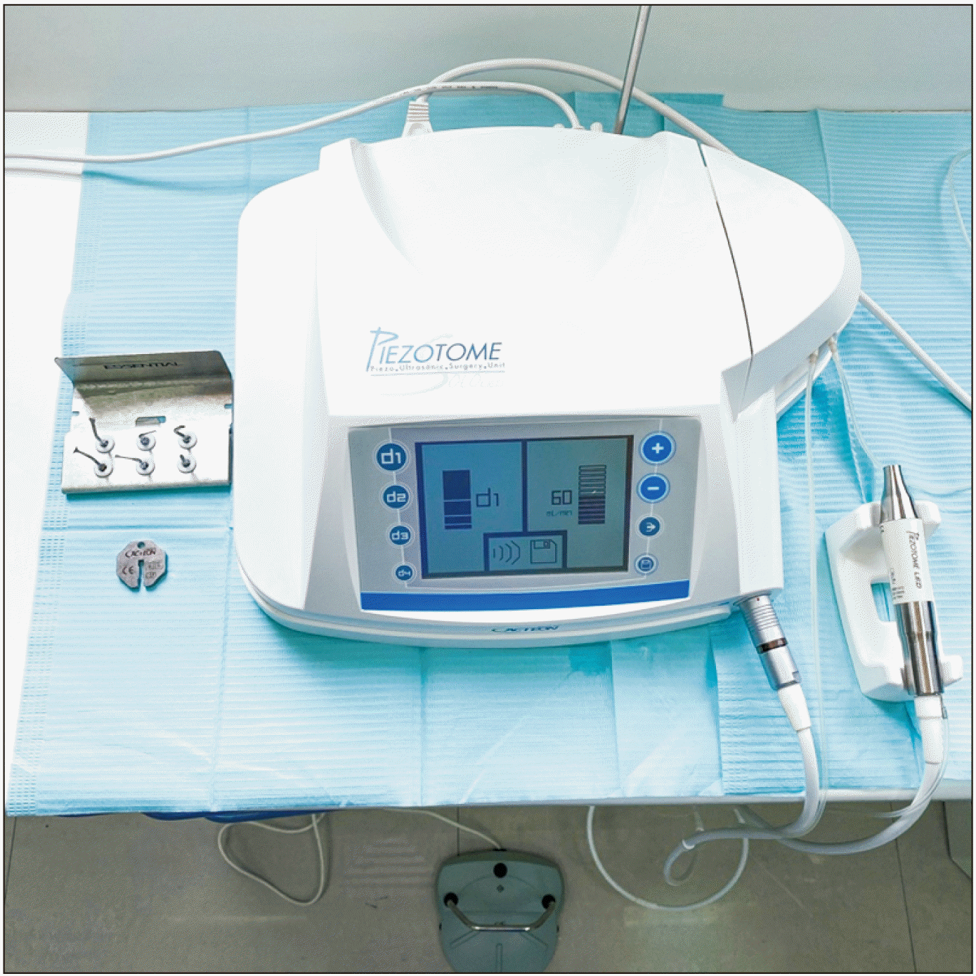
A Solo LED Piezotome Kit with essential tips (Satelec Acteon, Mérignac, France) (Fig. 3) was used for all piezotome group extraction procedures. Sodium chloride (0.9%) was used as an irrigation solution and cooling system. An LC2 tip was used for all the procedures and was inserted between the root surface and the bone parallel to the long axis of the tooth and then moved 3-4 mm toward the apex, severing the PDL with sweeping strokes.
The extraction steps that followed the periotome or piezotome step were similar for the two groups, in which suitable forceps were used to extract the tooth. The extraction site was dried with sterilized gauze and inspected for any degree of gingival laceration. A gauze pack was then placed on the extraction socket. All extractions were uneventful without any complications or associated fracture of the teeth or bone. The participants were given verbal and written postoperative instructions. The participants in both groups were instructed to take a 400 mg ibuprofen tablet if they felt any pain or discomfort after the surgery. Pain was evaluated using visual analog scale (VAS)20,21 at five different phases including I: during the procedure, II: later in the day, eight hours post-extraction after the resolution of the LA, III: on the 2nd day, IV: on the 3rd day, and V: on the 7th day. In addition, the number of participants who used analgesics after extraction was recorded in both groups, and the dosage was calculated later. All relevant data were collected by direct interviews and recorded using a previously prepared questionnaire.
3. Statistical analysis
The IBM SPSS Statistics for Windows (ver. 22; IBM, Armonk, NY, USA) was used for data entry and analysis. An independent sample t-test was used to compare the means of the two groups for quantitative variables and a chi-square test for categorical variables. Statistical significance was set at P≤0.05.
III. Results
The present trial had 48 samples that were equally and randomly allotted into periotome or piezotome groups. Clinical parameters were assessed using a validated questionnaire. All samples in both groups had either complete tooth structure or intact roots without crowns and had mobility ≤grade II. The mean clinical operation time was approximately five minutes for the periotome group and approximately eight minutes for the piezotome group, with a statistically significant difference (P=0.043).(Table 1)
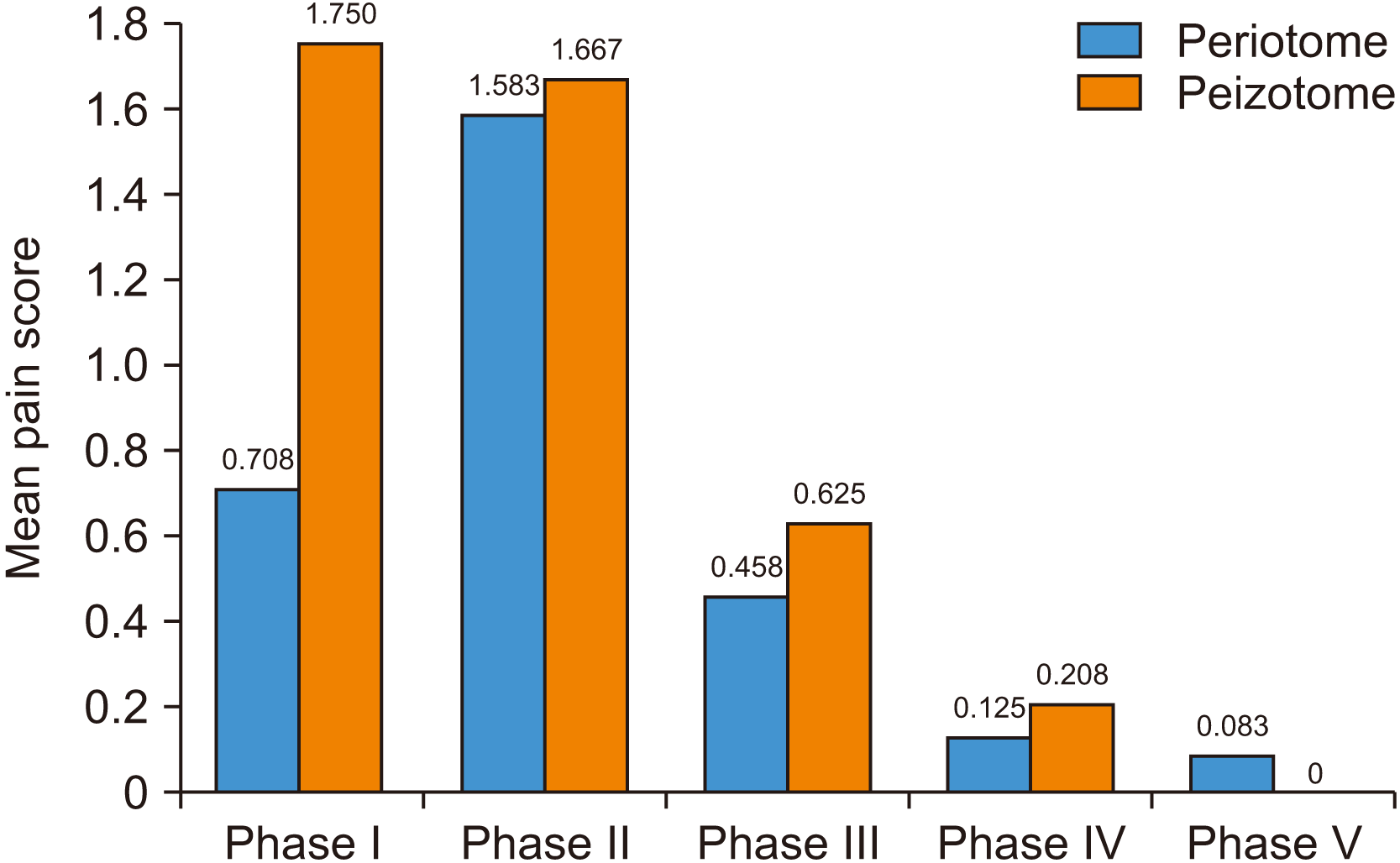
Gingival laceration was relatively reduced with use of the piezotome than with the periotome, but the differences were not statistically noteworthy.(Table 1) While comparing the mean pain scores in the five phases according to VAS, it was noted that the periotome group reported significantly (P=0.010) lower pain scores during the procedure in Phase I than the piezotome group. The piezotome group had higher pain scores than the periotome group in all phases except Phase V, which was on the 7th day after extraction, but these differences were statistically significant only for Phase I.(Table 2, Fig. 4)
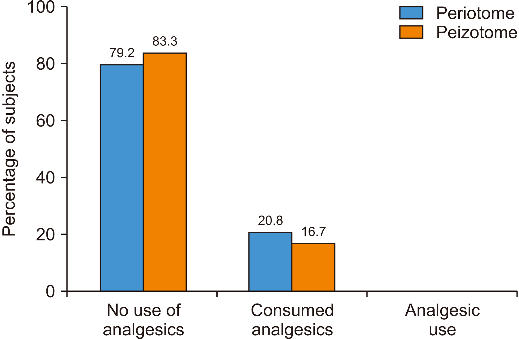
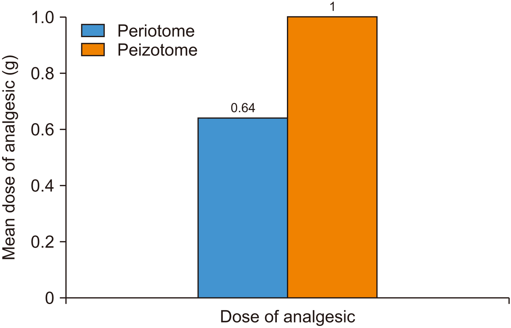
The present study shows that analgesics were used by 20.8% of participants in the periotome group and 16.7% of participants in the piezotome group; this difference was statistically insignificant.(Table 3, Fig. 5) Considering the mean dose of analgesics taken by patients, it was observed that participants in the piezotome group used 1 g of ibuprofen, while those in the periotome group used only 0.64 g, and no statistically significant difference was observed between the groups in this regard.(Table 3, Fig. 6)
IV. Discussion
The need to achieve a pain-free postoperative period calls for atraumatic dental extractions that induce minimal trauma during tooth removal, preserve the adjacent bone and gingiva, and provide the best healing conditions for the extraction socket. In the present study, extraction was performed by cutting the periodontal fibers using either a periotome or a piezotome before tooth mobilization. When these fibers are severed, extraction of the tooth with minimal lateral pressure is permitted by relatively simple movements using forceps22. The aim of the current clinical trial was to evaluate and compare the efficacy of the periotome and piezotome as aids for atraumatic extraction in terms of operative duration, presence or absence of gingival laceration, operative and postoperative pain levels, and analgesic intake pattern following the extraction procedure.
The first parameter under consideration was the operative duration recorded from the time of insertion of the instrument until complete removal of the tooth from the socket. According to Rakhshan23 and de Santana-Santos et al.24, longer surgeries result in more painful sockets in the case of third molar extractions, which was one of the reasons for inclusion of the “surgical duration” parameter in this study. Our research showed that the surgical duration in the the piezotome group was significantly longer than that in the periotome group, which is similar to the study by Melek and Noureldin25. Extended clinical time with the use of piezotomes has also been reported in studies performed by Arakji et al.16 and Goyal et al.26. As stated by Troedhan et al.27, surgery time could be initially longer, when purely working with ultrasonic surgical devices like piezotomes, although surgical time could be reduced after a certain learning curve. Sharma et al.8 also reported a shorter operation time with the use of periotomes, which is similar to the results of the periotome group in the present study. This points to the possibility of periotomes being advantageous to the operator in terms of time management.
All atraumatic extractions are expected to minimize gingival trauma. According to Sharma et al.8, the use of periotomes resulted in significantly less gingival laceration than the control group. This is consistent with the results of the current study, which showed that gingival laceration was less prevalent in the periotome group; however, the piezotome group exhibited a much better outcome in this regard, although it was not significantly different. The slightly better performance of the piezotomes could be due to the cavitation effect created by interaction between the irrigant solution and the oscillating tips, resulting in a clear surgical site during the procedure and allowing for greater operator precision13. In addition, nerves, blood vessels, and soft tissue are not injured by the microvibrations of the piezotome, which are optimally adjusted to target only mineralized tissue28.
After conventional tooth extraction, it is common for patients to experience pain once the effect of LA wears off, and the degree of severity varies between patients3,29. Breivik and Björnsson29, Bortoluzzi et al.30, and Rakhshan23 have mentioned that postoperative pain is often related to the degree of surgical trauma. Surgical extraction results in physical injury to the tissues and causes a sequential release of inflammatory mediators from mast cells, vasculature, and other cells depending on the extent of injury, and proximity to the nerve might produce more intense pain in some difficult cases10,23. Atraumatic extraction techniques using microsurgical instrumentation such as periotomes4 and piezotomes31 aim to reduce trauma and postoperative pain due to extraction. Studies by El-Abbasy832 and Srivastava et al.1 have shown that patients who had undergone surgical removal of impacted third molars by piezotomes experienced gradually diminishing moderate to mild pain during the first four days postoperatively, as interpreted from their mean VAS pain scores20. This is not in alignment with the present study, as much lower mean pain scores were reported by patients in the piezotome group, which showed a decreasing trend from mild to painless at one week after the extraction. This disagreement could be due to the simple extractions performed here compared to extraction of impacted third molars in the comparative studies. The mean pain scores indicative of mild to negligible postoperative pain, as expressed by the periotome group in the current study, are consistent with the findings of Sharma et al.8. In this trial, the mean pain scores were highest during the extraction procedure (Phase I) for the piezotome group, while they were maximal for the periotome group eight hours postoperatively (Phase II) and decreased gradually thereafter for both groups. The piezotome group had higher pain scores than the periotome group during the procedure and postoperatively until the third day (Phase IV). For Phase V recordings on the 7th postoperative day, pain was reported as totally absent only by the piezotome group. This was contradictory to the reports of a trial by Melek and Noureldin25, in which pain was minimal and was completely resolved by the third postoperative day in all patients in both the piezotome and periotome groups. Considering analgesic intake, the percentage of participants who took analgesics postoperatively was higher in the periotome group, while the mean dosage of analgesics was higher in the piezotome group, which could be a reflection of higher pain scores experienced by participants in the periotome group initially at eight hours postoperatively. The higher dosage of analgesics in the piezotome group might be due to the prolonged intensity of pain during phases III and IV. Troedhan et al.27 reported the use of a mean dosage of 3 g of ibuprofen following piezo-surgical removal of impacted third molars, which is not in agreement with the present results that indicate a lower mean dosage of only 1 g by the piezotome group. This is probably due to the lower complexity of the extractions in the current study. As mentioned earlier, considering the link between of surgery and postoperative pain, the present trial found that operative duration and dosage of analgesic used were both higher in the piezotome group than in the periotome group. Garcia Garcia et al.33 reported a correlation between operative time and analgesic use over the first 48 hours post-surgery. The duration of operation performed by a single surgeon could indicate the difficulty of the procedure, duration of tissue injury, and severity of pain34. In that study, all extractions were performed by a single operator, and the same findings are reflected in the present research.
V. Conclusion
Based on this study, it can be concluded that the present clinical trial favors the use of periotomes over piezotomes for atraumatic extractions due to a shorter operating time, lower postoperative VAS pain scores, and smaller dosage of analgesics, although the piezotomes exhibited superior ability in preventing gingival lacerations. Another noteworthy finding is that the periotome is a more economically viable and low-maintenance instrument than the piezotome. One limitation of this study was the inability to compare meaningfully and reasonably due to lack of similar studies in the scientific databases. For the same reasons, further research is suggested on a larger sample size considering more reliable parameters such as bone levels postoperatively among the two groups to justify and conclude whether the periotome could be preferred over the piezotome for atraumatic extractions.
 XML Download