Journal List > Korean J Physiol Pharmacol > v.27(1) > 1516080696

Kumar, Gaur, Yerrabelli, Sahoo, Vairappan, and Goud: Association between metabolic syndrome components and cardiac autonomic modulation in southern Indian adults with pre-metabolic syndrome: hyperglycemia is the major contributing factor

Abstract

Metabolic syndrome (MetS) involves multi-factorial conditions linked to an elevated risk of type 2 diabetes mellitus and cardiovascular disease. Pre-metabolic syndrome (pre-MetS) possesses two MetS components but does not meet the MetS diagnostic criteria. Although cardiac autonomic derangements are evident in MetS, there is little information on their status in pre-MetS subjects. In this study, we sought to examine cardiac autonomic functions in pre-MetS and to determine which MetS component is more responsible for impaired cardiac autonomic functions. A total of 182 subjects were recruited and divided into healthy controls (n=89) and pre-MetS subjects (n=93) based on inclusion and exclusion criteria. We performed biochemical profiles on fasting blood samples to detect pre-MetS. Using standardized protocols, we evaluated anthropometric data, body composition, baroreflex sensitivity (BRS), heart rate variability (HRV), and autonomic function tests (AFTs). We further examined these parameters in pre-MetS subjects for each MetS component. Compared to healthy controls, we observed a significant cardiac autonomic dysfunction (CAD) through reduced BRS, lower overall HRV, and altered AFT parameters in pre-MetS subjects, accompanied by markedly varied anthropometric, clinical and biochemical parameters. Furthermore, all examined BRS, HRV, and AFT parameters exhibited an abnormal trend and significant correlation toward hyperglycemia. This study demonstrates CAD in pre-MetS subjects with reduced BRS, lower overall HRV, and altered AFT parameters. Hyperglycemia was considered an independent determinant of alterations in all the examined BRS, HRV, and AFT parameters. Thus, hyperglycemia may contribute to CAD in pre-MetS subjects before progressing to MetS.

INTRODUCTION

Metabolic syndrome (MetS) is an amalgamation of predisposing factors, comprising insulin resistance (IR), raised blood pressure (BP), atherogenic dyslipidemia, abdominal obesity, pro-inflammatory state, and prothrombotic state [1]. Individuals with MetS have a greater risk of developing type 2 diabetes mellitus (T2DM), cardiovascular disease (CVD), and all-cause mortality. These dreadful consequences may be mitigated by controlling the MetS components with suitable interventions [2]. There is also evidence that MetS is a modest and applicable clinical tool for identifying high-risk individuals prone to T2D and CVD. Moreover, MetS components have been independently linked to T2D and CVD, making them therapeutic targets [3]. The prevalence of MetS is rising at pandemic proportions in developed and emerging nations [4]. The global prevalence of MetS among adults is estimated between 20% and 25% [5]. Based on the National Health and Nutrition Examination Survey reports from 2003 to 2012, the prevalence of MetS in the United States was 33%, and the prevalence increased with age [6]. Despite the devastating prevalence of MetS and its risk severity, determining the causative connection between MetS and CVD has been challenging. It is still open for debate whether they have shared underlying pathophysiology [7].
The leading risk factor for Mets is a condition called pre-MetS, which is often overlooked, asymptomatic and unnoticed. Pre-MetS is considered to have any two MetS components but does not fulfill the MetS diagnostic criterion [8]. Pre-MetS is not a specific condition but a set of risk factors grouped together, significantly increasing the risk of developing MetS later in life, and is probably a lifetime burden of CVD. Thus, detecting and treating pre-MetS at an early stage is critical. An ongoing debate is whether pre-MetS deserve targeted identification and clinical intervention. Several studies have shown that MetS causes cardiac autonomic dysfunction (CAD), which manifests as an imbalance between sympathetic and parasympathetic activity [9-12]. The components of MetS, including abdominal obesity, IR, and chronic inflammation, have been implicated as essential mechanisms linking hyperinsulinemia, sympathetic overactivity, and CAD [13].
Non-invasive evaluation of cardiac autonomic functions may include heart rate variability (HRV), autonomic function tests (AFTs), heart rate recovery, heart rate turbulence, sympathetic skin response, R-R interval variation, and baroreflex sensitivity (BRS) [14]. Spectral analysis of HRV (a measure of the variance in time between each heartbeat) can assess the cardiac autonomic modulation of the heart and provides vital insights into the pathogenesis of CAD. Lower HRV, especially lower vagal activity, has been interrelated to ischaemic heart disease, heart failure, and coronary heart disease [15]. Moreover, 12-year follow-up research found that having lower HRV increased the odds of developing MetS [16]. BRS measures arterial baroreflex function [17]. BRS assessment is a sensitive and effective method for evaluating CAD, with practical applications in a clinical setting. The factors that affect BRS include autonomic imbalance and arterial distensibility. Many studies have documented the implication of reduced BRS in obesity, triglyceridemia, MetS, hypertension, T2DM, myocardial infarction, coronary artery disease, and heart failure [12,18]. Classic AFTs, including heart rate (HR) and BP responses to standing, deep breathing, and isometric handgrip, provide a simple and non-invasive method for assessing the sympathovagal control of the cardiovascular system. A cohort study found a link between MetS and a greater risk of early CAD [19]. In the same cohort, they also looked at the prospective connections between individual components of MetS and detected an early deficit of cardiac autonomic functions.
We have recently found that CAD forms sympathovagal imbalance, as measured by reduced BRS, lower HRV, and high resting HR in patients with MetS [12]. Though umpteen studies have demonstrated CAD in MetS subjects, there is a paucity of research on assessing the cardiac autonomic functional status in pre-MetS subjects. We hypothesized that early cardiac autonomic functional derangements are already present in pre-MetS and carry some predictive power for significant cardiovascular co-morbidities and complications in the future. Early detection of CAD in pre-MetS can delay or even prevent the development of serious cardiovascular complications. We designed this study to confirm the hypothesis and compare electrocardiogram (ECG)-derived measurements of cardiac autonomic functions between apparently healthy control and pre-MetS subjects. Furthermore, we investigated which MetS component was responsible for impaired cardiac autonomic functions and tested whether measures of autonomic function, such as BRS, HRV, and AFT indices, were related to different components of pre-MetS.

METHODS

Study participants

We performed a cross-sectional observational study in the Department of Physiology, Puducherry Teaching Hospital, India. The Institutional Ethical Committee approved the study protocol for human studies at JIPMER, Puducherry (JIP/IEC/2018/0301). A power analysis was performed before the study using OpenEpi, version 3 (www.OpenEpi.com). Accordingly, when the effect size was considered moderate based on the previous report with a difference of 0.54 between two independent means of the log-transformed ratio of low-frequency to high-frequency power (LF/HF) of HRV, the sample size obtained was 156 subjects with 78 in each group, which have resulted from 95% confidence and 90% power [20]. However, we enrolled 182 subjects, with 89 subjects in the control group and 93 subjects in the study group. Before collecting the data, we obtained written informed consent from all the participants. The subjects aged 18 to 65 years of either gender volunteered to participate in this study.
The study group comprises pre-MetS subjects recruited based on predetermined eligibility criteria set by National Cholesterol Education Program Adult Treatment Panel III guidelines. Pre-MetS was detected using two of the following five conditions [21]. Elevated waist circumference (WC) ≥ 90 cm in men and 80 cm in women (South-Asian criteria); hyperglycemia with fasting plasma glucose (FPG) ≥ 100 mg/dl (5.6 mM) or previously diagnosed with T2D or on medication for T2D; atherogenic dyslipidemia with serum triglyceride (TG) ≥ 150 mg/dl (1.7 mM) and high-density-lipoprotein cholesterol (HDL-c) ≤ 40 mg/dl (1.03 mM) in men, ≤ 50 mg/dl (1.29 mM) in women or on specific treatment for dyslipidemia; raised BP with systolic BP ≥ 130 mmHg or diastolic BP ≥ 85 mmHg or receiving antihypertensive medication. The control group comprises age-matched healthy participants having normal clinical and biochemical (blood glucose and lipid) profiles. Patients with cardiovascular, neurological, psychiatric, endocrine, and other significant co-morbidities or using medication for a chronic condition were excluded from the study during the screening procedure.

Demographic, anthropometric, clinical, and biochemical data collection

Subjects were instructed to report to the Department of Physiology on their scheduled appointment date fixed during the screening process. A standard questionnaire was directed to gather data on health status, personal and family history of chronic diseases (hypertension, T2D, and CVD), smoking and alcohol intake, medication, and lifestyle details. Anthropometric measurements were performed using the World Health Organisation (WHO) guidelines [22]. The body mass (weight in kg) was assessed using a digital weighing machine. Height was measured with a wall-mounted stadiometer. Body mass index (BMI) was calculated using Quetelet’s index [23].
We used non-stretchable tape to measure WC (cm) around the abdomen at the level of the umbilicus (belly button) between the lower edge of the last palpable rib and the top of the iliac crest. In contrast, hip circumference (HC in cm) was measured as the largest diameter around the buttocks in a standing position. The ratio of waist-hip (WHR) and waist-height (WHtR) were calculated. The percentage of body fat (BF%) was assessed using a body fat analyzing device (Quadscan 4000; Bodystat, Isle of Man, UK) with a bioelectrical impedance analysis approach. Participants were asked to remove any metals and lie supine on the couch. Then electrodes were connected to the hand and foot in a tetrapolar pattern, which operates because the electrical conductivity of fat-free tissue is far greater than the fat mass.
BP was measured in the right arm using an Omron automated BP device after 10 min of rest in a sitting posture (SEM-1; Omron, Kyoto, Japan). The same observer obtained a minimum of three recordings at 1-min intervals. An average of three BP readings was taken. Fasting blood samples were drawn by vein puncture and processed for biochemical analysis of FPG, total cholesterol (TC), HDL-c, and TG using commercial kits adapted to clinical chemistry autoanalyzer (Olympus 400; Beckman Coulter, Orlando, FL, USA). Friedwalds’s formula assessed low-density lipoprotein cholesterol (LDL-c) and very low-density lipoproteins (VLDL) [24]. Homeostatic model assessment of IR (HOMA-IR) and other lipid profile-derived parameters were calculated (TC/HDL-c, TG/HDL-c, LDL/HDL-c, non-HDL-c/HDL-c, and atherogenic index of plasma (AIP: log10[TG/HDL-c])). We used commercially available enzyme-linked immunosorbent assay (ELISA) kits and followed the manufacturer’s instructions to measure insulin, high-sensitive C reactive protein (hs-CRP), and adiponectin (Calbiotech, El Cajon, CA, USA).

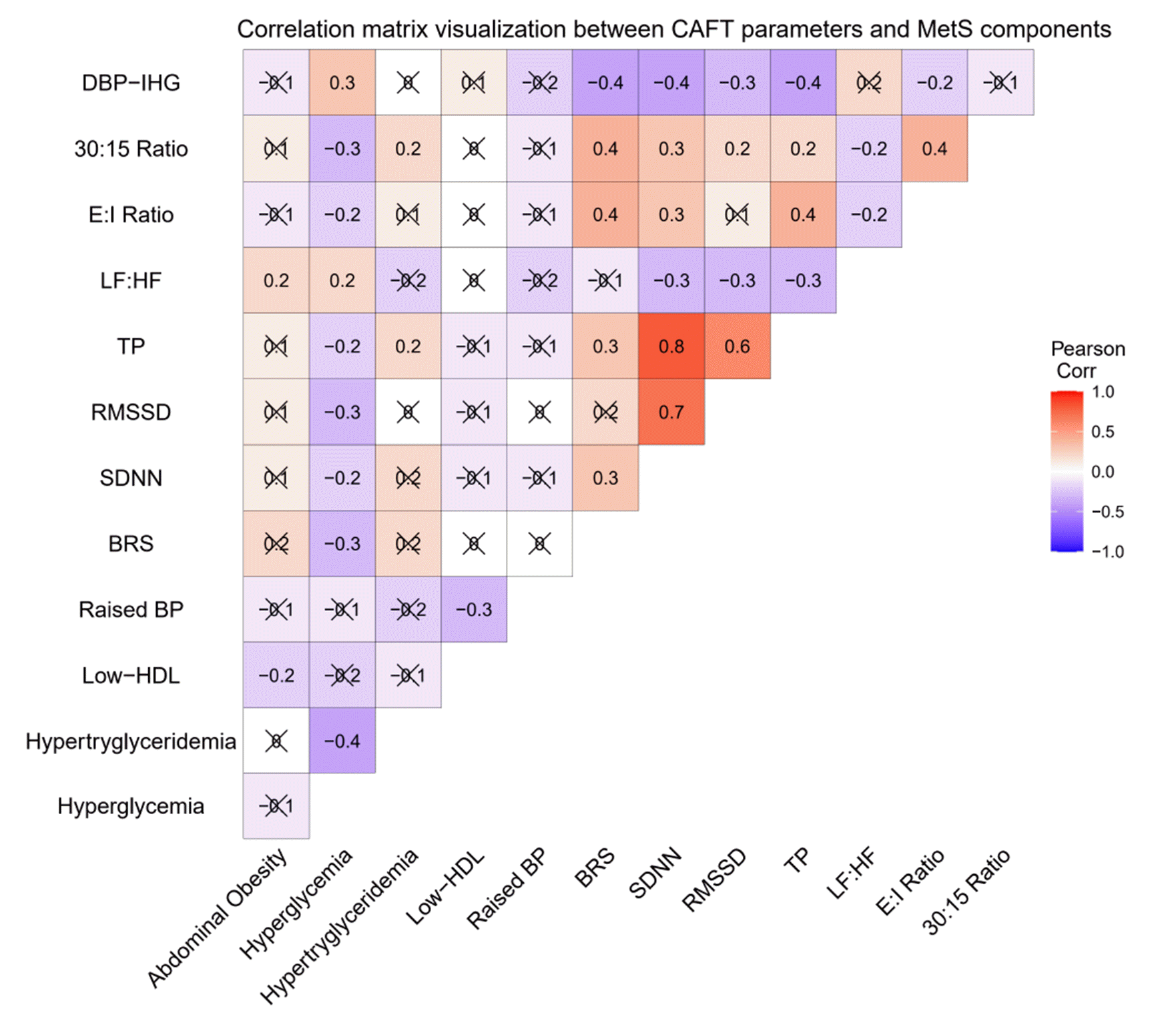
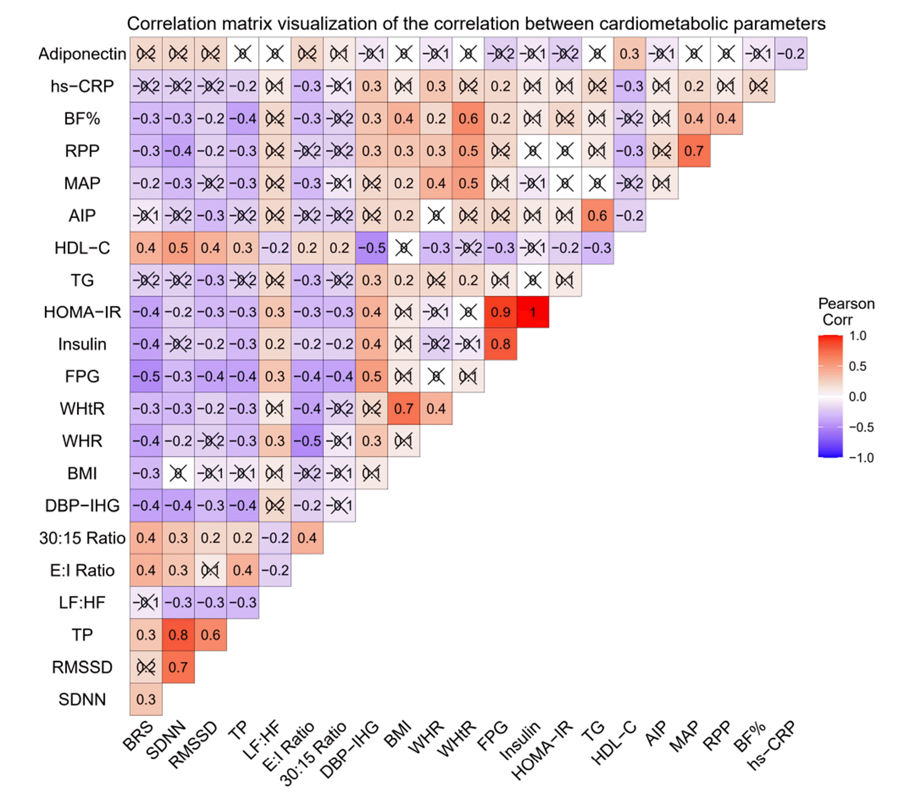
Measures of cardiac autonomic modulation

Short-term HRV

Following the standard guidelines published by the Task Force of the European and North American society for HRV, lead II electrocardiogram (ECG) was obtained for 5 min after 15 min of supine rest [25]. We used BIOPAC MP 150 data acquisition system (BIOPAC Inc., Goleta, CA, USA) to record the lead II ECG. We carefully analyzed artifacts and ectopic beats and removed them thoroughly using a windows-based computer software AcqKnowledge (version 4.2; BIOPAC Inc.). Both time domain and frequency domain indices (TDI and FDI) were analyzed by Kubios software (version 2.0; Biomedical Signal Analysis Group, University of Kuopio, Finland). FDI were depicted as spectral power such as total power (TP), very-low-frequency (VLF) power, HF, and LF power in both absolute powers (ms2) and normalized units (nu). The TDI encompassed standard deviation of normal to normal (SDNN) interval, square root of the mean squared differences of successive normal to normal intervals (RMSSD), adjacent RR interval differing more than 50 milliseconds (NN50), and its percentage (pNN50).

BRS

We assessed BRS noninvasively using Finapres hemodynamic cardiovascular monitor (Finometer; Finapres Medical Systems, Amsterdam, Netherlands) and BeatScope Easy computer program (Finapres Medical Systems). Finapres reconstructs brachial pressure from finger pressure through generalized waveform inverse modeling, and generalized level correction and measurements are based on the volume clamp method of Penaz and the Physiocal criteria of Wesseling [17]. The subjects were instructed to rest in a supine position. The brachial cuff was tied 2 cm above the cubital fossa around the midarm. A finger cuff with an infrared light-emitting diode was wrapped around the middle phalanx of the middle finger. Two sensors were installed for the height correction, one at heart level and the other at finger level. After connecting the cuff cables to the finometer, the recordings were obtained after 10 min of supine rest. The “return to flow calibration and the physiocal” was done during the initial 5 min of the recordings for level correction between the brachial and finger pressure. After this, 10 min of continuous recording was done. BRS was stated as the inter-beat-interval change, in ms, with instantaneous change in BP, in mmHg. Rate-Pressure Product (RPP) [26], a determinant of the myocardial oxygen consumption and workload, has been determined using the formula RPP = 10–2 × (basal heart rate [BHR] × systolic blood pressure [SBP]).

Conventional AFTs [27,28]

  • HR response to deep breathing: Baseline ECG recordings were taken in the supine position for 30 sec. Subjects were instructed to breathe slowly and deeply at six breaths per minute, with 5 sec of inspiratory and expiratory cycles. The E:I ratio is the measure of response calculated as a ratio of the longest RR interval during expiration (E) to the shortest RR interval during inspiration (I).

  • Lying to standing test: This test aimed to determine the effects of orthostasis (standing) on HR and BP. The baseline ECG and BP measurements were taken while the subject was lying down, and the subject was told to stand up within 3 sec. A continuous ECG was recorded, and the BP was monitored every 40 sec by an automatic BP monitor for five minutes. The 30/15 ratio is calculated as the ratio of the longest RR intervals around the 30th beat and the shortest RR intervals around the 15th beat in response to orthostasis.

  • Isometric handgrip test: The subjects were instructed to press the handgrip dynamometer (Inco, Ambala, India) for two minutes at 1/3rd of their maximum strength after obtaining their baseline BP value. Recordings of BP were taken during the first and second minutes of contraction. The measure of response is the difference between baseline and maximum rise in diastolic blood pressure (ΔDBPIHG). ΔDBPIHG is considered normal at ≥ 16 and abnormal at ≤ 10.

Statistical analysis

The SPSS software program (version 22) was used for all statistical analyses (IBM Corp., Armonk, NY, USA). The Kolmogorov–Smirnov test was used to determine the normality of data. Non-normal distribution data were reported as median (interquartile range), whereas normal distribution data were reported as mean ± SD. Student’s t-test for continuous parametric data and Mann–Whitney U-test for nonparametric data were used to compare the control and test groups. We provided percentages and frequencies for categorical data, and the chi-square test (χ2) determined the difference between groups. A point-biserial correlation was used to measure the strength and direction of the association between continuous variables (cardiac AFT parameters) and dichotomous variables (presence and absence of each MetS component). We used Pearson’s correlation coefficient analysis to assess the degree of correlation between cardiometabolic variables. The correlation heatmaps were created using the “ggcorrplot” R package (R Studio windows version 4.2.1). Multiple linear regression analysis was used to investigate the independent contribution of WhtR, FPG, TG, HDL-c, RPP, and BF% to BRS, HRV, and AFT parameters. All analyses were two-tailed, and the study employed p < 0.05 as a level of statistical significance.

RESULTS

Table 1 presents descriptive statistical values for the study participants’ demographic, anthropometric, and body composition data. The standard definition of MetS identified 93 participants with the pre-MetS. The study variables in pre-MetS subjects were compared to 89 healthy control subjects of the same age and gender. Participants in the pre-MetS group had similar age, gender distribution, physical activity status, and family history of hypertension compared with control group participants. Hyperglycemia is more common among pre-MetS participants (60.2%). A significantly higher prevalence of smoking, alcohol consumption, family history of T2D and CVD, hyperglycemia, high TG, low HDL-c, and raised BP was observed in the pre-MetS group (Table 1). Anthropometric and body composition data revealed the expected unfavorable phenotype in pre-MetS subjects, with substantially higher weight, BMI, WC, HC, WHR, and WHtR. Body composition data showed significantly higher body fat% and lower body lean% than control subjects (Table 1).
Table 2 shows the biochemical profile of the study participants. As expected, the pre-MetS subjects showed an impaired glycemic profile as significant hyperglycemia and IR than the control group. Though TC and LDL-c were not significantly different, other lipid profile markers (TG, VLDL, LDL/HDL, TC/HDL, TG/HDL, non-HDL/HDL, and AIP) were significantly higher in pre-MetS than controls, but HDL-c was markedly lower. Compared to controls, the pre-MetS group had altered adipokine secretion with substantially higher hs-CRP and lower adiponectin (Table 2).
Table 3 presents BP variability, HRV, and AFT parameters compared among the study participants. The cardiovascular variables BHR, SBP, DBP, mean arterial pressure (MAP), and RPP were considerably more significant in pre-MetS than in controls, while BRS was markedly lower. All the TDI of HRV were significantly lower in the pre-MetS group, including mean RR, SDNN, RMSSD, NN50, and pNN50, suggesting lower overall HRV with decreased parasympathetic activity. Among the FDI of HRV, we found lower TP, HF, LF, VLF, HFnu, and greater LFnu and LF/HF ratio in pre-MetS than controls, suggesting probable silent CAD and sympathovagal imbalance. Based on AFT parameters, we observed lower autonomic reactivity as significantly lower E:I ratio, 30:15 ratio, and higher ΔDBPIHG in pre-MetS subjects than controls.
Furthermore, we analyzed the impact of each diagnostic component of MetS on BRS, HRV, and AFTs in the pre-MetS group to investigate which criteria have impaired the autonomic modulation (Fig. 1). Among the MetS components analyzed, we found significant variations in BRS, SDNN, RMSSD, TP, LF/HF, E:I ratio, 30:15 ratio, and ΔDBPIHG related to the presence of hyperglycemia criterion. More precisely, hyperglycemia showed a strong correlation with reduced BRS, SDNN, RMSSD, TP, E:I ratio, 30:15 ratio, and increased LF:HF ratio and DBPIHG. Abdominal obesity was connected to a considerably higher LF/HF ratio of HRV. Surprisingly, hypertriglyceridemia was linked positively to TP of HRV and 30:15 ratio. No significant differences were observed in these BRS, HRV, and AFT parameters associated with the other 2 MetS criteria (low HDL-c and elevated BP).
Fig. 2 shows the CVD risk variables associated with the BRS, HRV, and AFT parameters. Hyperglycemia (raised FPG) and HOMA-IR were significantly associated with all the studied parameters, including BRS, SDNN, RMSSD, TP, LF/HF, E:I ratio, 30:15 ratio, and ΔDBPIHG. The BRS and ΔDBPIHG had the most substantial power among the studied parameters regarding correlation with FPG. The other CVD risk variables (WHR, WHtR, insulin, TG, HDL-c, and BF%) also showed a few associations with BRS, HRV, and AFT parameters. Multiple linear regression analysis assesses the independent association of BRS, HRV, and AFT parameters (as dependant variables) with MetS risk factors (independent variables) in the pre-MetS group (Table 4). Among the MetS risk factors, FPG levels were revealed to be an independent predictor of alterations in all the examined BRS, HRV, and AFT parameters.

DISCUSSION

Globally, the prevalence of MetS is increasing and becoming a significant threat in all age groups and should be monitored carefully. Furthermore, each component of MetS increases the jeopardy of T2D and CVD [29]. One possible mechanism underpinning the relationship between MetS and CVD events is an aberrant modulation in autonomic tone with sympathovagal imbalance [30]. Pre-MetS is the asymptomatic precursor of MetS and is expected to be associated with early CAD. Thus, pre-MetS is becoming a viable target for early management to avoid developing the MetS and its associated CAD in the future. HRV, BRS, and AFT measurements have been widely used in clinical settings as investigative and predictive tools to explore cardiac autonomic functional status.
The present study involved 182 study participants, categorized into pre-MetS and control groups based on the standard criteria for MetS. The following were the significant findings of the study: (1) Pre-MetS subjects were more prone to have CAD than the control group; (2) BRS, HRV, and AFT parameters were significantly different between the pre-MetS and control groups; (3) BRS, HRV, and AFT values were substantially different only in groups defined by the existence of the hyperglycemia criterion when the participants in pre-MetS were further evaluated for each diagnostic component of MetS; (4) All of the BRS, HRV, and AFT parameters studied had a significant association with FPG levels; and (5) The raised FPG levels were shown to be an independent determinant of all the BRS, HRV, and AFT parameters studied.

Anthropometric and body composition data in pre-MetS

In addition to significantly elevated WC, which is already regarded as a component of MetS, the study found weight, BMI, HC, WHR, WHtR, and BF% were significantly higher in pre-MetS subjects signifying the presence of abdominal obesity, predisposing to CVD risk. Among the anthropometric parameters, WHR, particularly WHtR, has been reported to be linked with CVD deaths [31]. Notably, the significant association of WHtR with BRS, SDNN, RMSSD, TP, and E:I ratio in pre-MetS subjects suggests an early CAD (Fig. 2). Moreover, a substantial rise in BF% with a concomitant reduction in body lean% in pre-MetS subjects might suggest a dysfunctional energy metabolism involving glucose and lipids due to increased adiposity. The substantial relationship between BF% and BRS, SDNN, RMSSD, TP, and E:I ratio might imply that excess adiposity contributes to early CAD (Fig. 2).

Glycemic parameters, lipid profiles, and circulating biomarkers in pre-MetS

The altered glycemic profile in pre-MetS subjects indicated an insulin-resistant condition with significantly higher FPG, insulin, and HOMA-IR (Table 2). Lipid profile abnormalities were observed in the pre-MetS group with markedly elevated TG, lipid to lipoprotein ratios, and lower HDL-c. The significant associations of FPG and HDL-c with the examined parameters of BRS, HRV, and AFTs and the independent contribution of FPG and HDL-c to altered BRS, HRV, and AFT parameters imply that the subjects in the pre-MetS group are at an increased CVD risk associated with CAD due to IR, and atherogenic dyslipidemia. Our recent findings support the theory that persistent low-grade inflammation and hypoadiponectinemia are vital in developing metabolic abnormalities in MetS patients [12]. Similarly, significantly higher hs-CRP and lower adiponectin levels in pre-MetS, as well as the association of hs-CRP with a few studied BRS, HRV, and AFT parameters, suggest an association of chronic inflammation with CAD in the current investigation.

Blood pressure variability (BPV) parameters in pre-MetS

Among the BPV parameters, BHR is the most direct and straightforward predictor of cardiac health. Low vagal potency and autonomic imbalance have been linked to a high resting HR [32]. Higher resting HR could indicate an imbalance in the heart’s sympathovagal control. Consequently, myocardial oxygenation and workload are determined by the RPP, and an increase in RPP has been identified as a CVD risk factor [26]. The pre-MetS group had a significant increase in BHR and RPP, along with systolic, diastolic, and mean arterial BP (SBP, DBP, and MAP), when compared to controls, indicating poor CV health and increased susceptibility to cardiac arrhythmias and other cardiovascular complications. Additionally, BRS, a strong determinant of cardiac mortality, was significantly lower in the pre-MetS group than in controls, confirming poor CV health and cardiac autonomic imbalance in these subjects. Pikkujämsä et al. [33] found impaired BRS and respiratory modulation of HR in hypertensive subjects with or without IR syndrome. These findings were backed up by our study findings in the pre-MetS group. In another study, Lindgren et al. [34] mentioned that BRS could precisely detect autonomic complications and disease prognosis and found a cardiovascular autonomic imbalance in MetS and IR subjects as a low BRS.

HRV indices in pre-MetS

The spectral analysis of HRV is a robust approach for evaluating cardio-vagal modulation and the type and severity of CAD in both health and illness. To date, no data on cardio-vagal modulation in pre-MetS has been penned. In the current investigation, we detected a strong and consistent link between pre-MetS and reduced HRV in the form of changes in the time and frequency domain measurements. The TP of HRV represents entire autonomic activity and cardio-vagal regulation. Reduced TP has recently been linked with sudden CVD morbidity and mortality [15,35]. Consequently, the pre-MetS group in this study demonstrated a significant drop in the TP, presumably increasing the risk of unfavorable CVD events. The lower TP was complemented by a lower SDNN, which indicates a lower overall HRV. The LF power of HRV represents both sympathetic and parasympathetic modulation, with the cardiac sympathetic drive being the most prominent. The pre-MetS group had considerably higher LFnu than the controls, indicating sympathetic dominance (Table 3). Inhibition of vagal activity was also observed in the pre-MetS group by a substantial drop in the parasympathetic drive indicators, including HF power and HFnu. In the pre-MetS group, lower HF power and HFnu were accompanied by lower RMSSD (an indicator of parasympathetic activity), confirming inadequate cardiac vagal regulation. The LF/HF ratio shows the sympathovagal balance, and an increased LF/HF ratio suggests a sympathovagal imbalance. In the present study, the pre-MetS group had a much higher LF/HF ratio, indicating a considerable sympathovagal imbalance in these subjects. These findings are also consistent with the previous cross-sectional studies by Liao et al. [36], Min et al. [37], and Koskinen et al. [38]. They revealed that MetS and its components were linked to CAD with reduced parasympathetic and increased sympathetic activity [36-38].

Classical AFTs in pre-MetS

Classical AFTs employ physiological exercises and noninvasively determine fast and dynamic changes in autonomic nervous activity. Parasympathetic function tests include HR response to deep breathing (E:I ratio) and standing (30:15 ratio), whereas sympathetic functions are assessed using the isometric handgrip (ΔDBPIHG) [27,28]. Reduced parasympathetic reactivity was observed as a considerable drop in the E:I ratio and the 30:15 ratio in pre-MetS subjects. Furthermore, the higher ΔDBPIHG in pre-MetS subjects suggests increased sympathetic reactivity. As a result of elevated sympathetic tone and reactivity and reduced vagal tone and reactivity in pre-MetS patients, the current study’s results may suggest CAD in these subjects. Endukuru et al. [12] and Keerthi et al. [39] reported similar findings and demonstrated impaired AFT parameters in MetS and prediabetes, respectively. Several multifaceted metabolic pathways involving glucotoxicity, lipotoxicity, altered insulin signaling, increased cytokine activity, and interstitial deposition of triacylglycerol result in increased glucose and free fatty acid circulation, increasing pancreatic insulin secretion, leading to increased sodium reabsorption, and enhanced sympathetic nervous system activity, which could underlie reduced BRS and lower HRV in pre-Met subjects [40]. Furthermore, pre-MetS subjects exhibit excess adiposity, atherogenic dyslipidemia, elevated BP, hypoadiponectinemia, and pro-inflammatory events, which may be linked to cardiac autonomic imbalance.

Cardiac autonomic modulation parameters in pre-MetS subjects according to the presence and absence of concurrent MetS components

Despite the well-recognized link between CAD and pre-MetS, we assessed BRS, HRV, and AFT parameters in pre-MetS subjects stratified by the presence and absence of concurrent MetS components. In the present study, we observed a significant correlation of hyperglycemia with lower BRS, SDNN, RMSSD, E:I ratio, 30:15 ratio, and higher LF/HF ratio and ΔDBPIHG in the pre-MetS subjects. We also observed significant differences in a few parameters of HRV in pre-MetS with abdominal obesity (Fig. 1). Surprisingly, hypertriglyceridemia was linked positively to TP of HRV and 30:15 ratio. However, no differences were observed in the other two MetS components (low HDL-c and elevated BP). Moreover, FPG levels could remain an independent predictor of reduced BRS, low HRV, and abnormal AFT parameters in pre-MetS subjects. Similar results were reported by Rasic et al. [41] and Balcioğlu et al. [42]. They investigated CAD in MetS and its components and found hyperglycemia as a reliable predictor for HRV [41,42]. Recent research suggested that hyperglycemia may cause CAD via increased formation of advanced glycation end products, endothelial dysfunction, and oxidative stress, all of which may lead to neuronal damage and subsequent autonomic impairment [43]. In addition, the association between hyperglycemia and CAD may be bidirectional. It has been suggested that CAD causes hyperglycemia via impaired insulin release by the pancreas, increased glucose production by the liver, impaired glucose uptake, and IR in skeletal muscles [43]. Hence, CAD might be a result of, as well as a precursor to hyperglycemia, and a vicious cycle of hyperglycemia and CAD may exist.

Strengths and limitations

The noteworthy observation of the current study is that pre-MetS is asymptomatic and persists for a long time, exposing subjects to premature CVD risks before they fully develop MetS and T2DM. Thus, early diagnosis of pre-MetS and its associated CAD is of great clinical importance, leading to more intense monitoring of at-risk people. Our study has other strengths: we enrolled pre-MetS subjects with no major co-morbidities. We assessed numerous measures of cardiac autonomic modulation to evaluate CAD in pre-MetS. Additionally, we analyzed the impact of individual components of MetS on BRS, HRV, and AFT parameters in the pre-MetS group to find out which MetS component contributes more to CAD. However, there are several limitations in the study that must be addressed. First, the cross-sectional research design may not demonstrate the cause-effect relationship and bidirectional association between autonomic impairments and metabolic disturbances of MetS. In the future, well-designed longitudinal studies with robust measures are needed to examine these relationships over time. The sample size was modest, and we have not involved other inflammatory markers and adipokines contributing to CAD and CVD risks in pre-MetS.
In conclusion, reduced BRS, lower HRV, and altered AFT parameters in the pre-MetS group revealed an early CAD compared to controls. Thus, it appears that CAD begins early in the pre-MetS stage. These findings confirmed the hypothesis that early cardiac autonomic functional derangements are present in the initial stages of pre-MetS and associated with progressive deterioration before the development of MetS and T2DM. Hyperglycemia potentiates CAD with a sympathovagal imbalance in pre-MetS subjects. These findings may reveal that pre-MetS subjects with hyperglycemia are more predisposed to CAD and CVD risk. Consequently, early diagnosis of CAD and screening for the presence of MetS components should be equally important. Further research should demonstrate whether early treatment of CAD and MetS components can improve CVD outcomes.

ACKNOWLEDGEMENTS

We are thankful to the study participants who contributed to the study.

Notes

FUNDING

This work was funded by Jawaharlal Institute of Postgraduate Medical Education and Research (JIPMER) in the form of an intramural Ph.D. research grant JIP/Res/Intramural/Phs-1/2018-19/98.

CONFLICTS OF INTEREST

The authors declare no conflicts of interest.

REFERENCES

1. Grundy SM, Brewer HB Jr, Cleeman JI, Smith SC Jr, Lenfant C. 2004; Definition of metabolic syndrome: report of the National Heart, Lung, and Blood Institute/American Heart Association conference on scientific issues related to definition. Circulation. 109:433–438. DOI: 10.1161/01.CIR.0000111245.75752.C6. PMID: 14744958. PMID: https://www.scopus.com/inward/record.uri?partnerID=HzOxMe3b&scp=0842277242&origin=inward.
2. Samson SL, Garber AJ. 2014; Metabolic syndrome. Endocrinol Metab Clin North Am. 43:1–23. DOI: 10.1016/j.ecl.2013.09.009. PMID: 24582089. PMCID: PMC5095229. PMID: https://www.scopus.com/inward/record.uri?partnerID=HzOxMe3b&scp=84896840279&origin=inward.
3. McNeill AM, Rosamond WD, Girman CJ, Golden SH, Schmidt MI, East HE, Ballantyne CM, Heiss G. 2005; The metabolic syndrome and 11-year risk of incident cardiovascular disease in the atherosclerosis risk in communities study. Diabetes Care. 28:385–390. DOI: 10.2337/diacare.28.2.385. PMID: 15677797. PMID: https://www.scopus.com/inward/record.uri?partnerID=HzOxMe3b&scp=12844262139&origin=inward.
4. Cornier MA, Dabelea D, Hernandez TL, Lindstrom RC, Steig AJ, Stob NR, Van Pelt RE, Wang H, Eckel RH. 2008; The metabolic syndrome. Endocr Rev. 29:777–822. DOI: 10.1210/er.2008-0024. PMID: 18971485. PMCID: PMC5393149. PMID: https://www.scopus.com/inward/record.uri?partnerID=HzOxMe3b&scp=57349096045&origin=inward.
5. Alberti KG, Zimmet P, Shaw J. 2005; The metabolic syndrome--a new worldwide definition. Lancet. 366:1059–1062. DOI: 10.1016/S0140-6736(05)67402-8. PMID: 16182882. PMID: https://www.scopus.com/inward/record.uri?partnerID=HzOxMe3b&scp=25144459980&origin=inward.
6. Aguilar M, Bhuket T, Torres S, Liu B, Wong RJ. 2015; Prevalence of the metabolic syndrome in the United States, 2003-2012. JAMA. 313:1973–1974. DOI: 10.1001/jama.2015.4260. PMID: 25988468. PMID: https://www.scopus.com/inward/record.uri?partnerID=HzOxMe3b&scp=84929501636&origin=inward.
7. Stagnaro S. 2007; Epidemiological evidence for the non-random clustering of the components of the metabolic syndrome: multicentre study of the Mediterranean Group for the Study of Diabetes. Eur J Clin Nutr. 61:1143–1144. DOI: 10.1038/sj.ejcn.1602668. PMID: 17299468. PMID: https://www.scopus.com/inward/record.uri?partnerID=HzOxMe3b&scp=34548583506&origin=inward.
8. Yin Q, Chen X, Li L, Zhou R, Huang J, Yang D. 2013; Apolipoprotein B/apolipoprotein A1 ratio is a good predictive marker of metabolic syndrome and pre-metabolic syndrome in Chinese adolescent women with polycystic ovary syndrome. J Obstet Gynaecol Res. 39:203–209. DOI: 10.1111/j.1447-0756.2012.01907.x. PMID: 22672648. PMID: https://www.scopus.com/inward/record.uri?partnerID=HzOxMe3b&scp=84875738636&origin=inward.
9. Thayer JF, Yamamoto SS, Brosschot JF. 2010; The relationship of autonomic imbalance, heart rate variability and cardiovascular disease risk factors. Int J Cardiol. 141:122–131. DOI: 10.1016/j.ijcard.2009.09.543. PMID: 19910061. PMID: https://www.scopus.com/inward/record.uri?partnerID=HzOxMe3b&scp=77952241896&origin=inward.
10. Stuckey MI, Tulppo MP, Kiviniemi AM, Petrella RJ. 2014; Heart rate variability and the metabolic syndrome: a systematic review of the literature. Diabetes Metab Res Rev. 30:784–793. DOI: 10.1002/dmrr.2555. PMID: 24816921. PMID: https://www.scopus.com/inward/record.uri?partnerID=HzOxMe3b&scp=84910662918&origin=inward.
11. Wulsin LR, Horn PS, Perry JL, Massaro JM, D'Agostino RB Sr. 2016; The contribution of autonomic imbalance to the development of metabolic syndrome. Psychosom Med. 78:474–480. DOI: 10.1097/PSY.0000000000000290. PMID: 26716816. PMID: https://www.scopus.com/inward/record.uri?partnerID=HzOxMe3b&scp=84952685378&origin=inward.
12. Endukuru CK, Gaur GS, Yerrabelli D, Sahoo J, Vairappan B. 2020; Impaired baroreflex sensitivity and cardiac autonomic functions are associated with cardiovascular disease risk factors among patients with metabolic syndrome in a tertiary care teaching hospital of South-India. Diabetes Metab Syndr. 14:2043–2051. DOI: 10.1016/j.dsx.2020.10.018. PMID: 33113471. PMID: https://www.scopus.com/inward/record.uri?partnerID=HzOxMe3b&scp=85093648588&origin=inward.
13. Jarczok MN, Li J, Mauss D, Fischer JE, Thayer JF. 2013; Heart rate variability is associated with glycemic status after controlling for components of the metabolic syndrome. Int J Cardiol. 167:855–861. DOI: 10.1016/j.ijcard.2012.02.002. PMID: 22386703. PMID: https://www.scopus.com/inward/record.uri?partnerID=HzOxMe3b&scp=84880917025&origin=inward.
14. Shahani BT, Day TJ, Cros D, Khalil N, Kneebone CS. 1990; RR interval variation and the sympathetic skin response in the assessment of autonomic function in peripheral neuropathy. Arch Neurol. 47:659–664. DOI: 10.1001/archneur.1990.00530060069021. PMID: 2161208. PMID: https://www.scopus.com/inward/record.uri?partnerID=HzOxMe3b&scp=0025339359&origin=inward.
15. Kudaiberdieva G, Görenek B, Timuralp B. 2007; Heart rate variability as a predictor of sudden cardiac death. Anadolu Kardiyol Derg. 7 Suppl 1:68–70. PMID: 17584685. PMID: https://www.scopus.com/inward/record.uri?partnerID=HzOxMe3b&scp=34447109483&origin=inward.
16. Wulsin LR, Horn PS, Perry JL, Massaro JM, D'Agostino RB. 2015; Autonomic imbalance as a predictor of metabolic risks, cardiovascular disease, diabetes, and mortality. J Clin Endocrinol Metab. 100:2443–2448. DOI: 10.1210/jc.2015-1748. PMID: 26047073. PMID: https://www.scopus.com/inward/record.uri?partnerID=HzOxMe3b&scp=84930813947&origin=inward.
17. Imholz BP, Wieling W, van Montfrans GA, Wesseling KH. 1998; Fifteen years experience with finger arterial pressure monitoring: assessment of the technology. Cardiovasc Res. 38:605–616. DOI: 10.1016/S0008-6363(98)00067-4. PMID: 9747429. PMID: https://www.scopus.com/inward/record.uri?partnerID=HzOxMe3b&scp=0032102644&origin=inward.
18. Skrapari I, Tentolouris N, Katsilambros N. 2006; Baroreflex function: determinants in healthy subjects and disturbances in diabetes, obesity and metabolic syndrome. Curr Diabetes Rev. 2:329–338. DOI: 10.2174/157339906777950589. PMID: 18220637. PMID: https://www.scopus.com/inward/record.uri?partnerID=HzOxMe3b&scp=33746912406&origin=inward.
19. Callaghan BC, Xia R, Banerjee M, de Rekeneire N, Harris TB, Newman AB, Satterfield S, Schwartz AV, Vinik AI, Feldman EL, Strotmeyer ES. 2016; Metabolic syndrome components are associated with symptomatic polyneuropathy independent of glycemic status. Diabetes Care. 39:801–807. DOI: 10.2337/dc16-0081. PMID: 26965720. PMCID: PMC4839175. PMID: https://www.scopus.com/inward/record.uri?partnerID=HzOxMe3b&scp=84964757867&origin=inward.
20. Assoumou HG, Pichot V, Barthelemy JC, Dauphinot V, Celle S, Gosse P, Kossovsky M, Gaspoz JM, Roche F. 2010; Metabolic syndrome and short-term and long-term heart rate variability in elderly free of clinical cardiovascular disease: the PROOF study. Rejuvenation Res. 13:653–663. DOI: 10.1089/rej.2010.1019. PMID: 20818933. PMID: https://www.scopus.com/inward/record.uri?partnerID=HzOxMe3b&scp=78651273905&origin=inward.
21. Alberti KG, Eckel RH, Grundy SM, Zimmet PZ, Cleeman JI, Donato KA, Fruchart JC, James WP, Loria CM, Smith SC Jr. 2009; Harmonizing the metabolic syndrome: a joint interim statement of the International Diabetes Federation Task Force on Epidemiology and Prevention; National Heart, Lung, and Blood Institute; American Heart Association; World Heart Federation; International Atherosclerosis Society; and International Association for the Study of Obesity. Circulation. 120:1640–1645. DOI: 10.1161/CIRCULATIONAHA.109.192644. PMID: 19805654. PMID: https://www.scopus.com/inward/record.uri?partnerID=HzOxMe3b&scp=70350245011&origin=inward.
22. World Health Organization. 1995. Physical status: the use of and interpretation of anthropometry, report of a WHO expert committee. World Health Organization;Geneva: https://apps.who.int/iris/handle/10665/37003. PMID: https://www.scopus.com/inward/record.uri?partnerID=HzOxMe3b&scp=34447109483&origin=inward.
23. Garrow JS, Webster J. 1985; Quetelet's index (W/H2) as a measure of fatness. Int J Obes. 9:147–153. PMID: https://www.scopus.com/inward/record.uri?partnerID=HzOxMe3b&scp=34447109483&origin=inward.
24. Fukuyama N, Homma K, Wakana N, Kudo K, Suyama A, Ohazama H, Tsuji C, Ishiwata K, Eguchi Y, Nakazawa H, Tanaka E. 2008; Validation of the Friedewald equation for evaluation of plasma LDL-cholesterol. J Clin Biochem Nutr. 43:1–5. DOI: 10.3164/jcbn.2008036. PMID: 18648653. PMCID: PMC2459246. PMID: https://www.scopus.com/inward/record.uri?partnerID=HzOxMe3b&scp=50349085281&origin=inward.
25. Heart rate variability. 1996; Standards of measurement, physiological interpretation, and clinical use. Task Force of the European Society of Cardiology and the North American Society of Pacing and Electrophysiology. Eur Heart J. 17:354–381. PMID: 8737210. PMID: https://www.scopus.com/inward/record.uri?partnerID=HzOxMe3b&scp=0029988782&origin=inward.
26. White WB. 1999; Heart rate and the rate-pressure product as determinants of cardiovascular risk in patients with hypertension. Am J Hypertens. 12(2 Pt 2):50S–55S. DOI: 10.1016/S0895-7061(98)00280-5. PMID: 10090295.
27. Novak P. 2011; Quantitative autonomic testing. J Vis Exp. (53):2502. DOI: 10.3791/2502. PMID: 21788940. PMCID: PMC3196175. PMID: https://www.scopus.com/inward/record.uri?partnerID=HzOxMe3b&scp=80355131581&origin=inward.
28. Ewing DJ, Martyn CN, Young RJ, Clarke BF. 1985; The value of cardiovascular autonomic function tests: 10 years experience in diabetes. Diabetes Care. 8:491–498. DOI: 10.2337/diacare.8.5.491. PMID: 4053936. PMID: https://www.scopus.com/inward/record.uri?partnerID=HzOxMe3b&scp=0021984563&origin=inward.
29. Kurl S, Laaksonen DE, Jae SY, Mäkikallio TH, Zaccardi F, Kauhanen J, Ronkainen K, Laukkanen JA. 2016; Metabolic syndrome and the risk of sudden cardiac death in middle-aged men. Int J Cardiol. 203:792–797. DOI: 10.1016/j.ijcard.2015.10.218. PMID: 26595786. PMID: https://www.scopus.com/inward/record.uri?partnerID=HzOxMe3b&scp=84952684375&origin=inward.
30. Grassi G, Seravalle G. 2006; Autonomic imbalance and metabolic syndrome: unravelling interactions, mechanisms and outcomes. J Hypertens. 24:47–49. DOI: 10.1097/01.hjh.0000198040.47128.4c. PMID: 16331100. PMID: https://www.scopus.com/inward/record.uri?partnerID=HzOxMe3b&scp=29144486187&origin=inward.
31. Ashwell M, Gunn P, Gibson S. 2012; Waist-to-height ratio is a better screening tool than waist circumference and BMI for adult cardiometabolic risk factors: systematic review and meta-analysis. Obes Rev. 13:275–286. DOI: 10.1111/j.1467-789X.2011.00952.x. PMID: 22106927. PMID: https://www.scopus.com/inward/record.uri?partnerID=HzOxMe3b&scp=84857194676&origin=inward.
32. Lee JF, Harrison ML, Christmas KM, Kim K, Hurr C, Brothers RM. 2014; Elevated resting heart rate and reduced orthostatic tolerance in obese humans. Clin Auton Res. 24:39–46. DOI: 10.1007/s10286-013-0222-x. PMID: 24292891. PMID: https://www.scopus.com/inward/record.uri?partnerID=HzOxMe3b&scp=84894276783&origin=inward.
33. Pikkujämsä SM, Huikuri HV, Airaksinen KE, Rantala AO, Kauma H, Lilja M, Savolainen MJ, Kesäniemi YA. 1998; Heart rate variability and baroreflex sensitivity in hypertensive subjects with and without metabolic features of insulin resistance syndrome. Am J Hypertens. 11:523–531. DOI: 10.1016/S0895-7061(98)00035-1. PMID: 9633787. PMID: https://www.scopus.com/inward/record.uri?partnerID=HzOxMe3b&scp=0345164360&origin=inward.
34. Lindgren K, Hagelin E, Hansén N, Lind L. 2006; Baroreceptor sensitivity is impaired in elderly subjects with metabolic syndrome and insulin resistance. J Hypertens. 24:143–150. DOI: 10.1097/01.hjh.0000198024.91976.c2. PMID: 16331112. PMID: https://www.scopus.com/inward/record.uri?partnerID=HzOxMe3b&scp=29144476760&origin=inward.
35. Brotman DJ, Golden SH, Wittstein IS. 2007; The cardiovascular toll of stress. Lancet. 370:1089–1100. Erratum in: Lancet. 2007;370:1828. DOI: 10.1016/S0140-6736(07)61305-1. PMID: 17822755. PMID: https://www.scopus.com/inward/record.uri?partnerID=HzOxMe3b&scp=34548670283&origin=inward.
36. Liao D, Sloan RP, Cascio WE, Folsom AR, Liese AD, Evans GW, Cai J, Sharrett AR. 1998; Multiple metabolic syndrome is associated with lower heart rate variability. The Atherosclerosis Risk in Communities Study. Diabetes Care. 21:2116–2122. DOI: 10.2337/diacare.21.12.2116. PMID: 9839103. PMID: https://www.scopus.com/inward/record.uri?partnerID=HzOxMe3b&scp=0031737372&origin=inward.
37. Min KB, Min JY, Paek D, Cho SI. 2008; The impact of the components of metabolic syndrome on heart rate variability: using the NCEP-ATP III and IDF definitions. Pacing Clin Electrophysiol. 31:584–591. DOI: 10.1111/j.1540-8159.2008.01045.x. PMID: 18439173. PMID: https://www.scopus.com/inward/record.uri?partnerID=HzOxMe3b&scp=42649116100&origin=inward.
38. Koskinen T, Kähönen M, Jula A, Mattsson N, Laitinen T, Keltikangas-Järvinen L, Viikari J, Välimäki I, Rönnemaa T, Raitakari OT. 2009; Metabolic syndrome and short-term heart rate variability in young adults. The cardiovascular risk in young Finns study. Diabet Med. 26:354–361. DOI: 10.1111/j.1464-5491.2009.02686.x. PMID: 19388964. PMID: https://www.scopus.com/inward/record.uri?partnerID=HzOxMe3b&scp=64249094842&origin=inward.
39. Keerthi GS, Pal P, Pal GK, Sahoo JP, idhar MG Sr, Balachander J. 2016; Attenuated baroreflex sensitivity in normotensive prediabetes and diabetes in Indian adults. Endocr Res. 41:89–97. DOI: 10.3109/07435800.2015.1076454. PMID: 26513377. PMID: https://www.scopus.com/inward/record.uri?partnerID=HzOxMe3b&scp=84945324501&origin=inward.
40. Li Z, Tang ZH, Zeng F, Zhou L. 2013; Associations between the severity of metabolic syndrome and cardiovascular autonomic function in a Chinese population. J Endocrinol Invest. 36:993–999. DOI: 10.3275/9005. PMID: 23770583. PMID: https://www.scopus.com/inward/record.uri?partnerID=HzOxMe3b&scp=84886404812&origin=inward.
41. Rasic-Milutinovic ZR, Milicevic DR, Milovanovic BD, Perunicic-Pekovic GB, Pencic BD. 2010; Do components of metabolic syndrome contribute to cardiac autonomic neuropathy in non-diabetic patients? Saudi Med J. 31:650–657. PMID: 20563363. PMID: https://www.scopus.com/inward/record.uri?partnerID=HzOxMe3b&scp=77958048542&origin=inward.
42. Balcıoğlu AS, Akıncı S, Çiçek D, Çoner A, Bal UA, Müderrisoğlu İH. 2016; Cardiac autonomic nervous dysfunction detected by both heart rate variability and heart rate turbulence in prediabetic patients with isolated impaired fasting glucose. Anatol J Cardiol. 16:762–769. DOI: 10.14744/AnatolJCardiol.2015.6654. PMCID: PMC5324936. PMID: 27025199. PMID: https://www.scopus.com/inward/record.uri?partnerID=HzOxMe3b&scp=84994324029&origin=inward.
43. Carnagarin R, Matthews VB, Herat LY, Ho JK, Schlaich MP. 2018; Autonomic regulation of glucose homeostasis: a specific role for sympathetic nervous system activation. Curr Diab Rep. 18:107. DOI: 10.1007/s11892-018-1069-2. PMID: 30232652. PMID: https://www.scopus.com/inward/record.uri?partnerID=HzOxMe3b&scp=85053450279&origin=inward.

Fig. 1

Graph illustrating the correlation between cardiac autonomic function test (CAFT) parameters and each component of metabolic syndrome (MetS) in pre-MetS subjects (n = 93).

We created the correlation heatmap using the R package “ggcorrplot”. The color gradients display pairwise correlation measured by Pearson’s correlation coefficient (r values). In a total of 93 pre-MetS subjects, there were 31 subjects with abdominal obesity, 56 subjects with hyperglycemia, 26 subjects with hypertriglyceridemia, 36 subjects with low HDL-c, and 24 subjects with elevated BP. A cross represents an absence of statistical significance (p-value > 0.05). The correlation is significant at 0.05, 0.01, and 0.001. For instance, hyperglycemia was statistically correlated with BRS, SDNN, RMSSD, TP, LF:HF, E:I ratio, 30:15 ratio, and DBPIHG. HDL-c, high-density-lipoprotein cholesterol; BP, blood pressure; BRS, baroreflex sensitivity; SDNN, standard deviation of normal to normal interval; RMSSD, square root of the mean squared differences of successive normal to normal intervals; TP, total power; LF, low-frequency power; HF, high-frequency power; E:I ratio, a ratio of maximum RR interval during expiration to minimum RR interval during inspiration following deep breathing; DBPIHG, diastolic BP above baseline following sustained handgrip.
kjpp-27-1-49-f1.tif
Fig. 2

Correlation matrix visualization depicting the correlation between cardiometabolic parameters in pre-MetS subjects (n = 93).

We created the correlation heatmap using the R package “ggcorrplot”. The color gradient indicates Pearson’s correlation coefficients (r values) based on pairwise correlations. Crosses indicate a lack of statistical significance (p-value > 0.05). Correlation is significant at 0.05, 0.01, and 0.001. A statistically significant correlation was seen, for instance, between BRS, SDNN, RMSSD, TP, LF:HF, E:I ratio, 30:15 ratio, DBPIHG, insulin, HOMA-IR, BF%, hs-CRP, and FPG. MetS, metabolic syndrome; BRS, baroreflex sensitivity; SDNN, standard deviation of normal to normal interval; RMSSD, square root of the mean squared differences of successive normal to normal intervals; TP, total power; LF, low-frequency power; HF, high-frequency power; E:I ratio, a ratio of maximum RR interval during expiration to minimum RR interval during inspiration following deep breathing; DBPIHG, diastolic BP above baseline following sustained handgrip; BMI, body mass index; WHR, ratio of waist-hip; WHtR, ratio of waist-height; FPG, fasting plasma glucose; HOMA-IR, homeostatic model assessment of insulin resistance; TG, triglyceride; HDL-c, high-density-lipoprotein cholesterol; AIP, atherogenic index of plasma; MAP, mean arterial pressure; RPP, rate pressure product; BF%, percentage of body fat; hs-CRP, high-sensitive C reactive protein.
kjpp-27-1-49-f2.tif
Table 1
Comparison of demographic, anthropometric, and body composition data between healthy control and pre-MetS subjects
Variable Total subjects
(n = 182)
Control subjects
(n = 89)
Pre-MetS subjects
(n = 93)
p-value
Demographic data
Age (y) 45.07 ± 7.47 44.43 ± 6.95 45.68 ± 7.91 0.260
Gender (Men/Women) 99/83 46/43 53/40 0.473
Smoking 31 (17.0) 6 (6.7) 25 (26.9) 0.001
Alcohol intake 54 (29.7) 16 (18.0) 38 (40.9) 0.001
Physically active 121 (66.5) 62 (69.7) 59 (63.4) 0.374
Family H/O HTN 49 (26.9) 21 (23.6) 28 (30.1) 0.322
Family H/O T2D 74 (40.7) 29 (32.6) 45 (48.4) 0.030
Family H/O CVD 16 (8.8) 2 (2.2) 14 (15.1) 0.002
Increased WC 57 (31.3) 26 (29.2) 31 (33.3) 0.549
Hyperglycemia 70 (38.5) 14 (15.7) 56 (60.2) 0.001
High TG 28 (15.4) 2 (2.2) 26 (28.0) 0.001
Low HDL-c 43 (23.6) 7 (7.9) 36 (38.7) 0.001
Hypertension 26 (14.3) 2 (2.2) 24 (25.8) 0.001
Anthropometric measures
Height (cm) 164.02 ± 8.99 164.50 ± 8.51 163.52 ± 9.45 0.464
Weight (kg) 68.06 ± 10.08 66.35 ± 10.74 69.69 ± 9.16 0.025
Body mass index (kg/m2) 25.65 (22.90–28.00) 24.70 (22.50–27.20) 26.60 (24.55–28.25) 0.001
Waist circumference (cm) 89.32 ± 9.29 86.65 ± 9.88 91.88 ± 7.94 0.001
Hip circumference (cm) 98.67 ± 9.06 97.29 ± 9.15 99.98 ± 8.82 0.045
Waist-to-hip ratio 0.92 (0.87–0.96) 0.89 (0.85–0.93) 0.95 (0.91–0.99) 0.001
Waist-to-height ratio 0.55 ± 0.081 0.52 ± 0.062 0.58 ± 0.086 0.001
Body composition
Body fat (%) 23.75 (18.4–30.3) 21.70 (16.55–29.55) 25.30 (20.75–34.50) 0.001
Body lean (%) 76.10 (69.1–81.6) 78.30 (70.40–83.0) 74.70 (65.50–79.25) 0.002

The values are expressed as mean ± SD, number only, number (%), or median (inter-quartile range). MetS, metabolic syndrome; H/O HTN, history of hypertension; H/O T2D, history of type 2 diabetes mellitus; H/O CVD, history of cardiovascular disease; WC, waist circumference; TG, triglycerides; HDL-c: high-density-lipoprotein cholesterol.

Table 2
Comparison of biochemical profile between healthy control subjects and pre-MetS subjects
Variable Total subjects
(n = 182)
Control subjects
(n = 89)
Pre-MetS subjects
(n = 93)
p-value
Glucose profile
FPG (mg/dl) 94.87 (81–109.19) 84.0 (75.0–94.9) 107 (90.0–121.38) 0.001
Insulin (μU/ml) 11.29 (7.79–16.20) 9.65 (6.35–14.21) 12.80 (9.24–17.90) 0.001
HOMA1-IR 2.54 (1.58–4.3) 1.93 (1.15– 2.93) 3.35 (2.03–5.33) 0.001
Lipid profile and lipid ratios (risk factors)
TC (mg/dl) 170.50 (154.0–191.0) 170.0 (153.50–187.60) 171.89 (154.50–195.0) 0.207
TG (mg/dl) 140.00 (123.0–156.7) 125.90 (112.0–138.67) 154.0 (139.57–178.08) 0.001
HDL-c (mg/dl) 43.32 ± 7.70 45.97 ± 5.43 40.78 ± 8.66 0.001
LDL-c (mg/dl) 101.98 (86.30–113.0) 101.96 (88.50–106.60) 102.00 (84.50–127.45) 0.137
VLDL-c (mg/dl) 23 (17.0–30.0) 21.0 (15.72–28.50) 24.0 (18.90–32.94) 0.034
LDL/HDL 2.26 (1.91–2.65) 2.17 (1.87–2.41) 2.50 (1.99–3.35) 0.001
TC/HDL 3.90 (3.41–4.41) 3.71 (3.32–4.03) 4.20 (3.6–5.1) 0.001
TG/HDL 2.90 (2.20–3.40) 2.70 (2.10–3.20) 3.00 (2.46–3.65) 0.003
Non-HDL/HDL 2.90 (2.41–3.44) 2.71 (2.32–3.03) 3.20 (2.51–4.08) 0.001
AIP 0.50 (0.40–0.60) 0.40 (0.30–0.50) 0.50 (0.41–0.64) 0.009
Biomarkers
hs-CRP (mg/l) 11.85 (8.20–14.15) 10.98 (7.02–13.73) 12.25 (9.30–14.53) 0.007
Adiponectin (µg/ml) 12.12 (8.58–18.37) 13.03 (9.15–20.45) 11.20 (8.31–16.05) 0.021

The values are expressed as median (inter-quartile range) or mean ± SD. MetS, metabolic syndrome; FPG, fasting plasma glucose; HOMA-IR, homeostatic model assessment of insulin resistance; TC, total cholesterol; TG, triglyceride; HDL-c, high-density-lipoprotein cholesterol; LDL-c, low-density-lipoprotein cholesterol; VLDL-c, very-low-density lipoprotein cholesterol; AIP, atherogenic index of plasma; hs-CRP, high-sensitive C reactive protein.

Table 3
Comparison of BPV parameters, HRV indices, and CAFT parameters between healthy control and pre-MetS subjects
Variable Total subjects
(n = 182)
Control subjects
(n = 89)
Pre-MetS subjects
(n = 93)
p-value
BPV parameters
BHR (per min) 72.0 (66.96–79.33) 70.78 (66.08–78.08) 73.0 (67.35–81.50) 0.032
SBP (mmHg) 118.44 ± 10.36 115.40 ± 9.64 121.36 ± 10.24 0.001
DBP (mmHg) 74.29 ± 9.29 71.98 ± 8.32 76.49 ± 9.66 0.001
MAP (mmHg) 90.39 ± 10.01 86.38 ± 7.82 94.22 ± 10.40 0.001
RPP (mmHg/min) 85.05 (75.32–99.52) 80.0 (72.50–89.75) 91.40 (79.25–108.85) 0.001
BRS (ms/mmHg) 15.70 ± 6.93 17.43 ± 7.17 14.03 ± 6.29 0.001
Time-domain indices of HRV
Mean RR (ms) 848.29 ± 116.08 869.43 ± 112.18 828.05 ± 116.74 0.016
SDNN (ms) 40.70 (28.47–56.27) 43.7 (31.80–59.40) 38.90 (26.05–48.25) 0.011
RMSSD (ms) 45.50 (33.75–56.0) 52.0 (38.15–59.70) 40.70 (23.55–49.20) 0.001
NN50 30.70 (6.0–66.0) 42.0 (16.0–67.0) 17 (2.5–64.5) 0.001
pNN50 (%) 8.40 (1.68–18.05) 12.20 (4.15–18.65) 4.9 (0.80–17.45) 0.001
Frequency-domain indices of HRV
TP (ms2) 1,302.50 (707.25–1868.25) 1,384.0 (965.0–2108.50) 1,090.0 (581.5–1689.5) 0.003
HF (ms2) 434.50 (186.5–676.50) 463.0 (243.0–688.50) 407.0 (120.0–683.0) 0.046
LF (ms2) 404.0 (193.0–663.0) 476.0 (231.0–664.50) 315.0 (157.5–652.10) 0.034
VLF (ms2) 354.0 (67.0–651.0) 403.0 (116.0–667.0) 169.0 (48.5–543.0) 0.007
HF (nu) 50.02 (36.58–56.93) 52.74 (37.43–64.02) 47.80 (36.56–55.67) 0.020
LF (nu) 53.52 (43.56–59.28) 47.19 (35.93–62.49) 55.27 (47.1–69.8) 0.001
LF/HF 1.20 (0.67–2.11) 0.88 (0.56–1.67) 1.44 (0.71–2.57) 0.001
Cardiac autonomic function test parameters
E:I ratio 1.36 (1.21–1.48) 1.41 (1.31–1.52) 1.28 (1.15–1.43) 0.001
30:15 ratio 1.30 (1.21–1.50) 1.40 (1.24–1.56) 1.26 (1.15–1.43) 0.001
ΔDBPIHG 16.0(12.75–23.0) 14 (12.0–21.0) 17 (14.0–26.0) 0.002

The values are expressed as median (inter-quartile range) or mean ± SD. BPV, blood pressure variability; HRV, heart rate variability; CAFT, cardiac autonomic function test; MetS, metabolic syndrome; BHR, basal heart rate; SBP, systolic blood pressure; DBP, diastolic blood pressure; MAP, mean arterial pressure; RPP, rate pressure product; BRS, baroreflex sensitivity; Mean RR, mean-RR interval; SDNN, standard deviation of normal to normal interval; RMSSD, square root of the mean squared differences of successive normal to normal intervals; NN50, the number of interval differences of successive NN intervals greater than 50 ms; PNN50, the proportion derived by dividing NN50 by the total number of NN intervals; TP, total power; HF, high-frequency power; HF nu, HF power in normalized units (HF / (TP–VLF) × 100); LF, low-frequency power; LF nu, LF power in normalized units (LF / (TP–VLF) × 100); VLF, very-low-frequency; LF/HF, a ratio of the low-frequency component to the high-frequency; E:I ratio, a ratio of maximum RR interval during expiration to minimum RR interval during inspiration following deep breathing; ΔDBPIHG, a maximum rise in diastolic BP above baseline following sustained handgrip.

Table 4
Multiple linear regression analysis to assess the independent association of BRS, HRV, and AFT parameters (as dependant variables) with MetS risk factors (as independent variables) in the pre-MetS group, adjusted for age, gender, smoking, and alcohol history
Independent variable Dependent variables in pre-MetS
BRS SDNN RMSSD TP LF/HF E:I
ratio
30:15
ratio
ΔDBPIHG
WHtR
Beta (standardized) –0.259 0.006 –0.108 –0.101 0.019 –0.270 –0.103 –0.013
p-value 0.024* 0.955 0.394 0.408 0.886 0.032* 0.429 0.910
FPG
Beta (standardized) –0.418 –0.134 –0.273 –0.250 0.222 –0.303 –0.353 0.325
p-value 0.001* 0.155 0.009* 0.013* 0.046* 0.003* 0.001* 0.001*
TG
Beta (standardized) –0.006 –0.050 –0.141 –0.063 0.102 –0.144 –0.105 0.195
p-value 0.945 0.583 0.161 0.519 0.346 0.148 0.314 0.031*
HDL
Beta (standardized) 0.225 0.383 0.210 0.135 –0.108 0.032 0.076 –0.288
p-value 0.020* 0.001* 0.051 0.193 0.346 0.759 0.492 0.003*
RPP
Beta (standardized) –0.010 –0.151 –0.034 –0.053 0.050 0.034 –0.032 –0.071
p-value 0.925 0.146 0.765 0.627 0.679 0.759 0.781 0.464
Body fat%
Beta (standardized) –0.003 –0.180 –0.013 –0.243 0.076 –0.083 0.031 –0.110
p-value 0.979 0.108 0.915 0.041* 0.561 0.487 0.805 0.308

The values with an asterisk (*) indicate a significant p value (p<0.05). BRS, baroreflex sensitivity; HRV, heart rate variability; AFT, autonomic function test; MetS, metabolic syndrome; SDNN, standard deviation of normal to normal interval; RMSSD, square root of the mean squared differences of successive normal to normal intervals; TP, total power; LF, low-frequency power; HF, high-frequency power; E:I ratio, a ratio of maximum RR interval during expiration to minimum RR interval during inspiration following deep breathing; ΔDBPIHG, a maximum rise in diastolic BP above baseline following sustained handgrip; WHtR, ratio of waist-height; FPG, fasting plasma glucose; TG, triglyceride; HDL, high-density-lipoprotein; RPP, rate pressure product.

TOOLS
Similar articles