Abstract
Carboplatin, an advanced anticancer drug with excellent efficacy against ovarian cancer, was developed to alleviate the side effects that often occur with cisplatin and other platinum-based compounds. Our study reports the in vitro characteristics, viability, and activity of cells expressing the inducible nitric oxide synthase (iNOS) gene after carboplatin was conjugated with polysuccinimide (PSI) and administered in combination with other widely used anticancer drugs. PSI, which has promising properties as a drug delivery material, could provide a platform for prolonging carboplatin release, regulating its dosage, and improving its side effects. The iNOS gene has been shown to play an important role in both cancer cell survival and inhibition. Herein, we synthesized a PSI-carboplatin conjugate to create a modified anticancer agent and confirmed its successful conjugation. To ensure its solubility in water, we further modified the structure of the PSI-carboplatin conjugate with 2-aminoethanol groups. To validate its biological characteristics, the ovarian cancer cell line SKOV-3 and normal ovarian Chinese hamster ovary cells were treated with the PSI-carboplatin conjugate alone and in combination with paclitaxel and topotecan, both of which are used in conventional chemotherapy. Notably, PSI-carboplatin conjugation can be used to predict changes in the genes involved in cancer growth and inhibition. In conclusion, combination treatment with the newly synthesized polymer-carboplatin conjugate and paclitaxel displayed anticancer activity against ovarian cancer cells but was not toxic to normal ovarian cancer cells, resulting in the development of an effective candidate anticancer drug without severe side effects.
Ovarian cancer is known to have the highest incidence rate among gynecological cancers in females. Most ovarian cancer cases are initially asymptomatic, and there is no proper screening method [1,2]. At the time of initial diagnosis, many ovarian cancer patients have already developed metastases to the local lymph nodes. The early 5-year survival rate is 90%, but the survival rate drops to as low as 20% when the tumor has metastasized to a distal site [3]. Carboplatin, the oldest chemical compound currently used for ovarian cancer treatment, is now very important clinically for ovarian cancer patients [4,5]. Carboplatin has been reported to be most effective for patients with ovarian cancer [6]. Currently, anticancer drugs such as paclitaxel, topotecan and carboplatin are used in the treatment of ovarian cancer [7-9]. All anticancer drugs are administered according to the patient’s diagnosis status and can be administered as a single or combined treatment [10]. Carboplatin was developed to have fewer side effects than cisplatin and is known as an anticancer drug that significantly reduces cisplatin side effects [11,12]. Importantly, materials that deliver anticancer drugs to cancer cells should be nontoxic, biocompatible and biodegradable [13-15]. The compound polysuccinimide (PSI) is a drug delivery material that is biocompatible and biodegradable and has the characteristics of and potential to be a drug conjugation material [16-18]. There are many ways to conjugate drugs to a delivery material, which depend on the characteristics of the material. The differences in material properties are closely related to the in vivo pharmacokinetics after drug delivery, and the biocompatibility and biodegradability of the material should be determined by the characteristics of the delivery material used [19]. In particular, the efficacy and effectiveness of the delivery of drugs that can alleviate certain diseases, such as cancer, are among the most important issues [20,21]. PSI can also be used independently, as it exhibits toxic effects to cervical and ovarian cancer cells along with antibacterial and hemolytic properties [22]. Inducible nitric oxide synthase (iNOS) and other nitric oxide synthases promote the production of nitric oxide from L-arginine. NO is a very important molecule in cell signaling and is known to play a crucial role in many biological processes. In addition, the iNOS gene has been noted to have two functions: tumor cell suppression and tumor cell promotion [23-25]. In general, it has been reported that when NO is expressed at high concentrations by the actions of iNOS, it is cytotoxic and can inhibit tumor growth, while low NO expression promotes tumor growth through cell protection and cell death suppression [26,27]. Tumor promotion induces heat shock protein 70 and p53 mutations, inhibits T-lymphocyte proliferation by NO secretion from the tumor, and inhibits the immune response by inactivating caspases 1, 2, 3, 4, 6, 7, and 8 via S-nitrosylation [28,29]. All of these functions are involved in tumor promotion. Tumor suppression plays a role in tumor death via increasing the expression level of p53 and inhibiting metastasis, cell necrosis and angiogenesis [30]. iNOS is readily detected in a variety of ovarian cancer cell lines. However, although iNOS gene expression is often observed in ovarian cancer cells, the role of iNOS in ovarian cancer growth, survival and resistance to platinum compounds is unclear. Nonetheless, while numerous other studies have described the advantages and disadvantages of iNOS expression, these studies have not conclusively shown the direct role of iNOS in ovarian cancer cells and tumors [31]. In this study, we investigated the mRNA and protein expression resulting from the iNOS gene in ovarian cancer cell lines. Additionally, there have been no reports on the conjugation of PSI with platinum compounds, such as carboplatin, but conjugated carboplatin could be a candidate anticancer drug that can solve the past problem of carboplatin tolerance.
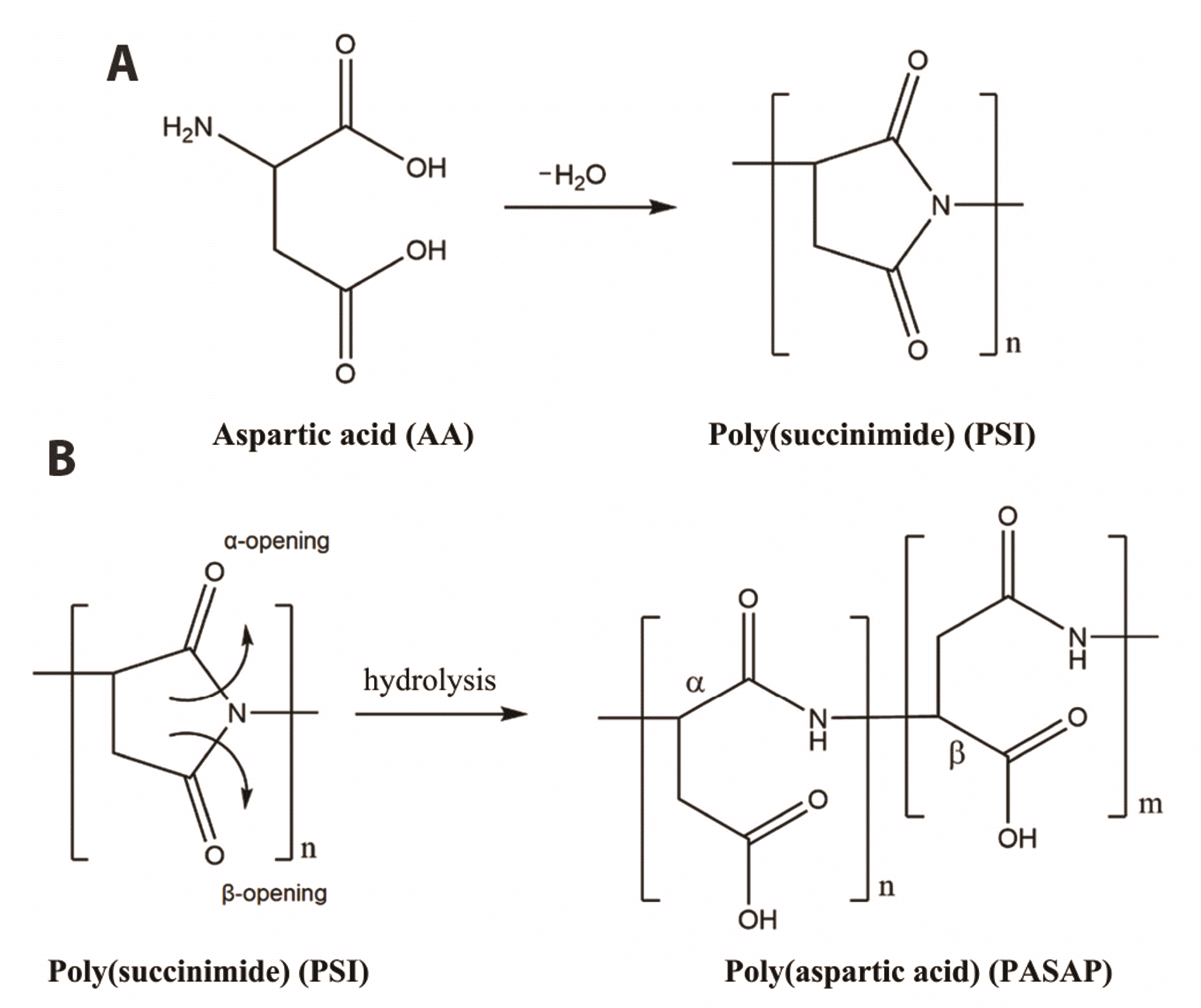
Synthesis of PSI from aspartic acid using thermal polycondensation: The derivatization of PSI to poly(aspartic acid) (PASP) can occur with molecules containing amine groups that serve as the nucleophile. L-Aspartic acid and o-phosphoric acid (Sigma-Aldrich, St. Louis, MO, USA) were added to a flask, and the solution was stirred (130 rpm) on a rotary evaporator immersed in an oil bath. The temperature was gradually increased to 180°C while the pressure was gradually reduced from atmospheric pressure to vacuum (5 mbar) over a period of 6.5 h. With the addition of acid and reducing the pressure, a higher rate of polymerization and a quicker reaction time were achieved than if the reaction were carried out at atmospheric pressure. During the synthesis, the mixture melted and became a completely translucent fluid at 180°C. The acid catalyst also suppressed color formation. After the liquid started to boil, the mixture hardened and formed a white and then off-white hard foam polymer. The material was then removed from the heat to harden.
PSI purification: Dimethylformamide (DMF) (VWR, Radnor, PA, USA) was added to the hardened foam for dissolution. A viscous brown solution was formed and then precipitated in a large amount (approximately 2,000 ml) of cold distilled water with strong stirring. Stirring was performed for no longer than 10 min due to PSI hydrolysis, and the polymer began to melt. The resulting white polymer was filtered through a glass filter. Washing and filtration steps were repeated several times to isolate PSI from the DMF and phosphoric acid (Sigma-Aldrich) until the pH reached at least 6. The filtered white polymer was placed in a 45°C drying oven for 3 days to dry.
Carboplatin-PASP conjugation: During the preparation of a 25% (w/w) PSI solution for conjugation, it has been proven that in alkaline solutions, the imide group of PSI undergoes a ring-opening reaction. During alkaline hydrolysis, poly(aspartamide), a water-soluble polymer, is produced with either α- or β-carboxylate groups in a β:α monomer ratio of approximately 3:1 after synthesis from aspartic acid. This ratio can be adjusted by changing the pH; a relatively high pH results in more β monomers than α monomers. Here, we used a commercially available chemotherapeutic agent, carboplatin (Sigma-Aldrich). Carboplatin is a platinum complex with two primary amine groups. For synthesis, PSI and carboplatin were added in a 5:1 molar ratio to produce a 5-fold grafted polymer; that is, every fifth PSI monomer was conjugated with a carboplatin molecule. The polymer was prepared with 30 mg of carboplatin dissolved in 4 ml of DMF. The dissolved carboplatin solutions were added to the appropriate amount of 25% PSI-DMF solution. Synthesis was carried out at 60°C with stirring (500 rpm) for 7 days.
1H NMR: All samples (5 mg) were dissolved in 0.6 ml of D2O. 1H NMR spectra were obtained using the Noesy1D sequence and analysis was carried out at 25°C. The 1H NMR measurement conditions were as follows: spectral width, 9,615.4 Hz; number of data points, 76,924; acquisition time, 4 sec; relaxation delay, 2 sec; and mixing time, 0.1 sec. The spectrum of the solvent water was also obtained to minimize the saturation power, and we used the presaturation method with a saturation delay of 1.5 sec and a saturation power of 6 at the saturation frequency. 1H NMR experiments were performed using a JEOL 400 MHz instrument.
Cell culture and cell viability assay: SKOV-3 (human ovarian cancer) cells were cultured in Dulbecco’s modified Eagle’s medium (Lonza, Rockland, ME, USA) supplemented with 5% fetal bovine serum (FBS; Gibco BRL, Grand Island, NY, USA), 10 μg/ml human transferrin (Sigma-Aldrich), and 3 × 10⁻⁸ M sodium selenite (Sigma-Aldrich). Chinese hamster ovary (CHO) cells were maintained in α-minimum essential medium (Lonza) containing 10% FBS (Gibco BRL) and 1% antibiotics (Lonza). Paclitaxel and topotecan (Sigma-Aldrich) were used as the anticancer drugs for combination therapy. Cell viability was determined using a 3‐(4,5‐dimethylthiazol‐2‐yl)‐5‐(3‐carboxymethoxyphenyl)‐2‐(4‐sulfophenyl)‐2H‐tetrazolium (MTS) assay. CHO and SKOV-3 cells were seeded at 5 × 10³ cells/well into a 96‐well plate and treated with drug for 24 h. The final concentrations of paclitaxel, topotecan and PSI-carboplatin were adjusted to 1 mg/ml. Cultured cells were added to 20 μl/well CellTiter 96 AQueous One Solution (Promega, Madison, WI, USA) and 100 µl of culture media for incubation at 37°C for 4 h. The absorbance was measured at 490 nm using a microplate reader, and cytotoxicity was calculated based on the obtained optical density (OD) values.
Real-time PCR: SKOV-3 cells were seeded at a density of 3 × 10⁵ cells/well in 6-well plates for drug treatment. Total RNA was isolated from the cells by using TRIzol Reagent (Invitrogen, Carlsbad, CA, USA) after 24 h of treatment. Complementary DNA was synthesized from 1 μg of total RNA using Superscript II RT (Invitrogen) according to the manufacturer’s instructions. Real-time PCR was performed for the relative quantification of Actin and iNOS mRNA levels by using SYBR Green PCR Master Mix (Applied Biosystems, Foster City, CA, USA). Primers were designed by using PrimerExpress software (Applied Biosystems). The primer sequences were as follows: hiNOS, 5’-CGCATGACCTTGGTGTTTGG-3’ and 5’-CATAGACCTTGGGCTTGCCA-3’; and hACTIN, 5’-CTGT CCACCTTCCAGCAGATGT-3’ and 5’-CGCAACTAAGTCATA GTCCGCC-3’.
Western blot analyses: Monoclonal rabbit anti-actin and goat anti-rabbit horseradish peroxidase (HRP)-conjugated antibodies were purchased from Santa Cruz Biotechnology (Santa Cruz, CA, USA). The monoclonal rabbit anti-iNOS antibody was purchased from Abcam (Cambridge, MA, USA). SKOV-3 cells were seeded at a density of 2 × 10⁶ cells/well in 100-mm plates for drug treatment. Whole cells were lysed in radioimmunoprecipitation assay (RIPA) buffer (10 mM Tris-HCl [pH 7.4], 0.15 M NaCl, 0.5% SDS, 1% NP-40, 1% Na deoxycholate, 1 mM EDTA, 1 mM phenylmethylsulfonyl fluoride, 1 μg/ml pepstatin, and 1 μg/ml leupeptin) 24 h after treatment, and the protein concentration was measured by a Bradford protein assay kit (Bio-Rad, Hercules, CA, USA). Proteins were separated on 10% SDS-polyacrylamide gels and transferred to polyvinylidene difluoride (PVDF) membranes. The membranes were blocked with 5% skim milk powder in Tris-buffered saline (TBS) (20 mM Tris-HCl, 137 mM NaCl [pH 7.4]) containing 0.1% Tween 20 (TBS-T buffer) for 1 h at room temperature (RT). Western blot analyses were performed with monoclonal anti-iNOS (Abcam) or monoclonal anti-actin (Santa Cruz Biotechnology) antibodies. Primary antibodies were added to TBS-T buffer at a 1:1,000 dilution and incubated for 90 min at RT prior to incubation with HRP-conjugated goat anti-rabbit antibody (1:5,000 dilution; Santa Cruz Biotechnology) for 1 h at RT. Proteins were detected by using an enhanced chemiluminescence detection system (GE Healthcare). The Western blot results were quantified using the ImageJ program (National Institutes of Health, Bethesda, MD, USA).
Statistical calculations were performed using the Analysis ToolPak in Microsoft Excel. For the cell viability assays, all measurements were performed in triplicate, and the mean ± SE of the OD values are presented in the bar graphs. Comparisons between the control and treatment groups were performed by using one-way ANOVA and subsequent Dunnett post-hoc tests. The cytotoxicity results are given in percentages (%) for data evaluation.
For real-time PCR analysis, all measurements were performed in triplicate, and the median values and interquartile ranges of the mRNA levels are presented in a scatter plot. Relative expression was calculated based on the ΔΔCt method, and iNOS levels were normalized to the level of the reference gene actin. Comparisons between the control and treatment groups were performed by using the Kruskal‒Wallis test and subsequent Dunn post hoc tests.
For Western blot quantification, all measurements were performed in triplicate. Protein levels were first normalized to the control treatment values and the iNOS levels were normalized to that of the reference gene (actin), and the results are expressed as the mean relative quantities (RQ values) ± SE in a bar graph. Comparisons between the control and treatment groups were performed by using one-way ANOVA and subsequent Dunnett post-hoc tests.
In all experiments, a calculated p-value of < 0.05 was considered a statistically significant finding and is represented with a “*” on the plots.
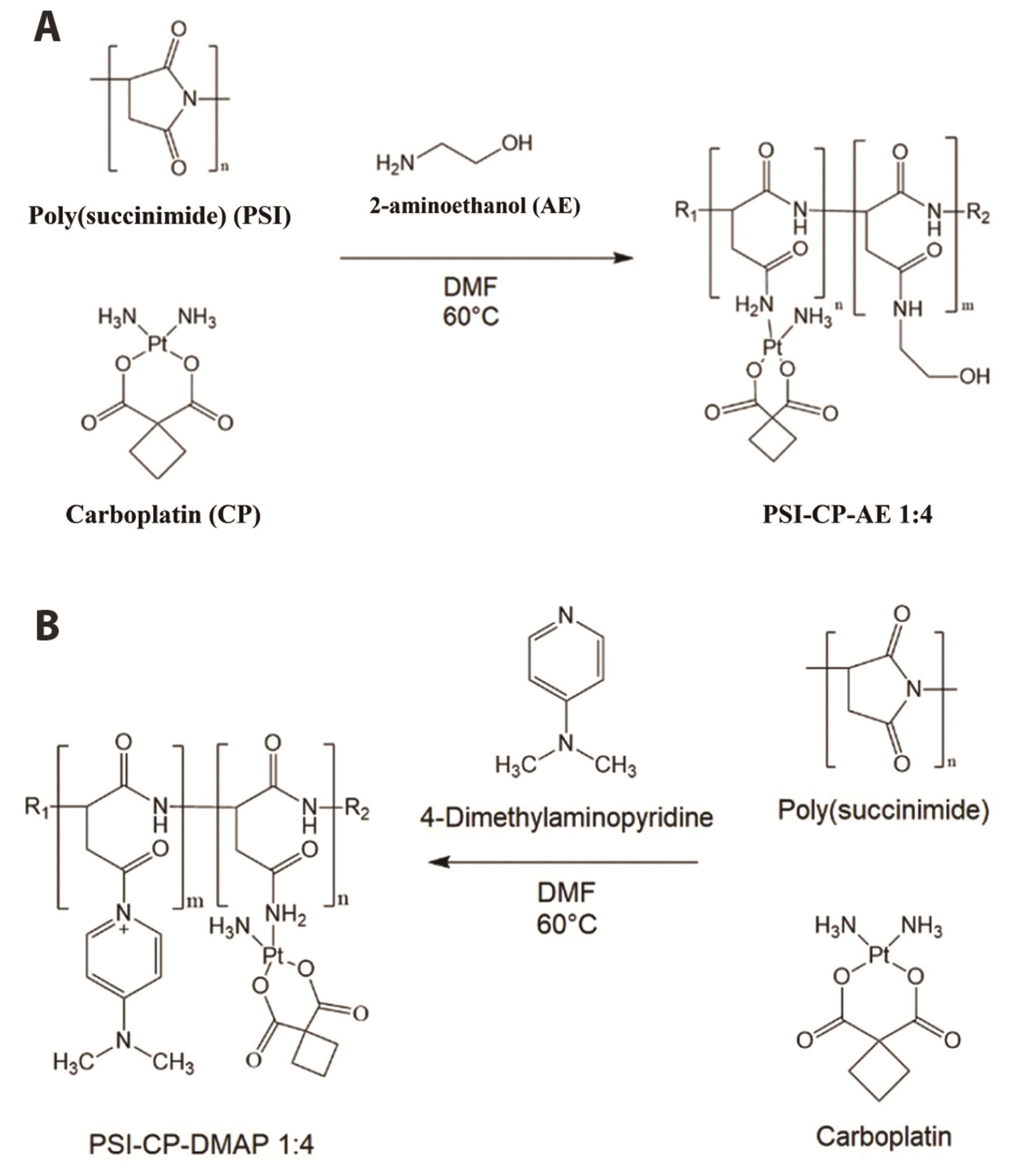
PSI was synthesized by thermal polycondensation in a mixture of L-aspartic acid monomers and an acid catalyst, o-phosphoric acid. In the nucleophilic reaction, water was removed from the molecule to produce reactive imide groups (Fig. 1A). PSI is a water-insoluble polyimide that serves as the base polymer for poly(aspartamide) via a ring-opening reaction (Fig. 1B). To increase water solubility and bioavailability, aminoethanol and 4-dimethylaminopyridine (DMAP) modifying groups were added to the free monomers on the PSI chain. The molecular ratio of modifying groups to carboplatin on the polymer was 4:1 (Fig. 2).
Aminoethanol modifying groups might help to improve the bioavailability of the conjugate. Thus, aminoethanol was added on the second day of the synthesis in ratios to occupy all of the free monomers on the PSI chain where carboplatin did not conjugate. The OH group of 2-aminoethanol increases the polarity of the molecule and thus its solubility. To separate the PSI from the remaining solvents, a dialysis membrane with a 14 kDa cutoff was used. The water was changed over 6 days of dialysis (the difference in the concentration drives smaller molecules out of the membrane) and resulted in clean PSI conjugates in the membrane by the end of the procedure. The solution was evaporated and lyophilized on a rotary evaporator until it was concentrated to approximately 2 ml. The temperature and pressure were adjusted to achieve fast and efficient evaporation (65 mbar, 90°C). Lyophilization resulted in ~100 mg of product (not measured). The polymer with the aminoethanol group had a yellowish, off-white color.
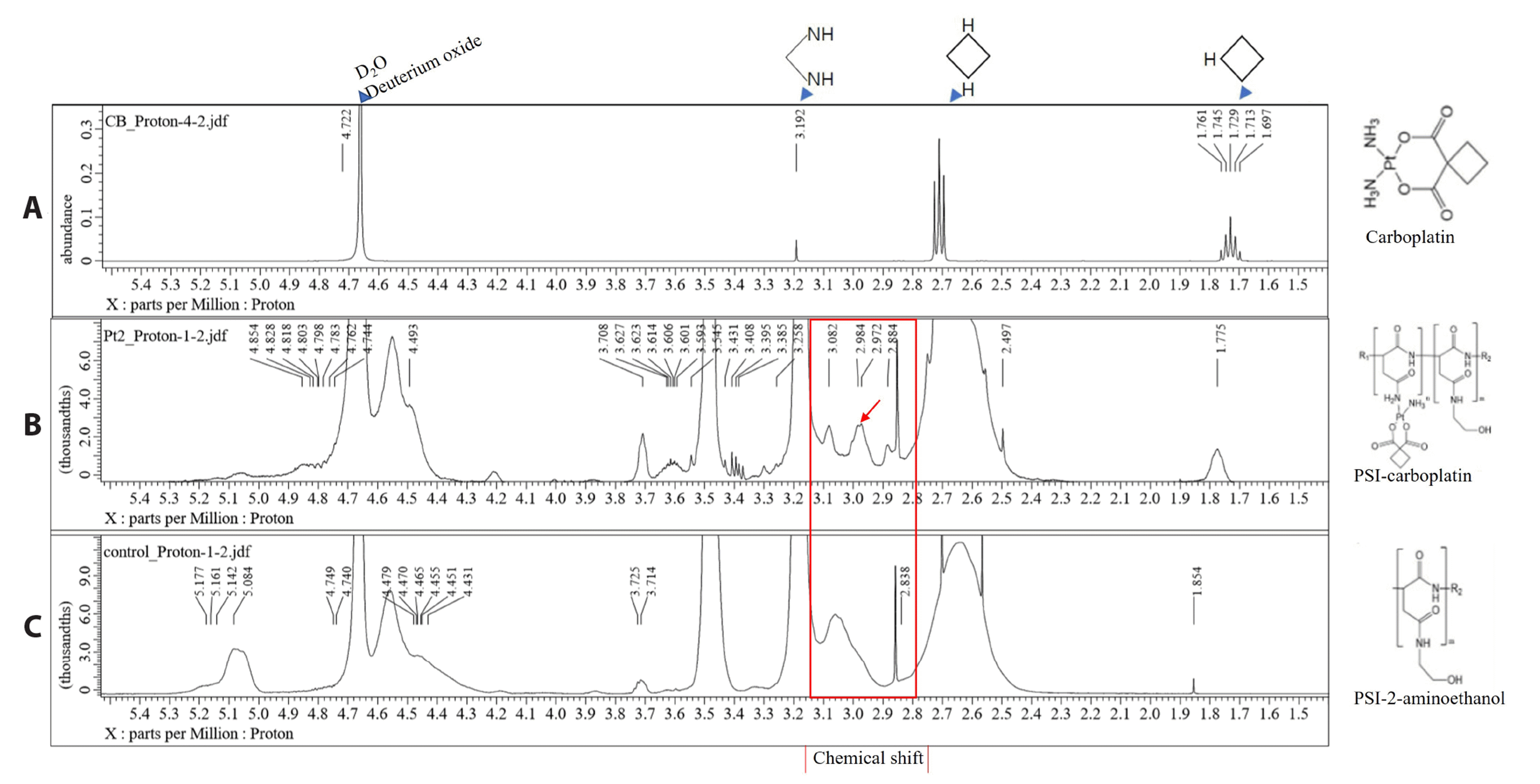
Spectrum A shows pure carboplatin, spectrum B is that of the PSI-carboplatin conjugate, and spectrum C is that of the PSI-aminoethanol conjugate. The functional groups in the NMR spectra are in the range of 2.5 to 3.0 ppm. Comparison of spectrum C with spectrum B confirmed the chemical shifts in spectrum B (Fig. 3).
Cell viability assays were performed with both SKOV-3 cells and CHO cells. This study was conducted to investigate the effects of PSI-carboplatin and other anticancer drugs on ovarian cancer cells and normal ovarian cells. The antitumor effect of paclitaxel and carboplatin in SKOV-3 cells was 75% and that of topotecan was 60%. In the PSI-carboplatin group, the antitumor effect was not different from that of the control group, whereas topotecan treatment with PSI-carboplatin showed a reduction in cell count of 60% (Fig. 4A). In CHO cells treated with paclitaxel, carboplatin or topotecan alone, the antitumor effect was less than 50%. The antitumor effect in the PSI-carboplatin group was not different from that of the control group, and no cytotoxicity was observed. In addition, compared to the control group, paclitaxel and PSI-carboplatin combined treatment did not display a change in cytotoxicity. However, the cytotoxicity of PSI-carboplatin with topotecan was less than 50% (Fig. 4B).
The mRNA expression of the iNOS gene was examined in SKOV-3 ovarian cancer cells. There was no difference in the iNOS mRNA expression level between the control group and the PSI-carboplatin, PSI-carboplatin with topotecan, carboplatin and topotecan treatment groups. The paclitaxel group exhibited a slight change in expression. However, the cells in the PSI-carboplatin with paclitaxel group strongly overexpressed iNOS (Fig. 5A).
iNOS protein expression was measured in SKOV-3 cancer cells (Fig. 5B). The iNOS expression level in the group treated with PSI alone did not differ significantly from that in the control group. Protein overexpression was observed in the following treatment groups in descending order: PSI-carboplatin, carboplatin, PSI-carboplatin-DMAP, topotecan, and PSI-carboplatin-topotecan. On the other hand, the PSI-carboplatin-paclitaxel-DMAP, PSI-carboplatin-paclitaxel, and paclitaxel groups had lower levels of iNOS protein expression than the control group (Fig. 5C).
Many anticancer drugs, such as those that are targeted and immunological in nature, are currently being studied and developed [32]. However, improved chemotherapeutic drugs have modified the characteristics of conventional anticancer drugs and can be developed more easily than novel drugs [33]. This method of drug modification also has the potential to overcome anticancer drug resistance, which is currently a problem in hospitals. In particular, polymers are biodegradable and have biological properties in vivo, so the development of anticancer drugs using these substances has the advantage of overcoming the problems of existing anticancer drugs [34,35]. Therefore, we investigated the anticancer effect and expression of the iNOS gene in ovarian cancer cells and normal cells after treatment with the inorganic platinum compound conjugated to PSI. We also examined the expression of the combination of the chemotherapy drugs conjugated with paclitaxel and topotecan, which have been prescribed to cancer patients, in conjunction with an adjunctive chemotherapeutic agent.
In the cell viability test, the carboplatin-conjugated polymers showed less cytotoxic effects than carboplatin. However, when we examined the cell survival rates, paclitaxel in combination with carboplatin-conjugated polymer treatment inhibited the growth of ovarian cancer cells. Toxicity tests in normal ovarian cancer cells showed that there was no effect on cell growth in the group treated with paclitaxel in combination with the carboplatin-conjugated polymer. In addition, iNOS mRNA was not expressed when cancer cells were treated with paclitaxel alone, but it was confirmed that the combination of carboplatin-conjugated polymer with paclitaxel resulted in its overexpression. iNOS protein expression was normal or elevated in all groups except for the paclitaxel group. There were also variations with different treatments, where no protein expression was observed compared to the mRNA levels.
Taken together, these results provide a basis for the development of anticancer drugs using the iNOS gene as candidate target for the development of novel ovarian cancer drugs. Further kinetic studies and animal experiments may be required to further investigate the mechanism of the carboplatin-conjugated polymers in vivo. Further research will also be needed to study the interactions between the polymer-carboplatin conjugate and paclitaxel and additional conjugated chemotherapeutic agents. The correlation between PSI-conjugated carboplatin and the iNOS gene in ovarian cancer cells and normal ovarian cells may be helpful in identifying the role of genes. Additional cell survival studies are needed to explain the low toxicity of PSI-carboplatin to SKOV-3 cells. The viability of normal ovarian cells decreased when they were treated with paclitaxel alone, but additional studies should be conducted on the lack of cytotoxicity when PSI-carboplatin was combined with paclitaxel. Additionally, more research to determine whether PSI-carboplatin inhibits the cytotoxicity of paclitaxel and additional studies with nonconjugated PSI-carboplatin may be needed. The interrelationships of PSI-carboplatin with paclitaxel will enable continuous research on the development of new anticancer drugs. As a result of the RNA and protein expression produced by the iNOS gene, this gene found in ovarian cancer cells may be an important factor in the treatment and diagnosis of cancer. From a clinical point of view, conjugated chemical drugs can affect treatment by influencing the generation of various resistant cell populations due to intratumoral heterogeneity. Such studies should be continued because conjugation with PSI-carboplatin platinum is an attempt to discover new binding interactions to improve anticancer agents by combining an organic compound with an inorganic compound. The PSI-carboplatin conjugate presented in this paper is an improved modified anticancer agent, but more research is needed to understand its significance. Therefore, we will continue to study the mechanism of action of PSI-carboplatin as an advanced anticancer drug so that we can become experts in the development of advanced cancer drugs for ovarian cancer patients. In addition, we hope that this and future research papers will be useful for ovarian cancer research. At this stage, many scientific phenomena have yet to be revealed, but research and development are expected to continue, even if the development of such anticancer drugs fails.
Therapy with the newly synthesized carboplatin-polymer conjugate and paclitaxel showed anticancer activity against ovarian cancer cells, and this study was successful in developing an effective candidate anticancer drug without serious side effects due to the low toxicity of the conjugate to normal ovarian cancer cells.
Although resistance and the problems of chemical anticancer drugs mentioned above were observed in the cell viability test, research with animal models should be conducted. Nevertheless, the nontoxicity to normal ovarian cancer cells demonstrates the possibility of further drug development. Additional studies on the molecular interactions between various cancer cell-associated signaling pathways are needed to further understand the relationship between iNOS and PSI-carboplatin.
Notes
FUNDING
This paper was supported by the Fund of the Biomedical Research Institute, Jeonbuk National University Hospital, the Research Base Construction Fund Support Program funded by Jeonbuk National University in 2021, and research funds for newly appointed professors of Jeonbuk National University in 2020.
REFERENCES
1. Reid BM, Permuth JB, Sellers TA. 2017; Epidemiology of ovarian cancer: a review. Cancer Biol Med. 14:9–32. DOI: 10.20892/j.issn.2095-3941.2016.0084. PMID: 28443200. PMCID: PMC5365187. PMID: https://www.scopus.com/inward/record.uri?partnerID=HzOxMe3b&scp=85017143290&origin=inward.
2. Kujawa KA, Lisowska KM. 2015; Ovarian cancer--from biology to clinic. Postepy Hig Med Dosw (Online). 69:1275–1290. Polish. DOI: 10.5604/17322693.1184451. PMID: 26671919. PMCID: PMC5365187. PMID: https://www.scopus.com/inward/record.uri?partnerID=HzOxMe3b&scp=84973411296&origin=inward.
3. Webb PM, Jordan SJ. 2017; Epidemiology of epithelial ovarian cancer. Best Pract Res Clin Obstet Gynaecol. 41:3–14. DOI: 10.1016/j.bpobgyn.2016.08.006. PMID: 27743768. PMID: https://www.scopus.com/inward/record.uri?partnerID=HzOxMe3b&scp=85008698217&origin=inward.
4. Karam A, Ledermann JA, Kim JW, Sehouli J, Lu K, Gourley C, Katsumata N, Burger RA, Nam BH, Bacon M, Ng C, Pfisterer J, Bekkers RLM, Casado Herráez A, Redondo A, Fujiwara H, Gleeson N, Rosengarten O, Scambia G, Zhu J, et al. 2017; Fifth Ovarian Cancer Consensus Conference of the Gynecologic Cancer InterGroup: first-line interventions. Ann Oncol. 28:711–717. DOI: 10.1093/annonc/mdx011. PMID: 28327917. PMID: https://www.scopus.com/inward/record.uri?partnerID=HzOxMe3b&scp=85021858496&origin=inward.
5. Potara M, Nagy-Simon T, Craciun AM, Suarasan S, Licarete E, Imre-Lucaci F, Astilean S. 2017; Carboplatin-loaded, Raman-encoded, chitosan-coated silver nanotriangles as multimodal traceable nanotherapeutic delivery systems and pH reporters inside human ovarian cancer cells. ACS Appl Mater Interfaces. 9:32565–32576. DOI: 10.1021/acsami.7b10075. PMID: 28872817. PMID: https://www.scopus.com/inward/record.uri?partnerID=HzOxMe3b&scp=85030162417&origin=inward.
6. Wang Y, Wang L, Chen G, Gong S. 2017; Carboplatin-complexed and cRGD-conjugated unimolecular nanoparticles for targeted ovarian cancer therapy. Macromol Biosci. 17:1600292. DOI: 10.1002/mabi.201600292. PMID: 27911475. PMCID: PMC5914160. PMID: https://www.scopus.com/inward/record.uri?partnerID=HzOxMe3b&scp=85006307295&origin=inward.
7. Coleman RL. 2002; Emerging role of topotecan in front-line treatment of carcinoma of the ovary. Oncologist. 7 Suppl 5:46–55. DOI: 10.1634/theoncologist.7-suppl_5-46. PMID: 12324633. PMID: https://www.scopus.com/inward/record.uri?partnerID=HzOxMe3b&scp=0036388369&origin=inward.
8. Zhang H, Jia L, Xu Y, Zhou XC, Kong B, Li D. 2012; Topotecan plus carboplatin and paclitaxel in first-line treatment of advanced ovarian cancer: a meta-analysis of randomized controlled trials. J Chemother. 24:67–73. DOI: 10.1179/1120009X12Z.0000000002. PMID: 22546760. PMID: https://www.scopus.com/inward/record.uri?partnerID=HzOxMe3b&scp=84866256593&origin=inward.
9. Skarlos DV, Aravantinos G, Kosmidis P, Athanassiadis A, Stathopoulos GP, Pavlidis N, Bafaloukos D, Karphathios S, Papakostas P, Bamia C, Fountzilas G. 1997; Paclitaxel with carboplatin versus paclitaxel with carboplatin alternating with cisplatin as first-line chemotherapy in advanced epithelial ovarian cancer: preliminary results of a Hellenic Cooperative Oncology Group study. Semin Oncol. 24(5 Suppl 15):S15-57–S15-61. PMID: 9346224. PMID: https://www.scopus.com/inward/record.uri?partnerID=HzOxMe3b&scp=9844262807&origin=inward.
10. Han ES, Wen W, Dellinger TH, Wu J, Lu SA, Jove R, Yim JH. 2018; Ruxolitinib synergistically enhances the anti-tumor activity of paclitaxel in human ovarian cancer. Oncotarget. 9:24304–24319. DOI: 10.18632/oncotarget.24368. PMID: 29849942. PMCID: PMC5966246.
11. Ho GY, Woodward N, Coward JI. 2016; Cisplatin versus carboplatin: comparative review of therapeutic management in solid malignancies. Crit Rev Oncol Hematol. 102:37–46. DOI: 10.1016/j.critrevonc.2016.03.014. PMID: 27105947. PMID: https://www.scopus.com/inward/record.uri?partnerID=HzOxMe3b&scp=84963800478&origin=inward.
12. Ledermann JA. 2017; Front-line therapy of advanced ovarian cancer: new approaches. Ann Oncol. 28(suppl_8):viii46–viii50. DOI: 10.1093/annonc/mdx452. PMID: 29232475. PMID: https://www.scopus.com/inward/record.uri?partnerID=HzOxMe3b&scp=85039553675&origin=inward.
13. Shenoi RA, Lai BF, Imran ul-haq M, Brooks DE, Kizhakkedathu JN. 2013; Biodegradable polyglycerols with randomly distributed ketal groups as multi-functional drug delivery systems. Biomaterials. 34:6068–6081. DOI: 10.1016/j.biomaterials.2013.04.043. PMID: 23688604. PMID: https://www.scopus.com/inward/record.uri?partnerID=HzOxMe3b&scp=84878532612&origin=inward.
14. Joye IJ, McClements DJ. 2016; Biopolymer-based delivery systems: challenges and opportunities. Curr Top Med Chem. 16:1026–1039. DOI: 10.2174/1568026615666150825143130. PMID: 26303423. PMID: https://www.scopus.com/inward/record.uri?partnerID=HzOxMe3b&scp=84959573045&origin=inward.
15. Tibbitt MW, Dahlman JE, Langer R. 2016; Emerging frontiers in drug delivery. J Am Chem Soc. 138:704–717. DOI: 10.1021/jacs.5b09974. PMID: 26741786. PMID: https://www.scopus.com/inward/record.uri?partnerID=HzOxMe3b&scp=84956787698&origin=inward.
16. Varga Z, Molnár K, Torma V, Zrínyi M. 2010; Kinetics of volume change of poly(succinimide) gels during hydrolysis and swelling. Phys Chem Chem Phys. 12:12670–12675. DOI: 10.1039/c0cp00527d. PMID: 20730186. PMID: https://www.scopus.com/inward/record.uri?partnerID=HzOxMe3b&scp=77957923369&origin=inward.
17. Sadeghi M, Hemmati S, Hamishehkar H. 2016; Synthesis of a novel superdisintegrant by starch derivatization with polysuccinimide and its application for the development of Ondansetron fast dissolving tablet. Drug Dev Ind Pharm. 42:769–775. DOI: 10.3109/03639045.2015.1075029. PMID: 26289005. PMID: https://www.scopus.com/inward/record.uri?partnerID=HzOxMe3b&scp=84968546844&origin=inward.
18. Juriga D, Laszlo I, Ludanyi K, Klebovich I, Chae CH, Zrinyi M. 2018; Kinetics of dopamine release from poly(aspartamide)-based prodrugs. Acta Biomater. 76:225–238. DOI: 10.1016/j.actbio.2018.06.030. PMID: 29940369. PMID: https://www.scopus.com/inward/record.uri?partnerID=HzOxMe3b&scp=85049303521&origin=inward.
19. Yendluri R, Lvov Y, de Villiers MM, Vinokurov V, Naumenko E, Tarasova E, Fakhrullin R. 2017; Paclitaxel encapsulated in halloysite clay nanotubes for intestinal and intracellular delivery. J Pharm Sci. 106:3131–3139. DOI: 10.1016/j.xphs.2017.05.034. PMID: 28600185. PMID: https://www.scopus.com/inward/record.uri?partnerID=HzOxMe3b&scp=85026483844&origin=inward.
20. Doppalapudi S, Jain A, Domb AJ, Khan W. 2016; Biodegradable polymers for targeted delivery of anti-cancer drugs. Expert Opin Drug Deliv. 13:891–909. DOI: 10.1517/17425247.2016.1156671. PMID: 26983898. PMID: https://www.scopus.com/inward/record.uri?partnerID=HzOxMe3b&scp=84961210141&origin=inward.
21. Nicolas J, Couvreur P. 2017; Polymer nanoparticles for the delivery of anticancer drug. Med Sci (Paris). 33:11–17. French. DOI: 10.1051/medsci/20173301003. PMID: 28120750. PMID: https://www.scopus.com/inward/record.uri?partnerID=HzOxMe3b&scp=85010748894&origin=inward.
22. Velazco-de-la-Garza J, Avérous L, Sosa-Santillán GdJ, Pollet E, Zugasti-Cruz A, Sierra-Rivera CA, Pérez-Aguilar NV, Oyervides-Muñoz E. 2020; Biological properties of novel polysuccinimide derivatives synthesized via quaternary ammonium grafting. Eur Polym J. 131:109705. https://doi.org/10.1016/j.eurpolymj.2020.109705. DOI: 10.1016/j.eurpolymj.2020.109705. PMID: https://www.scopus.com/inward/record.uri?partnerID=HzOxMe3b&scp=85083468448&origin=inward.
23. Jenkins DC, Charles IG, Thomsen LL, Moss DW, Holmes LS, Baylis SA, Rhodes P, Westmore K, Emson PC, Moncada S. 1995; Roles of nitric oxide in tumor growth. Proc Natl Acad Sci U S A. 92:4392–4396. DOI: 10.1073/pnas.92.10.4392. PMID: 7538668. PMCID: PMC41950. PMID: https://www.scopus.com/inward/record.uri?partnerID=HzOxMe3b&scp=0029079394&origin=inward.
24. Xie K, Huang S. 2003; Contribution of nitric oxide-mediated apoptosis to cancer metastasis inefficiency. Free Radic Biol Med. 34:969–986. DOI: 10.1016/S0891-5849(02)01364-3. PMID: 12684082. PMID: https://www.scopus.com/inward/record.uri?partnerID=HzOxMe3b&scp=0037446221&origin=inward.
25. Brüne B. 2003; Nitric oxide: NO apoptosis or turning it ON? Cell Death Differ. 10:864–869. DOI: 10.1038/sj.cdd.4401261. PMID: 12867993. PMID: https://www.scopus.com/inward/record.uri?partnerID=HzOxMe3b&scp=0042665423&origin=inward.
26. Forrester K, Ambs S, Lupold SE, Kapust RB, Spillare EA, Weinberg WC, Felley-Bosco E, Wang XW, Geller DA, Tzeng E, Billiar TR, Harris CC. 1996; Nitric oxide-induced p53 accumulation and regulation of inducible nitric oxide synthase expression by wild-type p53. Proc Natl Acad Sci U S A. 93:2442–2447. DOI: 10.1073/pnas.93.6.2442. PMID: 8637893. PMCID: PMC39816. PMID: https://www.scopus.com/inward/record.uri?partnerID=HzOxMe3b&scp=9244232804&origin=inward.
27. Rao CV. 2004; Nitric oxide signaling in colon cancer chemoprevention. Mutat Res. 555:107–119. DOI: 10.1016/j.mrfmmm.2004.05.022. PMID: 15476855. PMID: https://www.scopus.com/inward/record.uri?partnerID=HzOxMe3b&scp=5144230475&origin=inward.
28. Chazotte-Aubert L, Hainaut P, Ohshima H. 2000; Nitric oxide nitrates tyrosine residues of tumor-suppressor p53 protein in MCF-7 cells. Biochem Biophys Res Commun. 267:609–613. DOI: 10.1006/bbrc.1999.2003. PMID: 10631110. PMID: https://www.scopus.com/inward/record.uri?partnerID=HzOxMe3b&scp=0034685015&origin=inward.
29. Li J, Billiar TR, Talanian RV, Kim YM. 1997; Nitric oxide reversibly inhibits seven members of the caspase family via S-nitrosylation. Biochem Biophys Res Commun. 240:419–424. DOI: 10.1006/bbrc.1997.7672. PMID: 9388494. PMID: https://www.scopus.com/inward/record.uri?partnerID=HzOxMe3b&scp=0031576527&origin=inward.
30. Messmer UK, Brüne B. 1996; Nitric oxide-induced apoptosis: p53-dependent and p53-independent signalling pathways. Biochem J. 319(Pt 1):299–305. DOI: 10.1042/bj3190299. PMID: 8870682. PMCID: PMC1217768. PMID: https://www.scopus.com/inward/record.uri?partnerID=HzOxMe3b&scp=0029812216&origin=inward.
31. Kielbik M, Szulc-Kielbik I, Klink M. 2019; The potential role of iNOS in ovarian cancer progression and chemoresistance. Int J Mol Sci. 20:1751. DOI: 10.3390/ijms20071751. PMID: 30970628. PMCID: PMC6479373. PMID: https://www.scopus.com/inward/record.uri?partnerID=HzOxMe3b&scp=85064722293&origin=inward.
32. Morand S, Devanaboyina M, Staats H, Stanbery L, Nemunaitis J. 2021; Ovarian cancer immunotherapy and personalized medicine. Int J Mol Sci. 22:6532. DOI: 10.3390/ijms22126532. PMID: 34207103. PMCID: PMC8234871. PMID: 9cc32856714d4af4874eef616bd8726e. PMID: https://www.scopus.com/inward/record.uri?partnerID=HzOxMe3b&scp=85108078334&origin=inward.
33. Singh V, Kesharwani P. 2021; Dendrimer as a promising nanocarrier for the delivery of doxorubicin as an anticancer therapeutics. J Biomater Sci Polym Ed. 32:1882–1909. DOI: 10.1080/09205063.2021.1938859. PMID: 34078252. PMID: https://www.scopus.com/inward/record.uri?partnerID=HzOxMe3b&scp=85111069950&origin=inward.
34. Sun H, Yarovoy I, Capeling M, Cheng C. 2017; Polymers in the co-delivery of siRNA and anticancer drugs for the treatment of drug-resistant cancers. Top Curr Chem (Cham). 375:24. DOI: 10.1007/s41061-017-0113-z. PMID: 28176270. PMID: https://www.scopus.com/inward/record.uri?partnerID=HzOxMe3b&scp=85011843801&origin=inward.
35. Sanyakamdhorn S, Agudelo D, Tajmir-Riahi HA. 2017; Review on the targeted conjugation of anticancer drugs doxorubicin and tamoxifen with synthetic polymers for drug delivery. J Biomol Struct Dyn. 35:2497–2508. DOI: 10.1080/07391102.2016.1222971. PMID: 27598545. PMID: https://www.scopus.com/inward/record.uri?partnerID=HzOxMe3b&scp=84986212520&origin=inward.