

 PDF
PDF Citation
 Print
Print


INTRODUCTION
Hypothyroidism alone can damage cardiac contractility, lead to myocardial fibrosis, and eventually to heart failure [1,2]. Especially, myocardial fibrosis represents a poor prognosis [3]. When myocardial fibrosis occurs, not only do myocardial fibroblasts secrete much extracellular matrix due to overproduction of collagen, making the heart muscle stiffer and exacerbating the prognosis of many heart diseases, but the fibrotic heart tissue causes electrical and structural changes that predispose patients to arrhythmias, heart failure and ischemia [4]. Traditional long-term thyroid replacement therapy can quickly restore hormone levels in the body and gradually improve cardiac function, but not fibrotic situation [5]. In our previous research, H2S, a newly found functional gas, could ameliorate myocardial fibrosis in diabetic rats [6]. However, Whether H2S could treat myocardial fibrosis induced by hypothyroidism remains obscure.
Autophagy is a common physiological process that maintains the cell metabolic homeostasis in response to various cell stressors [7]. Moreover, hypothyroidism is characterized by enhanced thyrotropic stimulating hormone (TSH) level and reduced free thyroxine 4 (FT4) in serum, and TSH stimulation could suppress autophagy in primary mouse chondrocytes (PMCs) [8]. In addition, transforming growth factor-β1 (TGF-β1), a kind of unreplaceable transcription factor, takes a leading role in the occurrence and progression of myocardial fibrosis. SMAD family member 2 (Smad2), a receptor of TGF-β1, is the main molecule in the signal transduction pathway that mediates myocardial fibrosis [9,10]. However, whether autophagy and TGF-β1 signaling pathway are involved in hypothyroidism-induced myocardial fibrosis remain unknown.
In this study, we aimed at providing an effective drug component for treating hypothyroidism-induced myocardial fibrosis as a complement to thyroid replacement therapy. Furthermore, we explored the intrinsic mechanism for providing promising threptic targets.
Go to : 
METHODS
Animal models
We purchased eight-week-old male Sprague–Dawley (SD) rats from Changsha Slack Animal Experiment Center. After adaptive feeding for one week in a specific pathogen-free environment, we divided them into four groups: Control group, Hypothyroidism group (PTU group), PTU+H2S group (H2S intervention group), and H2S group (H2S control group). In the following 6 weeks, the rats in PTU group and PTU+H2S group were given PTU (propylthiouracil; Meilun Biological Company, Dalian, China) by gavage at a dose of 20 mg/100 g [1,5], while the other rats were treated with normal drinking water. In the following 4 weeks, the rats in PTU+H2S group and H2S group were intraperitoneally injected with NaHS (Sigma Aldrich, St. Louis, MO, USA) at the dose of 56 µmol/kg/d [11], while the other rats were treated with same amount of saline. Finally, the rats were sacrificed under chloral hydrate anesthesia (350 mg/kg). All protocol of animal work was authorized by the Animal Ethics Committee of the University of South China. In addition, all the animal experiments conformed to the Institutional and International ethical guidelines.
Enzyme-linked immunosorbent assay (ELISA)
We detected the concentrations of FT4 and TSH from rat serum using ELISA method according the protocol (Beyotime, Shanghai, China). After centrifuging the serum, the supernatant serum was taken to be added into 96-well plates (100 µl per well), shaken and incubated at 37°C for 2 h. Then 100 µl biotin-labelled antibody working solution was added into each well, incubated for 1 h, washed, shaken dry. Next, 100 µl horseradish peroxidase-labelled affinity solution was added, incubated again for 1 h, washed, the substrate solution was added sequentially, incubated for 30 min away from light. Finally, the reaction was terminated sequentially by adding the termination solution, and the absorbance of each well was measured at 450 nm within 5 min.
Echocardiography
All the data about the left ventricular function among all the rats were recorded by Visualsonics Vevo 2100 small animal echocardiography (Bio-Rad Laboratories, Hercules, CA, USA). Left ventricular end-systolic diameter (LVES), left ventricular end-diastolic diameter (LVED), left ventricular posterior wall thickness (LVPW), and interventricular septal thickness (IVSD) were recorded and left ventricular fractional shortening (LVFS) was calculated.
Masson staining
We operated Masson staining to observe the collagen deposition in myocardial interstitial tissue. The ventricular muscle specimens were fixed with 4% paraformaldehyde, subjected for ethanol gradient dehydration, and then processed in xylene transparency, paraffin embedding, sectioning (4-µm-thick sections), dewaxing and hydrating, hematoxylin dyeing, decolorizing, ponceau acid fuchsin solution dyeing, and differentiating in 1% phosphomolybdic acid aqueous solution. After observed under a light microscope, they were counter-stained with aniline blue dye solution, soaked in 1% glacial acetic acid, dehydrated in alcohol, and cleared in xylene. Finally, they were observed under a light microscope (Motic, Xiamen, China), after sealing.
Western blot
We extracted myocardial protein from left ventricular tissue in the ice-cold grinding bowl, and quantified them according to BCA protein assay kit protocol (Beyotime). Then 20 µg lysates were run on 10% SDS-PAGE gel and transferred to 0.45 µm PVDF membranes (Millipore, Billerica, MA, USA). Subsequently, blocked in 5% nonfat milk for one hour, the bands were then incubated with the specific first antibodies (anti-CSE, 1:1,000; anti-CBS, 1:1,000; anti-GAPDH, 1:1,000, anti-TIMP2, 1:1,000; anti-MMP13, 1:1,000; anti-Smad2, 1:500; anti-TGF-β1, 1:1,000; anti-p-Smad2, 1:1,000; anti-Atg4, 1:1,000; anti-Atg5, 1:1,000; anti-Beclin1 1:1,000; anti-P62, 1:1,000; anti LC3II/I, 1:1,000) at 4°C overnight. Then they were bound with horseradish peroxidase-conjugated (HRP) secondary antibodies (1:5,000) for one hour at room temperature. Finally, the bands were visualized by adding with enhanced chemiluminescence complied with the manufacturer’s instruction (Beyotime) and detected by using a Molecular Imager VersaDoc MP 5000 system (Bio-Rad Laboratories), analyzed by ImageJ software.
Statistical analysis
Data are presented as mean ± SD. Differences among multiple groups were performed by one-way ANOVA. Differences between two groups were compared using post-hoc least significant difference (LSD) test. SPSS 25.0 software (IBM Co., Armonk, NY, USA) was used for statistical analysis. p < 0.05 was considered statistically significant.
Go to :
RESULTS
FT4 and TSH levels in serum among all the groups
Before sacrificing all the rats, FT4 and TSH samples from the tail vein blood of each group of rats were detected. As shown in Table 1, as expected, FT4 levels were significantly reduced and TSH levels were obviously increased in PTU group (p < 0.05). However, compared to PTU group, FT4 and TSH levels were not significantly changed in PTU+H2S group. These data suggest that the animal models were successfully created but H2S is not able to restore the balance of thyroid hormone levels in such a short time.
H2S can improve the myocardial fibrosis of hypothyroidism rats
We performed Masson staining method to observe the collagen deposition in hypothyroidism-induced rats heart tissue. As shown in Fig. 1A, B, the collagen deposition content (blue part) in heart tissue of rats in PTU group is much larger than that in Control group (p < 0.05). Compared with that in PTU+H2S group, the collagen deposition content (blue part) is much smaller (p < 0.05). These results indicate that H2S is not able to actually alleviate the hypothyroidism-induced cardiac hypertrophy in heart tissue level.
 | Fig. 1Hydrogen sulfide (H2S) could ameliorate hypothyroidism-induced myocardial fibrosis and dysfunction.(A, B) Results of Masson staining in myocardial tissue among four groups. The red part represents the arrangement of myocardial tissue, and the blue part represents myocardial collagen deposition (A: magnification ×40). (C) Cardiac function was evaluated by M-mode echocardiography (a: Control group, b: PTU group, c: PTU+H2S group, d: H2S group). PTU, propylthiouracil. *p < 0.05 compared with Control group; #p < 0.05 compared with PTU group.
|
H2S can improve the cardiac function of hypothyroidism rats
In order to observe cardiac function among all the rats, we assessed the values of LVED, LVES, LVPW, and IVSD were measured and LVFS (%). As shown in Table 2 and Fig. 1C. The results from M-mode echocardiography showed that, compared to PTU group, PTU+H2S group had decreased values of LVED and LVES (p < 0.05) and increased values of LVFS (p < 0.05). While the values of LVPW and IVSD did not change differently between the two groups. These data suggest that H2S can improve the hypothyroidism-induced cardiac dysfunction.
Table 2
Contrast of echocardiographic parameters among four groups
| Groups | LVED (mm) | LVES (mm) | LVPW (mm) | IVSD (mm) | LVFS (%) |
|---|---|---|---|---|---|
| Control | 5.79 ± 0.15 | 3.43 ± 0.10 | 0.92 ± 0.01 | 0.91 ± 0.05 | 40.61 ± 2.83 |
| PTU | 6.54 ± 0.69* | 4.29 ± 0.46* | 0.82 ± 0.10* | 0.81 ± 0.08* | 34.25 ± 4.46* |
| PTU+H2S | 6.00 ± 0.07# | 3.68 ± 0.06# | 0.82 ± 0.09 | 0.81 ± 0.09 | 38.76 ± 1.39# |
| H2S | 6.17 ± 0.10 | 3.87 ± 0.46 | 1.03 ± 0.04 | 1.03 ± 0.04 | 37.15 ± 7.86 |
Values are expressed as mean ± SD. LVES, left ventricular end-systolic diameter; LVED, left ventricular end-diastolic diameter; LVPW, left ventricular posterior wall thickness; IVSD, interventricular septal thickness; LVFS, left ventricular fractional shortening; PTU, propylthiouracil; H2S, hydrogen sulfide. *p < 0.05 vs. Control group; #p < 0.05 vs. PTU group.
![]()
H2S has no significant effect on cardiac hypertrophy in hypothyroidism rats
In order to detect the effect of H2S on cardiac hypertrophy in hypothyroidism-induced rats. heart weight (HW), body weight (BW) and of all the rats were obtained and values of HW/BW were calculated. As shown in Table 3, the mean value of HW/BW in PTU+H2S group was not evidently superior to that in PTU group (p < 0.05). These results indicate that H2S is not able to actually alleviate the hypothyroidism-induced cardiac hypertrophy in heart tissue level.
Table 3
The values of heart weight (HW), body weight (BW) and ratio of HW and BW among four groups
| Groups | Number | BW (g) | HW (mg) | HW/BW (mg/g) |
|---|---|---|---|---|
| Control | 10 | 410.50 ± 7.66 | 959.90 ± 31.60 | 2.34 ± 0.06 |
| PTU | 8 | 262.75 ± 6.80* | 758.88 ± 62.92* | 2.89 ± 0.29* |
| PTU+H2S | 6 | 273.50 ± 14.11 | 716.17 ± 61.78 | 2.62 ± 0.12 |
| H2S | 10 | 407.6 ± 23.35 | 985.20 ± 65.95 | 2.42 ± 0.05 |
![]()
H2S can upregulate CSE, not CBS, protein expression level in hypothyroidism-induced myocardial fibrosis tissue in rats
As shown in Fig. 2A–C, We performed Western blot to view cystathionine-gamma-lyase (CSE) and cystathionine β-synthase (CBS) protein expressions. Compare to Control group, both of them were decreased in PTU group (p < 0.05). However, in PTU+H2S group, CSE, not CBS, protein expression level was upregulated compared with PTU group (p < 0.05). These data indicate that H2S can raise CSE expression in hypothyroidism-induced rat heart.
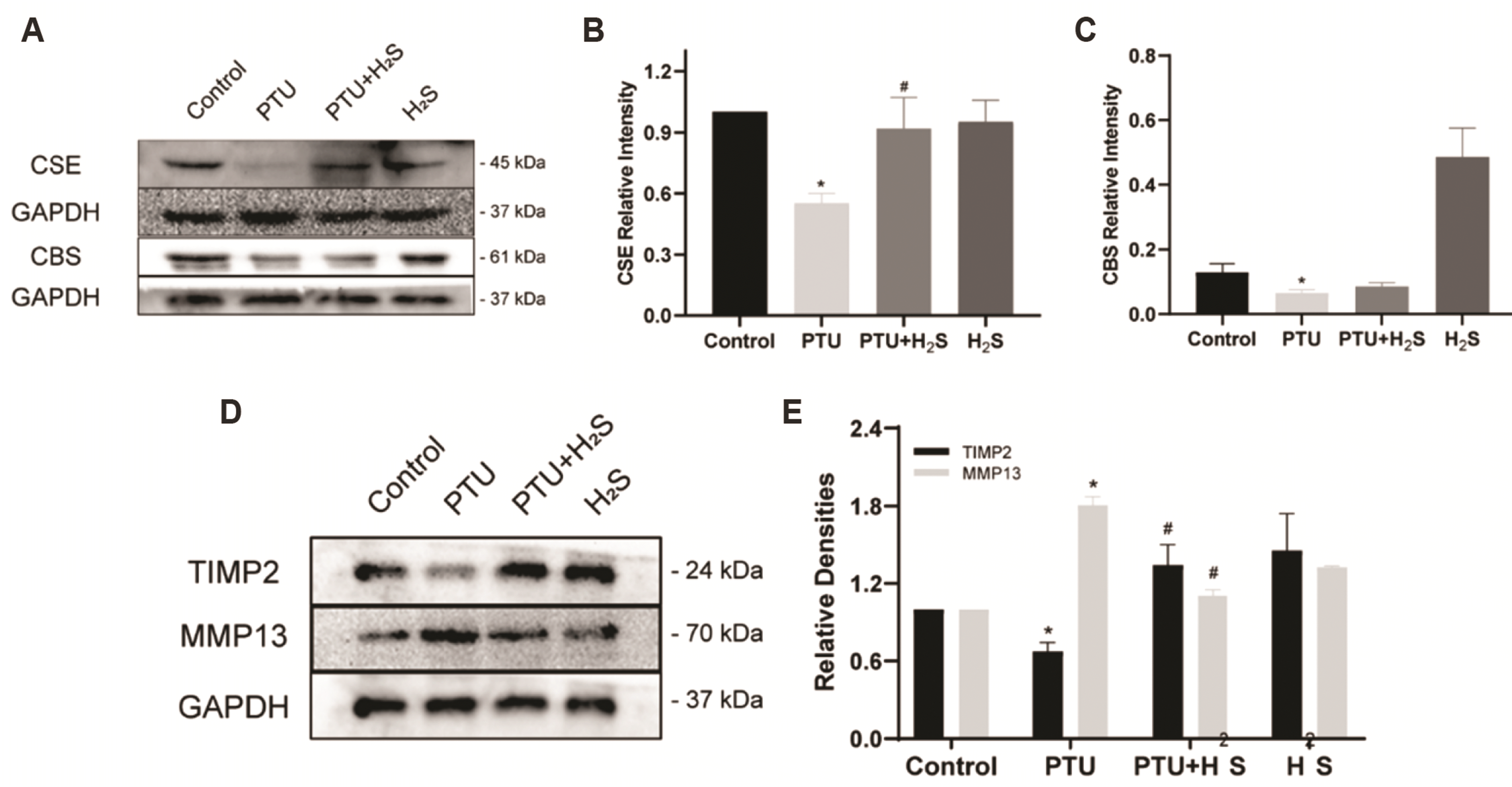 | Fig. 2Hydrogen sulfide (H2S) can enhance CSE protein expression and TIMP2/MMP13 protein expression ratio.(A–C) CSE and CBS protein expressions among four groups. (D, E) MMP13 and TIMP2 protein expressions among four groups. CSE, cystathionine-gamma-lyase; CBS, cystathionine β-synthase; MMP13, matrix metalloproteinase 13; TIMP2, tissue inhibitor of metalloproteinase 2; PTU, propylthiouracil. *p < 0.05 compared with Control group; #p < 0.05 compared with PTU group.
|
H2S can upregulate TIMP2/MMP13 protein expression ratio in hypothyroidism-induced myocardial fibrosis tissue
We employed Western blot to observe MMP13 and TIMP2 expressions in the heart tissue of rats in each group. As shown in Fig. 2D, E, MMP13 protein expression was upregulated (p < 0.05) and TIMP2 was downregulated (p < 0.05) in PTU group, while MMP13 was decreased (p < 0.05) and TIMP2 was increased (p < 0.05) in PTU+H2S group. These data suggest that H2S can up-regulate TIMP2/MMP13 ratio in fibrotic heart tissue of hypothyroidism-induced rats.
H2S can improve hypothyroidism-induced myocardial fibrosis by suppressing TGF-β1/Smad2 signal transduction pathway
We performed Western blot to determine TGF-β1, Smad2, and p-Smad2 expressions. As shown in Fig. 3A–C, these proteins expressions were upregulated in PTU group (p < 0.05) and downregulated in PTU+H2S group (p < 0.05). These data imply that inhibition of TGF-β1/Smad2 pathway is devoted to the process of H2S mitigating hypothyroidism-caused myocardial fibrosis.
 | Fig. 3Hydrogen sulfide (H2S) can inactivate the TGF-β1/Smad2 pathway activated by PTU and upregulate the autophagy downregulated by PTU.(A–C) TGF-β1 and Smad2, p-Smad2 protein expressions among four groups. (D–F) Atg4, Atg5, Beclin1, LC3II/I and P62 protein expressions among four groups. TGF-β1, transforming growth factor-β1; Smad2, SMAD family member 2; PTU, propylthiouracil. *p < 0.05 compared with Control group; #p < 0.05 compared with PTU group.
|
H2S can improve hypothyroidism-induced myocardial fibrosis by raising autophagy
We used Western blot to detect autophagy-related proteins expressions. As shown in Fig. 3D–F, Atg4, Atg5, Beclin1, LC3II/I expressions were reduced (p < 0.05) and P62 increased (p < 0.05) in PTU group, while Atg4, Atg5, Beclin1, LC3II/I expressions were upregulated (p < 0.05) and P62 downregulated (p < 0.05) in PTU+H2S group (p < 0.05). These data suggest that H2S can activate autophagy in hypothyroidism-induced fibrotic myocardial tissue in rats.
Go to :
DISCUSSION
It has been reported that hypothyroidism is highly related to various cardiovascular diseases, such as atrial fibrillation, myocardial fibrosis and heart failure [5,12,13]. However, classic long-term thyroid replacement therapy owns little ability to improve cardiac fibrotic situation [5]. H2S has been proven to possess the effect of anti-myocardial fibrosis after acute myocardial infarction in our previous experiments, and one of the main mechanisms is through upregulating TIMP2/MMP13 ratio [14]. So as to alleviate excessive damage of myocardial fibrous structure caused by overactivation of MMPs proteins [15]. Similar changes were found in our experiments, H2S alleviate the degree of hypothyroidism-induced myocardial fibrosis in SD rats, increase the TIMP2/MMP13 ratio and improve cardiac function. As well, H2S could relieve myocardial fibrosis, which usually comes from chronic inflammatory process, via exerting its anti-inflammatory and antioxidant effects [16,17]. Therefore, H2S could be a supplement for traditional thyroid replacement therapy to relieve hypothyroidism-induced myocardial fibrosis.
To further verify whether the improving effect is related to the endogenous H2S producing ability in heart tissue. In our experiment, we found that CSE protein expression level in heart tissue was recovered in the process of H2S alleviating hypothyroidism-induced myocardial fibrosis. Although, it was reported that knockdown of CSE gene did not significantly aggravate the chronic fibrotic response to myocardial infarction in mice [18], we assume the discrepancy is the results of the differences of animal modeling method and species. However, the fibrotic degree was alleviated after administration of NaHS, it was credited with decreasing inflammatory response by upregulating CSE expression [19]. Besides, we assumed that the CBS protein expression level was not altered in this improving process because CBS is not the main H2S producing enzyme in heart tissue, but brain tissue [20]. Hence, we can propose that impaired endogenous H2S generating ability was involved in the myocardial fibrosis process and restoration of this ability in heart tissue is involved in the process of exogenous H2S alleviating the myocardial fibrosis.
It has been reported that TSH stimulation could downregulate autophagy flux in PMCs [8]. Similarly, we found hypothyroidism could suppress autophagy in rat heart tissue in this experiment and H2S could ameliorate this pathological process. Also, activating autophagy could clear damaged organelles and extra collagen deposition to maintain cellular homeostasis so that severity of myocardial fibrosis and cardiac hypertrophy got alleviated [16,21,22]. H2S acts like a “switcher” in the autophagy process to regulates structure and function of myocardium in myocardial diseases [23]. On the other hand, some researchers showed that inhibition of excessive autophagy could improve cardiac fibrosis [24,25]. In these autophagic indicators, Beclin1 represents the initial stage of autophagy and interacts with multiple proteins in different stages [26,27]. Atg4 represents the autophagosome formation process [27], Atg5 represents the autophagosome elongation and closure [28], P62 acts as a cargo protein, loaded with the ubiquitylated target proteins, ready for delivery to lysosome degradation [29], which make it represent a biomarker of fusion and digestion in the autophagy process. As for LC3II/I, the transition from LC3I to LC3II, an autophagic membrane-bound form of LC3, plays a role the entire autophagic process [30,31]. Although the identification of autophagy is difficult to be defined in the field of myocardial fibrosis research [32], activating autophagy by H2S is an adaptive cellular energy metabolic processes in hypothyroidism-induced myocardial fibrosis. And the specific molecular regulating mechanism is needed to be further explored.
TGF-β1 signaling pathway, one of the commonly recognized key regulators on myocardial fibrosis, plays an essential role in the pathogenesis of myocardial fibrosiss [33-35]. It has been reported that H2S could act as an antioxidant against reactive oxygen species (ROS) in spontaneously hypertensive rats (SHR) [36], yet ROS biogenesis contributes to TGF-β1-mediated pro-fibrotic response [37]. In other experiments, administration of H2S could inhibit ROS production to mitigate TGF-β1 expression in injured renal caused by angiotensin converting enzyme (ACE) [38]. Addition of NaHS could ameliorate TGF-β expression through reducing ROS level to prevent lipopolysaccharide (LPS)-induced anxiety-like behavior deficits [39]. What is more, treatment of NaHS was able to decrease TGF-β1 expression level by repressing ROS generation so that hyperglycemia-mediated injured kidney got eased [40]. Thus, it could be speculated that H2S can inhibit TGF-β1 signal via the reduction of ROS. On the other hand, TGF-β1/Smad2 itself is a signaling pathway whose activation mediates a series of pathophysiological responses. Moreover, its stimulation could upregulate MMP2/9 level to aggravate myocardial fibrosis [41]. Thus it can be inferred that TGF-β1/Smad2 pathway activation could mediate down-regulation of TIMP2/MMP13. In our experiments, H2S could inhibit TGF-β1/Smad2 transduction signal pathway in PTU-induced myocardial fibrosis. Therefore, we can speculate that TGT-β1/Smad2 pathway is a potential therapeutic target and further suggest that H2S is a potential drug component for myocardial fibrosis.
In conclusion, our results not only demonstrate that H2S could relieve hypothyroidism-induced myocardial fibrosis by upregulating autophagy and inhibiting TGF-β1/Smad2 pathway, but also indicate that targeting TGF-β1/Smad2 pathway might be an effective strategy for treatment of myocardial fibrosis. Moreover, H2S could be a complement treatment for traditional hormone replacement therapy. To verify the authenticity of this inference, in our further experiments, further observe on the myocardial fibrosis and cardiac function of H2S on the basis of traditional hormone replacement therapy in different periods will be employed, and autophagy inhibitor and TGF-β1 Pathway activators will be adopted to verify its mechanism. When a mature H2S drug formulation is developed, we will apply for a clinical trial of H2S in patients with hypothyroidism.
Go to :
 XML Download