Abstract
The present case report describes an unusual variant of a missing medial tarsal artery (MTA) being replaced by the anterior medial malleolar artery (AMMA). The dorsalis pedis artery (DPA) did not give off any branches to the medial foot. The DPA coursed downward in the foot along with the superficial fibular nerve on the foot dorsum at the lateral side of the first metatarsal bone before entering the sole. In the medial malleolus, the AMMA arose from the anterior tibial artery and then ramified several branches, one of which descended to the medial talus. Thus, the possibility of a missing MTA being replaced by the AMMA should be considered by surgeons and radiologists when various surgeries are performed in the medial tarsal area.
Knowledge about the arterial variations around the ankle and foot is important for orthopedic and vascular surgeons and radiologists to prevent complications during surgical interventions [1,2]. The dorsalis pedis flap is one of the most commonly used foot flaps in traffic collisions, electrical burns, industrial injury, and ulcers. Variations in the origin, course, and branching pattern of the dorsalis pedis artery (DPA) have therefore been investigated for their clinical significance [1].
The arteries around the ankle and in the foot are the medial and lateral calcaneal arteries, medial and lateral plantar arteries, and the DPA. In the foot, arteries communicating between the DPA and the two plantar arteries give rise to multiple small perforators. The DPA gives rise to the tarsal, arcuate, and first dorsal metatarsal arteries [3].
The medial and lateral tarsal arteries arise as the DPA crosses the navicular bone. Two or three medial tarsal arteries (MTAs) ramify on the medial border of the foot and join the medial malleolar arterial network, whereas the lateral tarsal artery runs laterally under the extensor digitorum brevis, which supplies this muscle and the tarsal articulations, and anastomoses with branches of the arcuate, anterior lateral malleolar, and lateral plantar arteries, as well as the perorating fibular artery branch [3-5]. The MTA joins the anterior medial malleolar artery (AMMA) with the posterior medial malleolar artery and medial plantar artery branches [6]. However, there have been few descriptions of the coursing patterns and variations of the MTA. The present case report describes an unusual variant of a missing MTA being replaced by the AMMA.
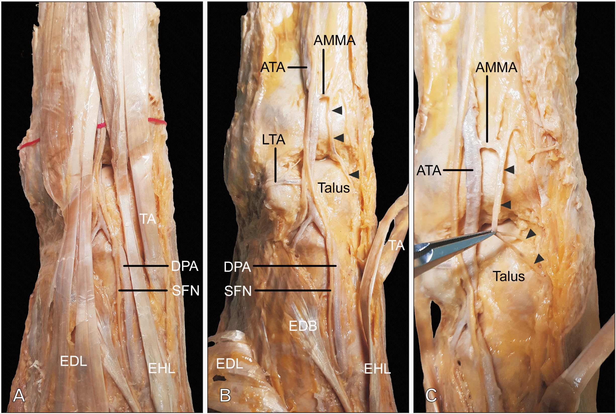
The MTA was found to be missing, and replaced by a branch of the AMMA in the right foot and ankle of a 77-year-old male cadaver with no history of trauma or surgical procedures to the ankle and foot. during routine dissection in a gross anatomy course (Fig. 1). The DPA did not give off any branches to the medial foot. The DPA coursed downward in the foot along with the superficial fibular nerve on the foot dorsum at the lateral side of the first metatarsal bone before entering the sole. In the medial malleolus, the AMMA arose from the anterior tibial artery and then ramified several branches, one of which descended to the medial talus. The lateral tarsal artery arose from the DPA just below the ankle joint and coursed on the lateral talus, giving off branches to supply the ankle joint and the lateral tarsal area.
In the left ankle and foot, both the MTA and lateral tarsal artery were present and the DPA was deviated laterally. The MTA was distinct and coursed downward obliquely on the medial tarsal dorsum.
The MTA was missing in the variant presented herein, and was replaced by a branch of the AMMA that descended to supply the area normally supplied by the MTA. The DPA coursed on the right side of the first metatarsal bone and did not give off branches that coursed on the metatarsal bone. The AMMA and the course of the DPA therefore appeared to replace the MTA.
The missing MTA replaced by the AMMA can be related with several clinical situations. Injury to the AMMA followed by fracture of the medial malleolus may cause ischemia in the medial foot and the medial malleolar arterial network. Fracture of the medial tarsal bones can result in injury to the distal part of the AMMA. Arterial variations including a missing MTA can be helpful during performing various surgeries including preoperative assessment using ultrasound at the flap in the medial tarsal area.
While the lateral tarsal and first metatarsal arteries are commonly used as flaps [7-10], the MTA has received less attention than the other DPA branches and there have been few studies of the anatomical features of the MTA. Ballmer et al. (1999) [6] found that the DPA gave two to four branches from its medial side, which were the MTA, and the average diameter at their origin was measured to be 0.4 mm (ranging from 0.2 to 0.7 mm). The branches given off at the cuneiform and navicular bone levels were the most prominent. When the artery that normally supplies an area or structure is missing, several changes have been reported adjacent to the normal location of the artery. Standring (2020) [3] suggested that the DPA could be larger than normal, to compensate for a small lateral plantar artery. Moriggl and Sturm (1996) [11] found that three missing regular thyroid arteries were replaced by an unusual lowest thyroid artery (thyroidea ima artery). Standring (2020) [3] also described that the sizes of the anterior jugular vein and external jugular vein have in inverse relationship in their sizes. Vas et al. (2022) [12] also presented a case of a replaced right hepatic artery that originated from the right distal renal artery.
Tsukuura and Yamamoto (2021) [13] stated that the medial tarsal area is a commonly used donor site for skin grafts to the digits and toes, with good color and texture matches as well as a concealable donor site scar. They also found that a medialis tarsus flap could be used as a true perforator flap with perforator-to-perforator anastomosis to reconstruct a soft tissue defect of the dorsal toe. The variant of the present study was located in the similar area. Thus, the possibility of a missing MTA being replaced by the AMMA should be considered by surgeons and radiologists when various surgeries are performed in the medial tarsal area.
Further research with larger numbers of specimens is necessary to determine the prevalence, course, branching pattern, and variations of the MTA and its connections with adjacent arteries in order to map arteries as preoperative data.
Acknowledgements
This work was supported by the National Research Foundation of Korea (NRF) grant funded by the Korea government (MSIT) (No. 2020R1C1C1003237).
Notes
References
1. Manjunatha HN. Hemamalini. 2021; Variations in the origin, course and branching pattern of dorsalis pedis artery with clinical significance. Sci Rep. 11:1448. DOI: 10.1038/s41598-020-80555-z. PMID: 33446776. PMCID: PMC7809105. PMID: 63c6925873f448a9bfa71b4f07055195.
2. Thunyacharoen S, Mahakkanukrauh C, Pattayakornkul N, Meetham K, Charumporn T, Mahakkanukrauh P. 2022; Anatomical variations of the dorsalis pedis artery in a Thai population. Int J Morphol. 40:137–42. DOI: 10.4067/S0717-95022022000100137.
3. Standring S. 2020. Gray's anatomy: the anatomical basis of clinical practice. 42nd ed. Elsevier;Amsterdam: DOI: 10.4067/s0717-95022022000100137.
4. Morris H. 1947. Morris' human anatomy: a complete systematic treatise. 10th ed. Blakiston;Philadelphia: DOI: 10.4067/s0717-95022022000100137.
5. Woodburne RT, Burkel WE. 1994. Essentials of human anatomy. 9th ed. Oxford University Press;New York: DOI: 10.4067/s0717-95022022000100137.
6. Ballmer FT, Hertel R, Noetzli HP, Masquelet AC. 1999; The medial malleolar network: a constant vascular base of the distally based saphenous neurocutaneous island flap. Surg Radiol Anat. 21:297–303. DOI: 10.1007/BF01631327. PMID: 10635091.
7. Wang T, Regmi S, Liu H, Pan J, Hou R. 2017; Free lateral tarsal artery perforator flap with functioning extensor digitorum brevis muscle for thenar reconstruction: a case report. Arch Orthop Trauma Surg. 137:273–6. DOI: 10.1007/s00402-016-2615-5. PMID: 28005165.
8. Vazales R, Masadeh S. 2020; First dorsal metatarsal artery flap for coverage of soft tissue defects of the distal foot: delayed technique, proximal and distally based fasciocutaneous and adipofascial variants. Clin Podiatr Med Surg. 37:765–73. DOI: 10.1016/j.cpm.2020.07.001. PMID: 32919603.
9. Bai L, Peng YB, Liu SB, Xie XX, Zhang XM. 2021; Anatomical basis of a pedicled cuboid bone graft based on the lateral tarsal artery for talar avascular necrosis. Surg Radiol Anat. 43:1703–9. DOI: 10.1007/s00276-021-02789-4. PMID: 34232369.
10. Abu-Ghname A, Lazo D, Chade S, Fioravanti A, Colicchio O, Alvarez D, Junior E, Maricevich M. 2022; The first dorsal metacarpal artery perforator free flap: The Comet Flap. Plast Reconstr Surg. 150:671e–4e. DOI: 10.1097/PRS.0000000000009403. PMID: 35791443.
11. Moriggl B, Sturm W. 1996; Absence of three regular thyroid arteries replaced by an unusual lowest thyroid artery (A. thyroidea ima): a case report. Surg Radiol Anat. 18:147–50. DOI: 10.1007/BF01795238. PMID: 8782323.
12. Vas D, Moreno Rojas J, Solà Garcia M. 2022; Sep. 13. Replaced right hepatic artery arising from the distal renal artery, a new variation. Surg Radiol Anat. [Epub]. https://doi.org/10.1007/s00276-022-03017-3. DOI: 10.1007/s00276-022-03017-3. PMID: 36097082.
13. Tsukuura R, Yamamoto T. 2021; Free medialis tarsus flap transfer for reconstruction of toe necrosis: a case report. Microsurgery. 41:671–5. DOI: 10.1002/micr.30780. PMID: 34156111.
Fig. 1
A MTA replaced by the AMMA. (A) The DPA coursed downward in the foot just lateral to the EHL tendon. (B) The DPA coursed downward along with the SFN on the dorsum of the foot on the lateral side of the first metatarsal bone. The MTA was found to be missing. The DPA did not give off any branches to the medial foot. The EDL tendon was reflected laterally, and the EHL and TA tendons were reflected medially. The AMMA arose from the ATA and ramified several branches, one (arrowheads) of which descended to the medial talus. EDB, extensor digitorum brevis. (C) One branch (arrowheads) of the AMMA that supplied the medial talus was indicated by an instrument. MTA, missing medial tarsal artery; AMMA, anterior medial malleolar artery; DPA, dorsalis pedis artery; EHL, extensor hallucis longus; SFN, superficial fibular nerve; EDL, extensor digitorum longus; TA, tibialis anterior; ATA, anterior tibial artery.


 PDF
PDF Citation
 Print
Print


 XML Download