

 PDF
PDF Citation
 Print
Print


Introduction
Between the inferior margin of the orbicularis oculi (OOc) and orbicularis oris (OOr) muscles, the zygomaticus major (Zmj), zygomaticus minor (Zmi), levator anguli oris (LAO), levator labii superioris (LLS), and levator labii superioris alaeque nasi (LLSAN) muscles are found [1]. In general, the OOr and OOc are considered independent muscles that work separately. However, variations have been reported in this area, e.g., different insertion patterns of Zmi [2, 3] and Zmj [4]. However, to our knowledge, variant muscle fibers connecting the inferior margin of the OOc to the OOr have not been reported. Here, we report such an anatomical variation.
Go to : 
Case Report
During routine dissection of the face, connecting fibers between the inferior margin of the OOc and the OOr were found in a cadaver of a 56-year-old at death male (Fig. 1). The skin just lateral to the nasolabial fold was removed to reveal the connecting fibers and the adjacent facial muscles passing beneath the nasolabial fold. The remaining skin was reflected to observe the course and attachments of the connecting fibers and facial muscles (Fig. 1A). Next, the remaining skin was removed to expose the entire muscles of the face (Fig. 1B). The inferior margin of the OOc was reflected superiorly to observe the course and attachment of the connecting fibers (Fig. 1C).
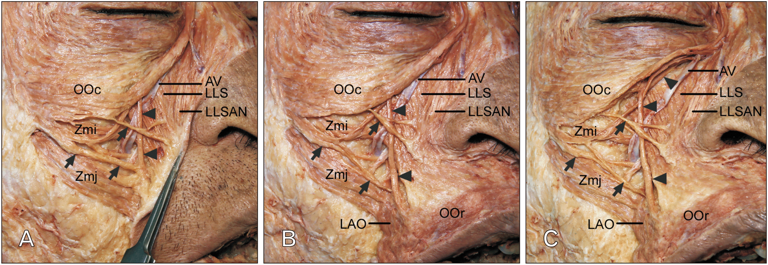 | Fig. 1Connecting fibers between the inferior margin of the OOc and OOr. (A) At the middle of the inferior margin of the OOc, the connecting fibers (arrowheads) descended perpendicularly and passed beneath the site between the middle and the lower thirds of the nasolabial fold. Adjacent to the connecting fibers, there were several fibers (arrows) from the OOc attaching along the nasolabial fold. Skin just lateral to the nasolabial fold was removed and the remaining skin was reflected to reveal the courses and attachments of the connecting fibers and the adjacent extending fibers from the OOc. (B) The connecting fibers (arrowheads) blended with the OOr between the inserting fibers of the LLS and LAO. The several fibers (arrows) from the OOc were toward the LLSAN, LLS, and Zmi. The remaining skin was removed to expose the entire muscles in the face. (C) The extending fibers of the OOc that divided the connecting fibers (arrowheads) were attached to the maxilla just above the origin site of the LLS. The inferior margin of the OOc were reflected superiorly to reveal the course and attachment of the connecting fibers. AV, angular vein; LAO, levator anguli oris; LLS, levator labii superioris; LLSAN, lavetor labii superioris alaeque nasi; OOc, orbicularis oculi; OOr, orbicularis oris; Zmi, zygomaticus minor; Zmj, zygomaticus major.
|
At the middle of the inferior margin of the OOc, some extending fibers of the OOc descended perpendicularly and passed beneath the site between the middle and the lower thirds of the nasolabial fold. Adjacent to the connecting fibers, there were several fibers from the OOc attaching along the nasolabial fold. These several fibers that extended from the OOc were attached to the dermis of the middle of the nasolabial fold or the site between the middle and lower thirds of the nasolabial fold. The connecting fibers coursed deep or superficial to these several fibers from the OOc and coursed superficial to the angular vein (Fig. 1A). The connecting fibers blended with the OOr between the inserting fibers of the LLS and LAO. The several fibers from the OOc were toward the LLSAN, LLS, and Zmi (Fig. 1B). The extending fibers of the OOc that divided the connecting fibers were attached to the maxilla just above the origin site of the LLS. The connecting fibers between the inferior margin of the OOc and OOr and the adjacent muscle fibers extending from the OOc were at the level of the dorsum of the nose to the level of the middle of the upper lip above the corner of the mouth (Fig. 1C). At the corresponding site of the contralateral side of the face, some of the middle inferior margin of the OOc extended to the Zmi obliquely and connecting fibers between the inferior fibers of the OOc and the OOr were not found. No other variations in the dissection field were identified. The authors state that every effort was made to follow all local and international ethical guidelines and laws that pertain to the use of human cadaveric donors in anatomical research [5].
Go to :
Discussion
The connecting fibers and their adjacent muscle fibers were attached along the nasolabial fold in the present study. The muscles of facial expression are often interdigitated with adjacent muscles [1, 6-8]. Additionally, peripheral fibers of the OOc overlie and occasionally blend with the muscles arising from the bones of the orbital rim [9].
The facial muscles responsible for producing the nasolabial fold have been discussed by several authors [4, 10-12]. Most authors have reported that the LLSAN inserted into the medial nasolabial fold, the LLS inserted into the middle third of the nasolabial fold, the Zmj inserted into the lateral nasolabial fold. Hur et al. [3] concluded that fibers extending from the OOc to the Zmi assist in deepening and elevating the nasolabial fold. In the present case, the connecting fibers passed deep to the site between the middle and lower thirds of the nasolabial fold, and their adjacent fibers that extended from the OOc were attached to the middle of the nasolabial fold or the site between the middle and lower thirds of the nasolabial fold. Contraction of the connecting fibers and adjacent muscle fibers extending from the OOc might also assist in deepening the nasolabial fold. In addition, the connecting fibers blended with the upper OOr where the LAO inserted at the corner of the mouth. Contraction of the connecting fibers might raise the corner of the mouth via contraction of the OOc.
The lower half of the orbital portion of the OOc raises the skin of the cheek, causing the wrinkles seen to radiate from the corner of the eye [13]. Thus, the connecting fibers and extending fibers from the OOc to the upper lip might assist in elevating the OOr. The OOc and OOr have different actions and distant sites. The OOc is a sphincteric muscle of the eyelids and plays an important role in facial expression, whereas contraction of the OOr causes narrowing of the mouth [1, 14]. The connecting fibers between the inferior margin of the OOc and OOr seen in the present case might assist in simultaneous movements of the eye and mouth.
Connecting fibers between the inferior margin of the OOc and OOr is a rare anatomical variation. Such a variant muscle of the face could possibly affect facial expression and when found unilaterally, this could result in asymmetry of facial expression.
Go to :
 XML Download